Loading
PDBj
MenuPDBj@FacebookPDBj@TwitterPDBj@YouTubewwPDB FoundationwwPDB
RCSB PDBPDBeBMRBAdv. SearchSearch help
Search by PDB author
7N7S
DownloadVisualize
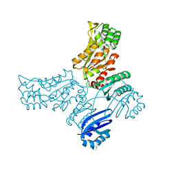
BU of 7n7s by Molmil
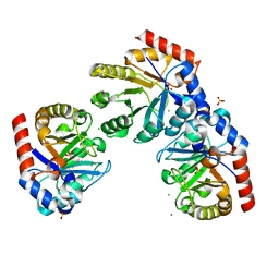
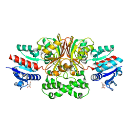
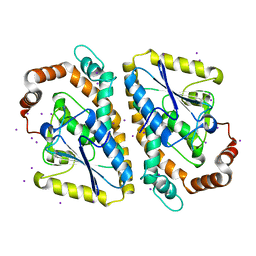
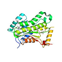
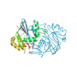
Crystal Structure of Hydroxymethylglutaryl-CoA reductase from Elizabethkingia anophelis NUHP1
Descriptor: 1,2-ETHANEDIOL, CITRIC ACID, Hydroxymethylglutaryl-CoA reductase, ...
Authors:Seattle Structural Genomics Center for Infectious Disease, Seattle Structural Genomics Center for Infectious Disease (SSGCID)
Deposit date:2021-06-11
Release date:2021-06-23
Last modified:2024-04-03
Method:X-RAY DIFFRACTION (2.4 Å)
Cite:Crystal Structure of Hydroxymethylglutaryl-CoA reductase from Elizabethkingia anophelis NUHP1
To be published
7N56
DownloadVisualize
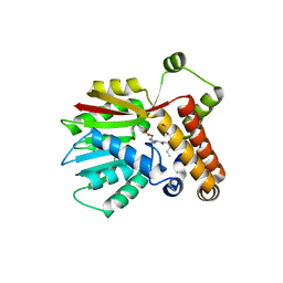
BU of 7n56 by Molmil
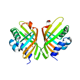
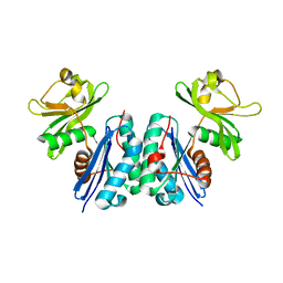
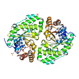
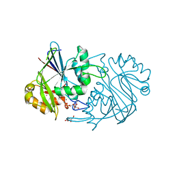
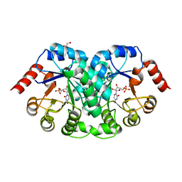
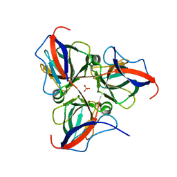
Crystal Structure of deoxyuridine 5'-triphosphate nucleotidohydrolase from Rickettsia prowazekii str. Madrid E
Descriptor: Deoxyuridine 5'-triphosphate nucleotidohydrolase, SULFATE ION
Authors:Seattle Structural Genomics Center for Infectious Disease (SSGCID)
Deposit date:2021-06-04
Release date:2021-07-07
Last modified:2023-10-18
Method:X-RAY DIFFRACTION (2.15 Å)
Cite:Crystal Structure of deoxyuridine 5'-triphosphate nucleotidohydrolase from Rickettsia prowazekii str. Madrid E
to be published
7S5N
DownloadVisualize
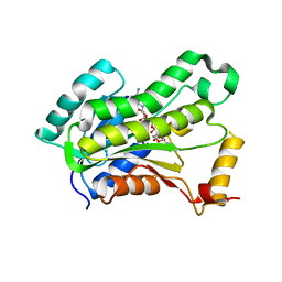
BU of 7s5n by Molmil
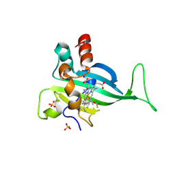
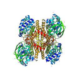
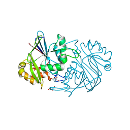
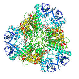
Crystal Structure of a Putative uncharacterized protein from Mycobacterium marinum
Descriptor: (4S)-2-METHYL-2,4-PENTANEDIOL, MymaA.17060.a, SULFATE ION
Authors:Seattle Structural Genomics Center for Infectious Disease (SSGCID)
Deposit date:2021-09-10
Release date:2021-09-22
Last modified:2024-04-03
Method:X-RAY DIFFRACTION (1.85 Å)
Cite:Crystal Structure of a Putative uncharacterized protein from Mycobacterium marinum
to be published
7S5M
DownloadVisualize
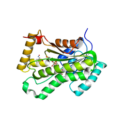
BU of 7s5m by Molmil
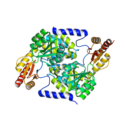
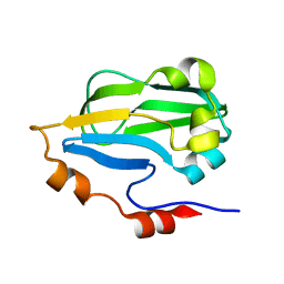
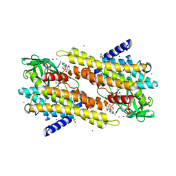
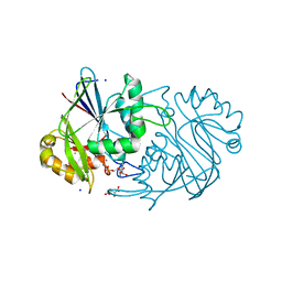
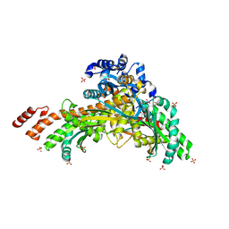
Crystal Structure of a 8-amino-7-oxononanoate synthase/2-amino-3-ketobutyrate coenzyme A ligase from Mycobacterium smegmatis
Descriptor: 8-amino-7-oxononanoate synthase
Authors:Seattle Structural Genomics Center for Infectious Disease (SSGCID)
Deposit date:2021-09-10
Release date:2021-09-22
Last modified:2023-10-18
Method:X-RAY DIFFRACTION (2.25 Å)
Cite:Crystal Structure of a 8-amino-7-oxononanoate synthase/2-amino-3-ketobutyrate coenzyme A ligase from Mycobacterium smegmatis
to be published
7SIR
DownloadVisualize
BU of 7sir by Molmil
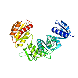
Crystal Structure of UDP-N-acetylmuramoylalanine-D-glutamate ligase from Acinetobacter baumannii AB5075-UW
Descriptor: FORMIC ACID, UDP-N-acetylmuramoylalanine--D-glutamate ligase
Authors:Seattle Structural Genomics Center for Infectious Disease, Seattle Structural Genomics Center for Infectious Disease (SSGCID)
Deposit date:2021-10-14
Release date:2021-11-03
Last modified:2023-10-18
Method:X-RAY DIFFRACTION (1.65 Å)
Cite:Crystal Structure of UDP-N-acetylmuramoylalanine-D-glutamate ligase from Acinetobacter baumannii AB5075-UW
to be published
7SBJ
DownloadVisualize
BU of 7sbj by Molmil
Crystal Structure of Ribulose-phosphate 3-epimerase from Stenotrophomonas maltophilia K279a
Descriptor: CHLORIDE ION, MAGNESIUM ION, Ribulose-phosphate 3-epimerase, ...
Authors:Seattle Structural Genomics Center for Infectious Disease (SSGCID)
Deposit date:2021-09-25
Release date:2021-10-27
Last modified:2023-10-18
Method:X-RAY DIFFRACTION (1.85 Å)
Cite:Crystal Structure of Ribulose-phosphate 3-epimerase from Stenotrophomonas maltophilia K279a
to be published
7SKB
DownloadVisualize
BU of 7skb by Molmil
Crystal Structure of aspartate-semialdehyde dehydrogenase from Acinetobacter baumannii
Descriptor: Aspartate-semialdehyde dehydrogenase, L(+)-TARTARIC ACID
Authors:Seattle Structural Genomics Center for Infectious Disease (SSGCID)
Deposit date:2021-10-20
Release date:2021-11-03
Last modified:2023-10-18
Method:X-RAY DIFFRACTION (1.8 Å)
Cite:Crystal Structure of aspartate-semialdehyde dehydrogenase from Acinetobacter baumannii
to be published
5BR9
DownloadVisualize
BU of 5br9 by Molmil
Crystal structure of an uncharacterized protein with similarity to peptidase YEAZ from Pseudomonas aeruginosa
Descriptor: Uncharacterized protein
Authors:Seattle Structural Genomics Center for Infectious Disease (SSGCID)
Deposit date:2015-05-29
Release date:2015-07-15
Last modified:2023-09-27
Method:X-RAY DIFFRACTION (2.35 Å)
Cite:Crystal structure of an uncharacterized protein with similarity to peptidase YEAZ from Pseudomonas aeruginosa
to be published
7S5O
DownloadVisualize
BU of 7s5o by Molmil
Crystal structure of Cytochrome c' beta from Nitrosomonas europaea ATCC 19718
Descriptor: ACETATE ION, Cytochrome_P460 domain-containing protein, HEME C, ...
Authors:Seattle Structural Genomics Center for Infectious Disease (SSGCID)
Deposit date:2021-09-11
Release date:2022-03-30
Last modified:2022-04-20
Method:X-RAY DIFFRACTION (1.8 Å)
Cite:Structural Characterization of Cytochrome c ' beta-Met from an Ammonia-Oxidizing Bacterium.
Biochemistry, 61, 2022
3HGB
DownloadVisualize
BU of 3hgb by Molmil
Crystal structure of glycine cleavage system protein H from Mycobacterium tuberculosis
Descriptor: Glycine cleavage system H protein
Authors:Seattle Structural Genomics Center for Infectious Disease (SSGCID)
Deposit date:2009-05-13
Release date:2009-05-26
Last modified:2023-09-06
Method:X-RAY DIFFRACTION (1.75 Å)
Cite:X-ray structure determination of the glycine cleavage system protein H of Mycobacterium tuberculosis using an inverse Compton synchrotron X-ray source.
J.Struct.Funct.Genom., 11, 2010
3NJB
DownloadVisualize
BU of 3njb by Molmil
Crystal structure of enoyl-coa hydratase from Mycobacterium smegmatis, iodide soak
Descriptor: Enoyl-CoA hydratase, IODIDE ION
Authors:Seattle Structural Genomics Center for Infectious Disease (SSGCID)
Deposit date:2010-06-17
Release date:2010-07-28
Last modified:2024-02-21
Method:X-RAY DIFFRACTION (2.2 Å)
Cite:SAD phasing using iodide ions in a high-throughput structural genomics environment.
J.Struct.Funct.Genom., 12, 2011
4WSH
DownloadVisualize
BU of 4wsh by Molmil
Crystal structure of Probable Uroporphyrinogen decarboxylase (UPD) (URO-D) from Pseudomonas aeruginosa
Descriptor: CHLORIDE ION, SULFATE ION, Uroporphyrinogen decarboxylase
Authors:Seattle Structural Genomics Center for Infectious Disease (SSGCID)
Deposit date:2014-10-27
Release date:2014-11-19
Last modified:2023-12-27
Method:X-RAY DIFFRACTION (1.95 Å)
Cite:Crystal structure of Probable Uroporphyrinogen decarboxylase (UPD) (URO-D) from Pseudomonas aeruginosa
to be published
3PFD
DownloadVisualize
BU of 3pfd by Molmil
Crystal structure of an Acyl-CoA dehydrogenase from Mycobacterium thermoresistibile bound to reduced flavin adenine dinucleotide solved by combined iodide ion SAD MR
Descriptor: Acyl-CoA dehydrogenase, DIHYDROFLAVINE-ADENINE DINUCLEOTIDE, IODIDE ION
Authors:Seattle Structural Genomics Center for Infectious Disease (SSGCID)
Deposit date:2010-10-28
Release date:2010-11-10
Last modified:2023-09-06
Method:X-RAY DIFFRACTION (2.1 Å)
Cite:SAD phasing using iodide ions in a high-throughput structural genomics environment.
J.STRUCT.FUNCT.GENOM., 12, 2011
3OIB
DownloadVisualize
BU of 3oib by Molmil
Crystal structure of a putative ACYL-COA Dehydrogenase from mycobacterium smegmatis, Iodide soak
Descriptor: 1,2-ETHANEDIOL, Acyl-CoA dehydrogenase, DIHYDROFLAVINE-ADENINE DINUCLEOTIDE, ...
Authors:Seattle Structural Genomics Center for Infectious Disease (SSGCID)
Deposit date:2010-08-18
Release date:2010-09-01
Last modified:2024-05-22
Method:X-RAY DIFFRACTION (2.1 Å)
Cite:SAD phasing using iodide ions in a high-throughput structural genomics environment.
J Struct Funct Genomics, 12, 2011
3P96
DownloadVisualize
BU of 3p96 by Molmil
Crystal structure of Phosphoserine phosphatase SerB from Mycobacterium avium, native form
Descriptor: 1,2-ETHANEDIOL, CHLORIDE ION, MAGNESIUM ION, ...
Authors:Seattle Structural Genomics Center for Infectious Disease (SSGCID)
Deposit date:2010-10-15
Release date:2010-11-10
Last modified:2024-02-21
Method:X-RAY DIFFRACTION (2.05 Å)
Cite:SAD phasing using iodide ions in a high-throughput structural genomics environment.
J.Struct.Funct.Genom., 12, 2011
7MCJ
DownloadVisualize
BU of 7mcj by Molmil
Crystal Structure of S-adenosylmethionine-dependent methyltransferase UmaA from Mycobacterium tuberculosis in complex with compound 8918
Descriptor: 1,2-ETHANEDIOL, CHLORIDE ION, MAGNESIUM ION, ...
Authors:Seattle Structural Genomics Center for Infectious Disease (SSGCID)
Deposit date:2021-04-02
Release date:2022-04-13
Last modified:2023-10-18
Method:X-RAY DIFFRACTION (1.8 Å)
Cite:Crystal Structure of S-adenosylmethionine-dependent methyltransferase UmaA from Mycobacterium tuberculosis in complex with compound 8918
to be published
5VRM
DownloadVisualize
BU of 5vrm by Molmil
CRYSTAL STRUCTURE OF THE INHA FROM MYCOBACTERIUM TUBERCULOSIS IN COMPLEX WITH AN12855, EBSI 4333.
Descriptor: Enoyl-[acyl-carrier-protein] reductase [NADH], [[(2~{R},3~{S},4~{R},5~{R})-5-(3-aminocarbonylpyridin-1-ium-1-yl)-3-oxidanyl-4-[[1-oxidanyl-6-[4-(trifluoromethyl)phenoxy]-3~{H}-2,1$l^{4}-benzoxaborol-1-yl]oxy]oxolan-2-yl]methoxy-oxidanyl-phosphoryl] [(2~{R},3~{S},4~{R},5~{R})-5-(6-aminopurin-9-yl)-3,4-bis(oxidanyl)oxolan-2-yl]methyl hydrogen phosphate
Authors:Abendroth, J, Edwards, T.E, Lorimer, D.
Deposit date:2017-05-11
Release date:2018-05-16
Last modified:2024-03-13
Method:X-RAY DIFFRACTION (2.5 Å)
Cite:Discovery of a cofactor-independent inhibitor ofMycobacterium tuberculosisInhA.
Life Sci Alliance, 1, 2018
5VRN
DownloadVisualize
BU of 5vrn by Molmil
CRYSTAL STRUCTURE OF THE INHA FROM MYCOBACTERIUM TUBERCULOSIS IN COMPLEX WITH AN12855, EBSI 4333.
Descriptor: Enoyl-[acyl-carrier-protein] reductase [NADH], [[(2~{R},3~{S},4~{R},5~{R})-5-(3-aminocarbonylpyridin-1-ium-1-yl)-4-[[5-[4-cyano-2-[(~{E})-hydroxyiminomethyl]phenoxy]-1-oxidanyl-3~{H}-2,1$l^{4}-benzoxaborol-1-yl]oxy]-3-oxidanyl-oxolan-2-yl]methoxy-oxidanyl-phosphoryl] [(2~{R},3~{S},4~{R},5~{R})-5-(6-aminopurin-9-yl)-3,4-bis(oxidanyl)oxolan-2-yl]methyl hydrogen phosphate
Authors:Abendroth, J, Edwards, T.E, Lorimer, D.
Deposit date:2017-05-11
Release date:2018-05-16
Last modified:2024-03-13
Method:X-RAY DIFFRACTION (2.55 Å)
Cite:Discovery of a cofactor-independent inhibitor ofMycobacterium tuberculosisInhA.
Life Sci Alliance, 1, 2018
5VRL
DownloadVisualize
BU of 5vrl by Molmil
CRYSTAL STRUCTURE OF THE INHA FROM MYCOBACTERIUM TUBERCULOSIS IN COMPLEX WITH AN12855, EBSI 4333.
Descriptor: (~{N}~{E})-~{N}-[[2-[[2-ethylsulfonyl-1,1-bis(oxidanyl)-3,4-dihydro-2,3,1$l^{4}-benzodiazaborinin-7-yl]oxy]-5-(trifluoromethyl)phenyl]methylidene]hydroxylamine, Enoyl-[acyl-carrier-protein] reductase [NADH]
Authors:Abendroth, J, Edwards, T.E, Lorimer, D.
Deposit date:2017-05-11
Release date:2018-05-16
Last modified:2024-03-13
Method:X-RAY DIFFRACTION (2.65 Å)
Cite:Discovery of a cofactor-independent inhibitor ofMycobacterium tuberculosisInhA.
Life Sci Alliance, 1, 2018
3PY6
DownloadVisualize
BU of 3py6 by Molmil
Crystal Structure of a Beta-Lactamase-Like Protein from Brucella Melitensis bound to GMP
Descriptor: Beta-lactamase-like, D(-)-TARTARIC ACID, GUANOSINE-5'-MONOPHOSPHATE, ...
Authors:Seattle Structural Genomics Center for Infectious Disease (SSGCID)
Deposit date:2010-12-11
Release date:2010-12-22
Last modified:2023-09-13
Method:X-RAY DIFFRACTION (1.7 Å)
Cite:Crystal Structure of a Beta-Lactamase-Like Protein from Brucella Melitensis bound to GMP
Acta Crystallogr.,Sect.F, 67, 2011
3PY5
DownloadVisualize
BU of 3py5 by Molmil
Crystal structure of a beta-lactamase-like protein from brucella melitensis bound to AMP
Descriptor: ADENOSINE MONOPHOSPHATE, Beta-lactamase-like, L(+)-TARTARIC ACID, ...
Authors:Seattle Structural Genomics Center for Infectious Disease (SSGCID)
Deposit date:2010-12-11
Release date:2010-12-29
Last modified:2023-09-13
Method:X-RAY DIFFRACTION (1.7 Å)
Cite:BrabA.11339.a: anomalous diffraction and ligand binding guide towards the elucidation of the function of a `putative beta-lactamase-like protein from Brucella melitensis.
Acta Crystallogr.,Sect.F, 67, 2011
3MD7
DownloadVisualize
BU of 3md7 by Molmil
Crystal structure of a beta-lactamase-like protein bound to GMP from brucella melitensis
Descriptor: Beta-lactamase-like, GUANOSINE-5'-MONOPHOSPHATE, L(+)-TARTARIC ACID, ...
Authors:Seattle Structural Genomics Center for Infectious Disease (SSGCID)
Deposit date:2010-03-30
Release date:2010-04-21
Last modified:2024-04-03
Method:X-RAY DIFFRACTION (1.27 Å)
Cite:BrabA.11339.a: anomalous diffraction and ligand binding guide towards the elucidation of the function of a `putative beta-lactamase-like protein from Brucella melitensis.
Acta Crystallogr.,Sect.F, 67, 2011
3QH8
DownloadVisualize
BU of 3qh8 by Molmil
Crystal structure of a beta-lactamase-like protein bound to AMP from brucella melitensis, long wavelength synchrotron data
Descriptor: 1,2-ETHANEDIOL, ADENOSINE MONOPHOSPHATE, Beta-lactamase-like, ...
Authors:Seattle Structural Genomics Center for Infectious Disease (SSGCID)
Deposit date:2011-01-25
Release date:2011-02-09
Last modified:2023-09-13
Method:X-RAY DIFFRACTION (1.6 Å)
Cite:Crystal structure of a Beta-Lactamase-Like Protein bound to AMP from Brucella Melitensis, long wavelength synchrotron data
Acta Crystallogr.,Sect.F, 67, 2011
8CSO
DownloadVisualize
BU of 8cso by Molmil
Crystal Structure of Orotidine 5'-phosphate decarboxylase from Klebsiella pneumoniae in complex with Uridine-5'-monophosphate
Descriptor: 1,2-ETHANEDIOL, Orotidine 5'-phosphate decarboxylase, URIDINE-5'-MONOPHOSPHATE
Authors:Seattle Structural Genomics Center for Infectious Disease, Seattle Structural Genomics Center for Infectious Disease (SSGCID)
Deposit date:2022-05-13
Release date:2022-06-08
Last modified:2023-10-18
Method:X-RAY DIFFRACTION (2.6 Å)
Cite:Crystal Structure of Orotidine 5'-phosphate decarboxylase from Klebsiella pneumoniae in complex with Guanosine-5'-monophosphate
to be published
8D1X
DownloadVisualize
BU of 8d1x by Molmil
Crystal Structure of aminopeptidase A from Neisseria gonorrhoeae
Descriptor: ACETATE ION, CHLORIDE ION, D(-)-TARTARIC ACID, ...
Authors:Seattle Structural Genomics Center for Infectious Disease, Seattle Structural Genomics Center for Infectious Disease (SSGCID)
Deposit date:2022-05-27
Release date:2022-06-08
Last modified:2023-10-18
Method:X-RAY DIFFRACTION (2.8 Å)
Cite:Crystal Structure of aminopeptidase A from Neisseria gonorrhoeae
to be published

224201

PDB entries from 2024-08-28

PDB statisticsPDBj update infoContact PDBjnumon