Loading
PDBj
MenuPDBj@FacebookPDBj@TwitterPDBj@YouTubewwPDB FoundationwwPDB
RCSB PDBPDBeBMRBAdv. SearchSearch help
Search by PDB author
7QLM
DownloadVisualize
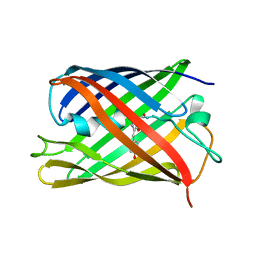
BU of 7qlm by Molmil
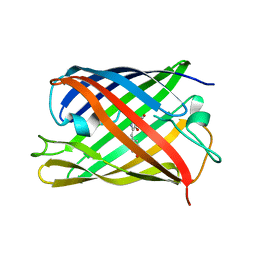
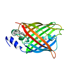
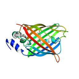
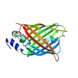
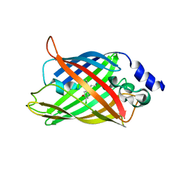
rsKiiro trans chromophore dark structure by SFX
Descriptor: rsKiiro
Authors:van Thor, J.J.
Deposit date:2021-12-20
Release date:2023-09-27
Last modified:2023-11-15
Method:X-RAY DIFFRACTION (1.3 Å)
Cite:Optical control of ultrafast structural dynamics in a fluorescent protein.
Nat.Chem., 15, 2023
7QLO
DownloadVisualize
BU of 7qlo by Molmil
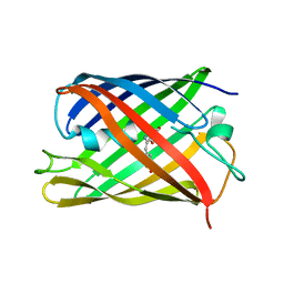
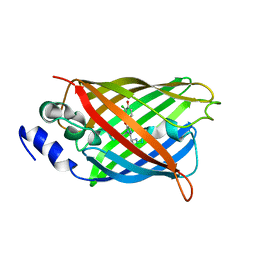
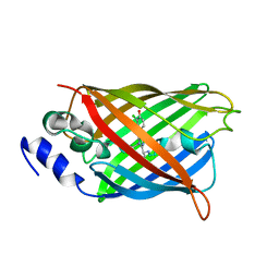
rsKiiro pump dump probe structure by TR-SFX
Descriptor: rsKiiro
Authors:van Thor, J.J.
Deposit date:2021-12-20
Release date:2023-10-18
Last modified:2023-11-15
Method:X-RAY DIFFRACTION (1.31 Å)
Cite:Optical control of ultrafast structural dynamics in a fluorescent protein.
Nat.Chem., 15, 2023
7QLN
DownloadVisualize
BU of 7qln by Molmil
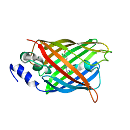
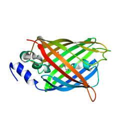
rsKiiro pump probe structure by TR-SFX
Descriptor: rsKiiro
Authors:van Thor, J.J.
Deposit date:2021-12-20
Release date:2023-10-18
Last modified:2023-11-15
Method:X-RAY DIFFRACTION (1.5 Å)
Cite:Optical control of ultrafast structural dynamics in a fluorescent protein.
Nat.Chem., 15, 2023
8A6O
DownloadVisualize
BU of 8a6o by Molmil
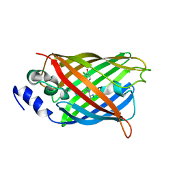
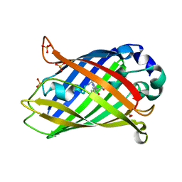
Room temperature rsEGFP2 with a chlorinated chromophore 600 fs after Photoexcitation
Descriptor: Green fluorescent protein
Authors:Fadini, A, van Thor, J.
Deposit date:2022-06-18
Release date:2023-07-19
Last modified:2024-02-07
Method:X-RAY DIFFRACTION (1.63 Å)
Cite:Serial Femtosecond Crystallography Reveals that Photoactivation in a Fluorescent Protein Proceeds via the Hula Twist Mechanism.
J.Am.Chem.Soc., 145, 2023
8A6S
DownloadVisualize
BU of 8a6s by Molmil
Room temperature rsEGFP2 with a chlorinated chromophore 1 microsecond after Photoexcitation
Descriptor: Green fluorescent protein
Authors:Fadini, A, van Thor, J.
Deposit date:2022-06-19
Release date:2023-07-19
Last modified:2024-02-07
Method:X-RAY DIFFRACTION (1.63 Å)
Cite:Serial Femtosecond Crystallography Reveals that Photoactivation in a Fluorescent Protein Proceeds via the Hula Twist Mechanism.
J.Am.Chem.Soc., 145, 2023
8A6N
DownloadVisualize
BU of 8a6n by Molmil
Room temperature rsEGFP2 with a chlorinated chromophore 300 fs after Photoexcitation
Descriptor: Green fluorescent protein
Authors:Fadini, A, van Thor, J.
Deposit date:2022-06-18
Release date:2023-07-19
Last modified:2024-02-07
Method:X-RAY DIFFRACTION (1.63 Å)
Cite:Serial Femtosecond Crystallography Reveals that Photoactivation in a Fluorescent Protein Proceeds via the Hula Twist Mechanism.
J.Am.Chem.Soc., 145, 2023
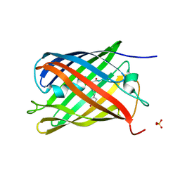
8A83
DownloadVisualize
BU of 8a83 by Molmil
rsEGFP2 with a chlorinated chromophore in the fluorescent ON-state in a crystal dehydrated after illumination
Descriptor: Green fluorescent protein, SULFATE ION
Authors:Fadini, A, van Thor, J, Chang, J.
Deposit date:2022-06-21
Release date:2023-07-19
Last modified:2024-02-07
Method:X-RAY DIFFRACTION (1.81 Å)
Cite:Serial Femtosecond Crystallography Reveals that Photoactivation in a Fluorescent Protein Proceeds via the Hula Twist Mechanism.
J.Am.Chem.Soc., 145, 2023
8A6R
DownloadVisualize
BU of 8a6r by Molmil
Room temperature rsEGFP2 with a chlorinated chromophore 100 ps after Photoexcitation
Descriptor: Green fluorescent protein
Authors:Fadini, A, van Thor, J.
Deposit date:2022-06-18
Release date:2023-07-19
Last modified:2024-02-07
Method:X-RAY DIFFRACTION (1.63 Å)
Cite:Serial Femtosecond Crystallography Reveals that Photoactivation in a Fluorescent Protein Proceeds via the Hula Twist Mechanism.
J.Am.Chem.Soc., 145, 2023
8A6G
DownloadVisualize
BU of 8a6g by Molmil
Room temperature rsEGFP2 with a chlorinated chromophore in the non-fluorescent OFF-state
Descriptor: Green fluorescent protein
Authors:Fadini, A, van Thor, J.
Deposit date:2022-06-17
Release date:2023-07-19
Last modified:2024-02-07
Method:X-RAY DIFFRACTION (1.63 Å)
Cite:Serial Femtosecond Crystallography Reveals that Photoactivation in a Fluorescent Protein Proceeds via the Hula Twist Mechanism.
J.Am.Chem.Soc., 145, 2023
8A6P
DownloadVisualize
BU of 8a6p by Molmil
Room temperature rsEGFP2 with a chlorinated chromophore 900 fs after photoexcitation
Descriptor: Green fluorescent protein
Authors:Fadini, A, van Thor, J.
Deposit date:2022-06-18
Release date:2023-07-19
Last modified:2024-02-07
Method:X-RAY DIFFRACTION (1.63 Å)
Cite:Serial Femtosecond Crystallography Reveals that Photoactivation in a Fluorescent Protein Proceeds via the Hula Twist Mechanism.
J.Am.Chem.Soc., 145, 2023
8A6Q
DownloadVisualize
BU of 8a6q by Molmil
Room temperature rsEGFP2 with a chlorinated chromophore 5 ps after photoexcitation
Descriptor: Green fluorescent protein
Authors:Fadini, A, van Thor, J.
Deposit date:2022-06-18
Release date:2023-07-19
Last modified:2024-02-07
Method:X-RAY DIFFRACTION (1.63 Å)
Cite:Serial Femtosecond Crystallography Reveals that Photoactivation in a Fluorescent Protein Proceeds via the Hula Twist Mechanism.
J.Am.Chem.Soc., 145, 2023
8A7V
DownloadVisualize
BU of 8a7v by Molmil
Room temperature rsEGFP2 in its OFF-state obtained with SFX
Descriptor: Green fluorescent protein
Authors:Fadini, A, van Thor, J.
Deposit date:2022-06-21
Release date:2023-07-19
Last modified:2024-02-07
Method:X-RAY DIFFRACTION (1.46 Å)
Cite:Serial Femtosecond Crystallography Reveals that Photoactivation in a Fluorescent Protein Proceeds via the Hula Twist Mechanism.
J.Am.Chem.Soc., 145, 2023
6KG9
DownloadVisualize
BU of 6kg9 by Molmil
Solution structure of CaDoc0917 from Clostridium acetobutylicum
Descriptor: And cellulose-binding endoglucanase family 9 CelL ortholog dockerin domain, CALCIUM ION
Authors:Feng, Y, Yao, X.
Deposit date:2019-07-11
Release date:2020-07-08
Last modified:2024-05-01
Method:SOLUTION NMR
Cite:Discovery and mechanism of a pH-dependent dual-binding-site switch in the interaction of a pair of protein modules.
Sci Adv, 6, 2020
6KGF
DownloadVisualize
BU of 6kgf by Molmil
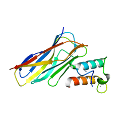
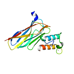
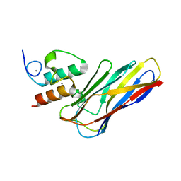
Crystal structure of CaDoc0917(R16D)-CaCohA2 complex at pH 8.2
Descriptor: And cellulose-binding endoglucanase family 9 CelL ortholog dockerin domain, CALCIUM ION, Probably cellulosomal scaffolding protein, ...
Authors:Feng, Y, Yao, X.
Deposit date:2019-07-11
Release date:2020-07-08
Last modified:2023-11-22
Method:X-RAY DIFFRACTION (2.3 Å)
Cite:Discovery and mechanism of a pH-dependent dual-binding-site switch in the interaction of a pair of protein modules.
Sci Adv, 6, 2020
6KGC
DownloadVisualize
BU of 6kgc by Molmil
Crystal structure of CaDoc0917(R49D)-CaCohA2 complex at pH 5.4
Descriptor: And cellulose-binding endoglucanase family 9 CelL ortholog dockerin domain, CALCIUM ION, Probably cellulosomal scaffolding protein, ...
Authors:Feng, Y, Yao, X.
Deposit date:2019-07-11
Release date:2020-07-08
Last modified:2023-11-22
Method:X-RAY DIFFRACTION (1.6 Å)
Cite:Discovery and mechanism of a pH-dependent dual-binding-site switch in the interaction of a pair of protein modules.
Sci Adv, 6, 2020
6KGD
DownloadVisualize
BU of 6kgd by Molmil
Crystal structure of CaDoc0917(R49D)-CaCohA2 complex at pH 8.0
Descriptor: And cellulose-binding endoglucanase family 9 CelL ortholog dockerin domain, CALCIUM ION, Probably cellulosomal scaffolding protein, ...
Authors:Feng, Y, Yao, X.
Deposit date:2019-07-11
Release date:2020-07-08
Last modified:2023-11-22
Method:X-RAY DIFFRACTION (1.65 Å)
Cite:Discovery and mechanism of a pH-dependent dual-binding-site switch in the interaction of a pair of protein modules.
Sci Adv, 6, 2020
6KGE
DownloadVisualize
BU of 6kge by Molmil
Crystal structure of CaDoc0917(R16D)-CaCohA2 complex at pH 5.5
Descriptor: And cellulose-binding endoglucanase family 9 CelL ortholog dockerin domain, CALCIUM ION, Probably cellulosomal scaffolding protein, ...
Authors:Feng, Y, Yao, X.
Deposit date:2019-07-11
Release date:2020-07-08
Last modified:2023-11-22
Method:X-RAY DIFFRACTION (2 Å)
Cite:Discovery and mechanism of a pH-dependent dual-binding-site switch in the interaction of a pair of protein modules.
Sci Adv, 6, 2020
6KG8
DownloadVisualize
BU of 6kg8 by Molmil
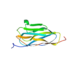
Solution structure of CaCohA2 from Clostridium acetobutylicum
Descriptor: Probably cellulosomal scaffolding protein, secreted cellulose-binding and cohesin domain
Authors:Feng, Y, Yao, X.
Deposit date:2019-07-11
Release date:2020-07-08
Last modified:2024-05-15
Method:SOLUTION NMR
Cite:Discovery and mechanism of a pH-dependent dual-binding-site switch in the interaction of a pair of protein modules.
Sci Adv, 6, 2020
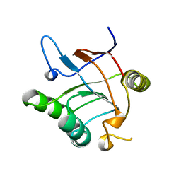
1VK1
DownloadVisualize
BU of 1vk1 by Molmil
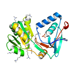
Conserved hypothetical protein from Pyrococcus furiosus Pfu-392566-001
Descriptor: Conserved hypothetical protein, DI(HYDROXYETHYL)ETHER, PHOSPHATE ION, ...
Authors:Shah, A, Liu, Z.J, Tempel, W, Chen, L, Lee, D, Yang, H, Chang, J, Zhao, M, Ng, J, Rose, J, Brereton, P.S, Izumi, M, Jenney Jr, F.E, Poole II, F.L, Shah, C, Sugar, F.J, Adams, M.W.W, Richardson, D.C, Richardson, J.S, Wang, B.C, Southeast Collaboratory for Structural Genomics (SECSG)
Deposit date:2004-04-13
Release date:2004-08-10
Last modified:2023-12-27
Method:X-RAY DIFFRACTION (1.2 Å)
Cite:(NZ)CH...O contacts assist crystallization of a ParB-like nuclease.
Bmc Struct.Biol., 7, 2007
2LCC
DownloadVisualize
BU of 2lcc by Molmil
Solution structure of RBBP1 chromobarrel domain
Descriptor: AT-rich interactive domain-containing protein 4A
Authors:Gong, W, Feng, Y.
Deposit date:2011-04-28
Release date:2012-02-08
Last modified:2024-05-15
Method:SOLUTION NMR
Cite:Structural insight into recognition of methylated histone tails by retinoblastoma-binding protein 1.
J.Biol.Chem., 2012
2MAM
DownloadVisualize
BU of 2mam by Molmil
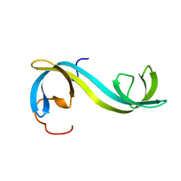
Solution structure of the interdigitated double Tudor domain of RBBP1
Descriptor: AT-rich interactive domain-containing protein 4A
Authors:Gong, W, Feng, Y.
Deposit date:2013-07-15
Release date:2014-01-15
Last modified:2024-05-15
Method:SOLUTION NMR
Cite:Retinoblastoma-binding protein 1 has an interdigitated double Tudor domain with DNA binding activity.
J.Biol.Chem., 289, 2014
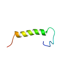
2MVN
DownloadVisualize
BU of 2mvn by Molmil
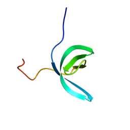
Solution structure of eEF1Bdelta CAR domain in TCTP-bound state
Descriptor: Elongation factor 1-delta
Authors:Wu, H, Feng, Y.
Deposit date:2014-10-09
Release date:2015-02-04
Last modified:2024-05-15
Method:SOLUTION NMR
Cite:Evolutionarily Conserved Binding of Translationally Controlled Tumor Protein to Eukaryotic Elongation Factor 1B.
J.Biol.Chem., 290, 2015
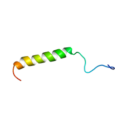
2MVM
DownloadVisualize
BU of 2mvm by Molmil
Solution structure of eEF1Bdelta CAR domain
Descriptor: Elongation factor 1-delta
Authors:Wu, H, Feng, Y.
Deposit date:2014-10-09
Release date:2015-02-04
Last modified:2024-05-15
Method:SOLUTION NMR
Cite:Evolutionarily Conserved Binding of Translationally Controlled Tumor Protein to Eukaryotic Elongation Factor 1B.
J.Biol.Chem., 290, 2015
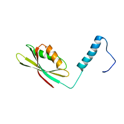
2N51
DownloadVisualize
BU of 2n51 by Molmil
NMR structure of the C-terminal region of human eukaryotic elongation factor 1B
Descriptor: Elongation factor 1-delta
Authors:Wu, H, Feng, Y.
Deposit date:2015-07-02
Release date:2016-05-18
Last modified:2024-05-15
Method:SOLUTION NMR
Cite:The C-terminal region of human eukaryotic elongation factor 1B delta.
J.Biomol.Nmr, 64, 2016
<12


221716

PDB entries from 2024-06-26

PDB statisticsPDBj update infoContact PDBjnumon