Loading
PDBj
MenuPDBj@FacebookPDBj@TwitterPDBj@YouTubewwPDB FoundationwwPDB
RCSB PDBPDBeBMRBAdv. SearchSearch help
Search by PDB author
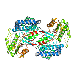
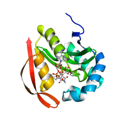
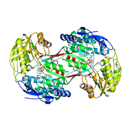
4I3T
DownloadVisualize
BU of 4i3t by Molmil
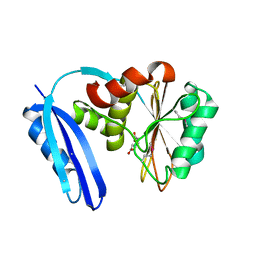
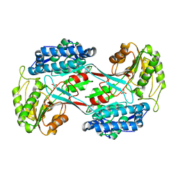
Structure of phosphonoacetaldehyde dehydrogenase in the apo state
Descriptor: Aldehyde dehydrogenase (NAD+), PHOSPHATE ION
Authors:Nair, S.K, Agarwal, V.
Deposit date:2012-11-26
Release date:2013-11-27
Last modified:2024-02-28
Method:X-RAY DIFFRACTION (2.1 Å)
Cite:Structure and function of phosphonoacetaldehyde dehydrogenase: the missing link in phosphonoacetate formation.
Chem.Biol., 21, 2014
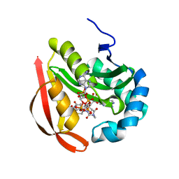
4I3X
DownloadVisualize
BU of 4i3x by Molmil
Structure of phosphonoacetaldehyde dehydrogenase in complex with phosphonoacetate and cofactor NAD+
Descriptor: Aldehyde dehydrogenase (NAD+), NICOTINAMIDE-ADENINE-DINUCLEOTIDE, PHOSPHONOACETIC ACID
Authors:Nair, S.K, Agarwal, V.
Deposit date:2012-11-26
Release date:2013-11-27
Last modified:2024-02-28
Method:X-RAY DIFFRACTION (2.07 Å)
Cite:Structure and function of phosphonoacetaldehyde dehydrogenase: the missing link in phosphonoacetate formation.
Chem.Biol., 21, 2014
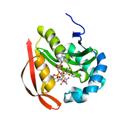
4I3W
DownloadVisualize
BU of 4i3w by Molmil
Structure of phosphonoacetaldehyde dehydrogenase in complex with gylceraldehyde-3-phosphate and cofactor NAD+
Descriptor: Aldehyde dehydrogenase (NAD+), GLYCERALDEHYDE-3-PHOSPHATE, NICOTINAMIDE-ADENINE-DINUCLEOTIDE
Authors:Nair, S.K, Agarwal, V.
Deposit date:2012-11-26
Release date:2013-11-27
Last modified:2024-11-06
Method:X-RAY DIFFRACTION (2.24 Å)
Cite:Structure and function of phosphonoacetaldehyde dehydrogenase: the missing link in phosphonoacetate formation.
Chem.Biol., 21, 2014
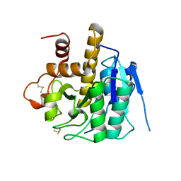
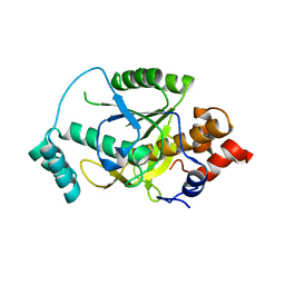
4H6V
DownloadVisualize
BU of 4h6v by Molmil
Structure of Patellamide maturation protease PatA
Descriptor: Subtilisin-like protein
Authors:Nair, S.K, Agarwal, V.
Deposit date:2012-09-19
Release date:2012-10-03
Last modified:2017-11-15
Method:X-RAY DIFFRACTION (1.7 Å)
Cite:Structures of cyanobactin maturation enzymes define a family of transamidating proteases.
Chem.Biol., 19, 2012
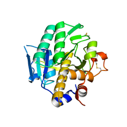
4H6W
DownloadVisualize
BU of 4h6w by Molmil
Structure of Prenylagaramide maturation protease PagA
Descriptor: N-terminal cyanobactin protease
Authors:Nair, S.K, Agarwal, V.
Deposit date:2012-09-19
Release date:2012-10-03
Last modified:2024-10-30
Method:X-RAY DIFFRACTION (2.45 Å)
Cite:Structures of cyanobactin maturation enzymes define a family of transamidating proteases.
Chem.Biol., 19, 2012
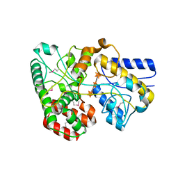
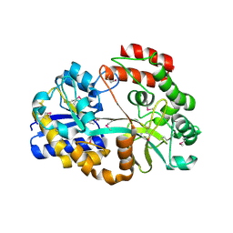
4G68
DownloadVisualize
BU of 4g68 by Molmil
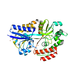
Biochemical and structural insights into xylan utilization by the thermophilic bacteriumcaldanaerobius polysaccharolyticus
Descriptor: ABC transporter, beta-D-xylopyranose-(1-4)-beta-D-xylopyranose-(1-4)-alpha-D-xylopyranose
Authors:Agarwal, V, Nair, S.K.
Deposit date:2012-07-18
Release date:2012-08-22
Last modified:2020-07-29
Method:X-RAY DIFFRACTION (1.8 Å)
Cite:Biochemical and Structural Insights into Xylan Utilization by the Thermophilic Bacterium Caldanaerobius polysaccharolyticus.
J.Biol.Chem., 287, 2012
4H6X
DownloadVisualize
BU of 4h6x by Molmil
Structure of Patellamide maturation protease PatG
Descriptor: Thiazoline oxidase/subtilisin-like protease
Authors:Nair, S.K, Agarwal, V.
Deposit date:2012-09-19
Release date:2012-10-03
Last modified:2017-11-15
Method:X-RAY DIFFRACTION (2 Å)
Cite:Structures of cyanobactin maturation enzymes define a family of transamidating proteases.
Chem.Biol., 19, 2012
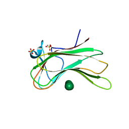
3OEB
DownloadVisualize
BU of 3oeb by Molmil
Crystal structure of the Q121E mutant of C.polysaccharolyticus CBM16-1 bound to mannopentaose
Descriptor: CALCIUM ION, S-layer associated multidomain endoglucanase, SULFATE ION, ...
Authors:Agarwal, V, Nair, S.K.
Deposit date:2010-08-12
Release date:2010-08-25
Last modified:2023-09-06
Method:X-RAY DIFFRACTION (1.55 Å)
Cite:Mutational insights into the roles of amino acid residues in ligand binding for two closely related family 16 carbohydrate binding modules.
J.Biol.Chem., 285, 2010
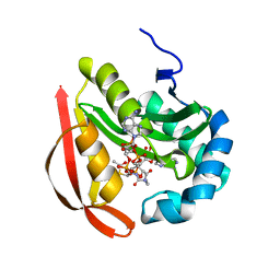
3R96
DownloadVisualize
BU of 3r96 by Molmil
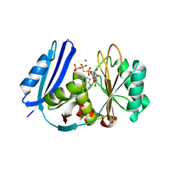
Crystal structure of Microcin C7 self immunity acetyltransferase MccE in complex with Acetyl-CoA and AMP
Descriptor: ACETYL COENZYME *A, ADENOSINE MONOPHOSPHATE, MccE protein
Authors:Nair, S.K, Agarwal, V.
Deposit date:2011-03-24
Release date:2011-04-20
Last modified:2024-02-21
Method:X-RAY DIFFRACTION (1.3 Å)
Cite:Structural Basis for Microcin C7 Inactivation by the MccE Acetyltransferase.
J.Biol.Chem., 286, 2011
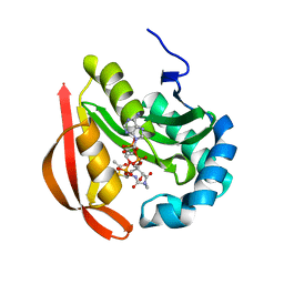
3R95
DownloadVisualize
BU of 3r95 by Molmil
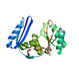
Crystal structure of Microcin C7 self immunity acetyltransferase MccE in complex with Acetyl-CoA
Descriptor: ACETYL COENZYME *A, MccE protein
Authors:Nair, S.K, Agarwal, V.
Deposit date:2011-03-24
Release date:2011-04-20
Last modified:2024-02-21
Method:X-RAY DIFFRACTION (1.6 Å)
Cite:Structural Basis for Microcin C7 Inactivation by the MccE Acetyltransferase.
J.Biol.Chem., 286, 2011
3R9F
DownloadVisualize
BU of 3r9f by Molmil
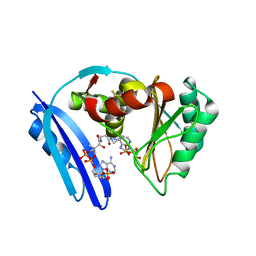
Crystal structure of Microcin C7 self immunity acetyltransferase MccE in complex with Coenzyme A and Glutamyl sulfamoyl adenosine (ESA)
Descriptor: COENZYME A, MccE protein, O5'-(L-GLUTAMYL-SULFAMOYL)-ADENOSINE
Authors:Nair, S.K, Agarwal, V.
Deposit date:2011-03-25
Release date:2011-04-20
Last modified:2024-02-21
Method:X-RAY DIFFRACTION (1.2 Å)
Cite:Structural Basis for Microcin C7 Inactivation by the MccE Acetyltransferase.
J.Biol.Chem., 286, 2011
3R9G
DownloadVisualize
BU of 3r9g by Molmil
Crystal structure of Microcin C7 self immunity acetyltransferase MccE in complex with Coenzyme A and processed Microcin C7 antibiotic
Descriptor: 5'-O-[(R)-[(N-acetyl-L-alpha-aspartyl)amino](3-aminopropoxy)phosphoryl]adenosine, COENZYME A, MccE protein
Authors:Nair, S.K, Agarwal, V.
Deposit date:2011-03-25
Release date:2011-04-27
Last modified:2024-02-21
Method:X-RAY DIFFRACTION (1.35 Å)
Cite:Structural Basis for Microcin C7 Inactivation by the MccE Acetyltransferase.
J.Biol.Chem., 286, 2011
3R9E
DownloadVisualize
BU of 3r9e by Molmil
Crystal structure of Microcin C7 self immunity acetyltransferase MccE in complex with coenzyme A and aspartyl sulfamoyl adenosine (DSA)
Descriptor: 5'-O-(L-alpha-aspartylsulfamoyl)adenosine, COENZYME A, MccE protein
Authors:Nair, S.K, Agarwal, V.
Deposit date:2011-03-25
Release date:2011-04-20
Last modified:2024-02-21
Method:X-RAY DIFFRACTION (1.25 Å)
Cite:Structural Basis for Microcin C7 Inactivation by the MccE Acetyltransferase.
J.Biol.Chem., 286, 2011
4R9F
DownloadVisualize
BU of 4r9f by Molmil
CpMnBP1 with Mannobiose Bound
Descriptor: MBP1, beta-D-mannopyranose-(1-4)-beta-D-mannopyranose
Authors:Chekan, J.R, Agarwal, V, Nair, S.K.
Deposit date:2014-09-04
Release date:2014-10-29
Last modified:2024-11-06
Method:X-RAY DIFFRACTION (1.4 Å)
Cite:Structural and Biochemical Basis for Mannan Utilization by Caldanaerobius polysaccharolyticus Strain ATCC BAA-17.
J.Biol.Chem., 289, 2014
4R9G
DownloadVisualize
BU of 4r9g by Molmil
CpMnBP1 with Mannotriose Bound
Descriptor: MBP1, ZINC ION, beta-D-mannopyranose-(1-4)-beta-D-mannopyranose-(1-4)-alpha-D-mannopyranose
Authors:Chekan, J.R, Agarwal, V, Nair, S.K.
Deposit date:2014-09-04
Release date:2014-10-29
Last modified:2024-02-28
Method:X-RAY DIFFRACTION (2.2 Å)
Cite:Structural and Biochemical Basis for Mannan Utilization by Caldanaerobius polysaccharolyticus Strain ATCC BAA-17.
J.Biol.Chem., 289, 2014
5TVA
DownloadVisualize
BU of 5tva by Molmil
A. aeolicus BioW with AMP and CoA
Descriptor: 6-carboxyhexanoate--CoA ligase, ADENOSINE MONOPHOSPHATE, COENZYME A
Authors:Estrada, P, Manandhar, M, Dong, S.-H, Deveryshetty, J, Agarwal, V, Cronan, J.E, Nair, S.K.
Deposit date:2016-11-08
Release date:2016-12-07
Last modified:2024-10-16
Method:X-RAY DIFFRACTION (2.25 Å)
Cite:The pimeloyl-CoA synthetase BioW defines a new fold for adenylate-forming enzymes.
Nat. Chem. Biol., 13, 2017
5TV5
DownloadVisualize
BU of 5tv5 by Molmil
BioW from Aquifex aeoulicus
Descriptor: 6-carboxyhexanoate--CoA ligase
Authors:Estrada, P, Manandhar, M, Dong, S.-H, Deveryshetty, J, Agarwal, V, Cronan, J.E, Nair, S.K.
Deposit date:2016-11-08
Release date:2016-12-07
Last modified:2017-05-31
Method:X-RAY DIFFRACTION (2.5 Å)
Cite:The pimeloyl-CoA synthetase BioW defines a new fold for adenylate-forming enzymes.
Nat. Chem. Biol., 13, 2017
5TV8
DownloadVisualize
BU of 5tv8 by Molmil
A. aeolicus BioW with AMP-CPP and pimelate
Descriptor: 6-carboxyhexanoate--CoA ligase, DIPHOSPHOMETHYLPHOSPHONIC ACID ADENOSYL ESTER, MAGNESIUM ION, ...
Authors:Estrada, P, Manandhar, M, Dong, S.-H, Deveryshetty, J, Agarwal, V, Cronan, J.E, Nair, S.K.
Deposit date:2016-11-08
Release date:2016-12-07
Last modified:2017-05-31
Method:X-RAY DIFFRACTION (2.55 Å)
Cite:The pimeloyl-CoA synthetase BioW defines a new fold for adenylate-forming enzymes.
Nat. Chem. Biol., 13, 2017
5TV6
DownloadVisualize
BU of 5tv6 by Molmil
A. aeolicus BioW with pimelate
Descriptor: 6-carboxyhexanoate--CoA ligase, PIMELIC ACID
Authors:Estrada, P, Manandhar, M, Dong, S.-H, Deveryshetty, J, Agarwal, V, Cronan, J.E, Nair, S.K.
Deposit date:2016-11-08
Release date:2016-12-07
Last modified:2024-10-30
Method:X-RAY DIFFRACTION (2.456 Å)
Cite:The pimeloyl-CoA synthetase BioW defines a new fold for adenylate-forming enzymes.
Nat. Chem. Biol., 13, 2017
3UQ4
DownloadVisualize
BU of 3uq4 by Molmil
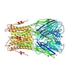
X-ray structure of a pentameric ligand gated ion channel from Erwinia chrysanthemi (ELIC) mutant F247L (F16L)
Descriptor: Gamma-aminobutyric-acid receptor subunit beta-1, SODIUM ION
Authors:Gonzalez-Gutierrez, G, Lukk, T, Agarwal, V, Papke, D, Nair, S.K, Grosman, C.
Deposit date:2011-11-19
Release date:2012-04-04
Last modified:2023-09-13
Method:X-RAY DIFFRACTION (3.5 Å)
Cite:Mutations that stabilize the open state of the Erwinia chrisanthemi ligand-gated ion channel fail to change the conformation of the pore domain in crystals.
Proc.Natl.Acad.Sci.USA, 109, 2012
3UQ5
DownloadVisualize
BU of 3uq5 by Molmil
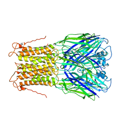
X-ray structure of a pentameric ligand gated ion channel from Erwinia chrysanthemi (ELIC) mutant L240A F247L (L9A F16L) in the presence of 10 mM cysteamine
Descriptor: Gamma-aminobutyric-acid receptor subunit beta-1, SODIUM ION
Authors:Gonzalez-Gutierrez, G, Lukk, T, Agarwal, V, Papke, D, Nair, S.K, Grosman, C.
Deposit date:2011-11-19
Release date:2012-04-04
Last modified:2023-09-13
Method:X-RAY DIFFRACTION (4.2 Å)
Cite:Mutations that stabilize the open state of the Erwinia chrisanthemi ligand-gated ion channel fail to change the conformation of the pore domain in crystals.
Proc.Natl.Acad.Sci.USA, 109, 2012
3UQ7
DownloadVisualize
BU of 3uq7 by Molmil
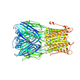
X-ray structure of a pentameric ligand gated ion channel from Erwinia chrysanthemi (ELIC) mutant L240S F247L (L9S F16L) in presence of 10 mM cysteamine
Descriptor: Gamma-aminobutyric-acid receptor subunit beta-1
Authors:Gonzalez-Gutierrez, G, Lukk, T, Agarwal, V, Papke, D, Nair, S.K, Grosman, C.
Deposit date:2011-11-19
Release date:2012-04-04
Last modified:2023-09-13
Method:X-RAY DIFFRACTION (3.8 Å)
Cite:Mutations that stabilize the open state of the Erwinia chrisanthemi ligand-gated ion channel fail to change the conformation of the pore domain in crystals.
Proc.Natl.Acad.Sci.USA, 109, 2012
6POO
DownloadVisualize
BU of 6poo by Molmil
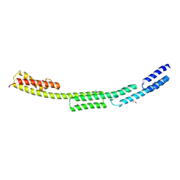
Novel structure of the N-terminal helical domain of BibA, a group B streptococcus immunogenic bacterial adhesin
Descriptor: BibA
Authors:Manne, K, Narayana, S.V.
Deposit date:2019-07-04
Release date:2020-08-12
Last modified:2024-10-23
Method:X-RAY DIFFRACTION (3.03 Å)
Cite:Novel structure of the N-terminal helical domain of BibA, a group B streptococcus immunogenic bacterial adhesin.
Acta Crystallogr D Struct Biol, 76, 2020
8TB1
DownloadVisualize
BU of 8tb1 by Molmil
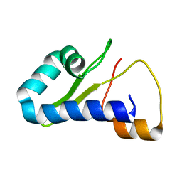
Solution NMR structure of a RiPP proteusin precursor protein
Descriptor: NHLP leader peptide family natural product, tumor homing peptide 1 (TH1) substrate chimera
Authors:McShan, A.C, Vinayak, A, Nguyen, N.A.
Deposit date:2023-06-28
Release date:2024-02-14
Last modified:2024-05-01
Method:SOLUTION NMR
Cite:Disordered regions in proteusin peptides guide post-translational modification by a flavin-dependent RiPP brominase.
Nat Commun, 15, 2024
4KWC
DownloadVisualize
BU of 4kwc by Molmil
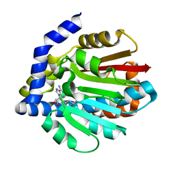
Structure of the plantazolicin methyltransferase BpumL in complex with SAH
Descriptor: BpumL, S-ADENOSYL-L-HOMOCYSTEINE
Authors:Hao, Y, Nair, S.K.
Deposit date:2013-05-23
Release date:2013-07-24
Last modified:2024-02-28
Method:X-RAY DIFFRACTION (1.994 Å)
Cite:Structural and functional insight into an unexpectedly selective N-methyltransferase involved in plantazolicin biosynthesis.
Proc.Natl.Acad.Sci.USA, 110, 2013

227111

PDB entries from 2024-11-06

PDB statisticsPDBj update infoContact PDBjnumon