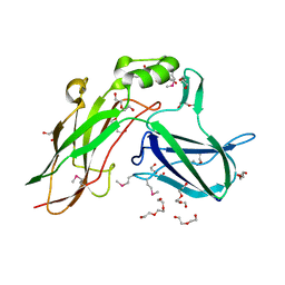
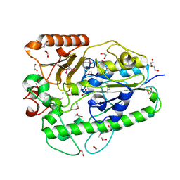
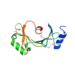
Loading
PDBj
MenuPDBj@FacebookPDBj@TwitterPDBj@YouTubewwPDB FoundationwwPDB
RCSB PDBPDBeBMRBAdv. SearchSearch help
Search by PDB author
4EIU
DownloadVisualize
BU of 4eiu by Molmil
Crystal structure of a DUF3823 family protein (BACUNI_03093) from Bacteroides uniformis ATCC 8492 at 1.90 A resolution
Descriptor: DI(HYDROXYETHYL)ETHER, TRIETHYLENE GLYCOL, uncharacterized hypothetical protein
Authors:Joint Center for Structural Genomics (JCSG)
Deposit date:2012-04-05
Release date:2012-05-30
Last modified:2023-02-01
Method:X-RAY DIFFRACTION (1.9 Å)
Cite:Crystal structure of a hypothetical protein (BACUNI_03093) from Bacteroides uniformis ATCC 8492 at 1.90 A resolution
To be published
4RWV
DownloadVisualize
BU of 4rwv by Molmil
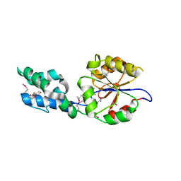
Crystal structure of PIP3 bound human nuclear receptor LRH-1 (Liver Receptor Homolog 1, NR5A2) in complex with a co-regulator DAX-1 (NR0B1) peptide at 1.86 A resolution
Descriptor: (2S)-3-{[(R)-{[(1S,2S,3R,4S,5S,6S)-2,6-dihydroxy-3,4,5-tris(phosphonooxy)cyclohexyl]oxy}(hydroxy)phosphoryl]oxy}propane -1,2-diyl dihexadecanoate, 1,2-ETHANEDIOL, 2-AMINO-2-HYDROXYMETHYL-PROPANE-1,3-DIOL, ...
Authors:Joint Center for Structural Genomics (JCSG), Partnership for Stem Cell Biology, Partnership for Stem Cell Biology (STEMCELL)
Deposit date:2014-12-05
Release date:2015-01-21
Last modified:2023-12-06
Method:X-RAY DIFFRACTION (1.859 Å)
Cite:Crystal structure of a Homo sapiens hepatocytic transcription factor hB1F-2 (B1F2) in complex with nuclear receptor subfamily 0 group B member 1 (NR0B1, residues 140-154) from human at 1.86 A resolution
To be published
4QAN
DownloadVisualize
BU of 4qan by Molmil
Crystal structure of a cystatin-like protein (RUMGNA_02398) from Ruminococcus gnavus ATCC 29149 at 2.10 A resolution
Descriptor: MAGNESIUM ION, hypothetical protein
Authors:Joint Center for Structural Genomics (JCSG)
Deposit date:2014-05-05
Release date:2014-07-23
Last modified:2023-02-01
Method:X-RAY DIFFRACTION (2.1 Å)
Cite:Crystal structure of a hypothetical protein (RUMGNA_02398) from Ruminococcus gnavus ATCC 29149 at 2.10 A resolution
To be published
4N3V
DownloadVisualize
BU of 4n3v by Molmil
Crystal structure of a bile-acid 7-alpha dehydratase (CLOHIR_00079) from Clostridium hiranonis DSM 13275 at 1.89 A resolution with product added
Descriptor: 1,2-ETHANEDIOL, Bile acid 7-alpha dehydratase, BaiE, ...
Authors:Joint Center for Structural Genomics (JCSG)
Deposit date:2013-10-07
Release date:2013-12-04
Last modified:2023-09-20
Method:X-RAY DIFFRACTION (1.89 Å)
Cite:Crystal structure of a bile-acid 7-alpha dehydratase (CLOHIR_00079) from Clostridium hiranonis DSM 13275 at 1.89 A resolution with product added
To be published
3KOP
DownloadVisualize
BU of 3kop by Molmil
Crystal structure of Protein with a cyclophilin-like fold (YP_831253.1) from Arthrobacter sp. FB24 at 1.90 A resolution
Descriptor: Uncharacterized protein
Authors:Joint Center for Structural Genomics (JCSG)
Deposit date:2009-11-13
Release date:2009-11-24
Last modified:2019-07-17
Method:X-RAY DIFFRACTION (1.9 Å)
Cite:Crystal structure of Protein with a cyclophilin-like fold (YP_831253.1) from Arthrobacter sp. FB24 at 1.90 A resolution
To be published
3B7F
DownloadVisualize
BU of 3b7f by Molmil
Crystal structure of a putative glycosyl hydrolase with bnr repeats (reut_b4987) from ralstonia eutropha jmp134 at 2.20 A resolution
Descriptor: 1,2-ETHANEDIOL, CHLORIDE ION, Glycosyl hydrolase, ...
Authors:Joint Center for Structural Genomics (JCSG)
Deposit date:2007-10-30
Release date:2007-11-13
Last modified:2023-01-25
Method:X-RAY DIFFRACTION (2.2 Å)
Cite:Crystal structure of Domain of Unknown Function with a 7-Bladed Beta-Propeller Fold (YP_299179.1) from Ralstonia eutropha JMP134 at 2.20 A resolution
To be published
3BA3
DownloadVisualize
BU of 3ba3 by Molmil
Crystal structure of Pyridoxamine 5'-phosphate oxidase-like protein (NP_783940.1) from Lactobacillus plantarum at 1.55 A resolution
Descriptor: 1,2-ETHANEDIOL, Pyridoxamine 5'-phosphate oxidase-like protein, SULFATE ION
Authors:Joint Center for Structural Genomics (JCSG)
Deposit date:2007-11-07
Release date:2007-11-20
Last modified:2023-01-25
Method:X-RAY DIFFRACTION (1.55 Å)
Cite:Crystal structure of Pyridoxamine 5'-phosphate oxidase-like protein (NP_783940.1) from Lactobacillus plantarum at 1.55 A resolution
To be published
3K50
DownloadVisualize
BU of 3k50 by Molmil
Crystal structure of Putative S41 protease (YP_211611.1) from Bacteroides fragilis NCTC 9343 at 2.00 A resolution
Descriptor: CHLORIDE ION, GLYCEROL, Putative S41 protease
Authors:Joint Center for Structural Genomics (JCSG)
Deposit date:2009-10-06
Release date:2009-10-13
Last modified:2023-02-01
Method:X-RAY DIFFRACTION (2 Å)
Cite:Crystal structure of Putative S41 protease (YP_211611.1) from Bacteroides fragilis NCTC 9343 at 2.00 A resolution
To be published
3B5Q
DownloadVisualize
BU of 3b5q by Molmil
Crystal structure of a putative sulfatase (NP_810509.1) from Bacteroides thetaiotaomicron VPI-5482 at 2.40 A resolution
Descriptor: 1,2-ETHANEDIOL, 4-(2-HYDROXYETHYL)-1-PIPERAZINE ETHANESULFONIC ACID, CHLORIDE ION, ...
Authors:Joint Center for Structural Genomics (JCSG)
Deposit date:2007-10-26
Release date:2007-11-13
Last modified:2023-01-25
Method:X-RAY DIFFRACTION (2.4 Å)
Cite:Crystal structure of a putative sulfatase (NP_810509.1) from Bacteroides thetaiotaomicron VPI-5482 at 2.40 A resolution
To be published
3JZL
DownloadVisualize
BU of 3jzl by Molmil
CRYSTAL STRUCTURE OF A PUTATIVE CYSTATHIONINE BETA-LYASE INVOLVED IN ALUMINUM RESISTANCE (LMOF2365_1314) FROM LISTERIA MONOCYTOGENES STR. 4B F2365 AT 1.91 A RESOLUTION
Descriptor: DI(HYDROXYETHYL)ETHER, GLYCEROL, Putative cystathionine beta-lyase involved in aluminum resistance
Authors:Joint Center for Structural Genomics (JCSG)
Deposit date:2009-09-23
Release date:2009-10-06
Last modified:2023-02-01
Method:X-RAY DIFFRACTION (1.91 Å)
Cite:Crystal structure of putative cystathionine beta-lyase involved in aluminum resistance (YP_013912.1) from Listeria monocytogenes 4b F2365 at 1.91 A resolution
To be published
3K0Z
DownloadVisualize
BU of 3k0z by Molmil
Crystal structure of Putative polyketide cyclase (NP_977253.1) from BACILLUS CEREUS ATCC 10987 at 1.91 A resolution
Descriptor: 2-[N-CYCLOHEXYLAMINO]ETHANE SULFONIC ACID, Putative polyketide cyclase
Authors:Joint Center for Structural Genomics (JCSG)
Deposit date:2009-09-25
Release date:2009-11-03
Last modified:2023-02-01
Method:X-RAY DIFFRACTION (1.91 Å)
Cite:Crystal structure of Putative polyketide cyclase (NP_977253.1) from BACILLUS CEREUS ATCC 10987 at 1.91 A resolution
To be published
3BHQ
DownloadVisualize
BU of 3bhq by Molmil
CRYSTAL STRUCTURE OF A PUTATIVE TETR-FAMILY TRANSCRIPTIONAL REGULATOR (MLR_4833) FROM MESORHIZOBIUM LOTI MAFF303099 AT 1.54 A RESOLUTION
Descriptor: 1,2-ETHANEDIOL, CHLORIDE ION, Transcriptional regulator
Authors:Joint Center for Structural Genomics (JCSG)
Deposit date:2007-11-28
Release date:2007-12-11
Last modified:2023-01-25
Method:X-RAY DIFFRACTION (1.54 Å)
Cite:Crystal structure of TetR-family transcriptional regulator (NP_105615.1) from Mesorhizobium loti at 1.54 A resolution
To be published
3BO9
DownloadVisualize
BU of 3bo9 by Molmil
Crystal structure of putative nitroalkan dioxygenase (TM0800) from Thermotoga maritima at 2.71 A resolution
Descriptor: 1,2-ETHANEDIOL, NONAETHYLENE GLYCOL, PHOSPHATE ION, ...
Authors:Joint Center for Structural Genomics (JCSG)
Deposit date:2007-12-17
Release date:2007-12-25
Last modified:2023-01-25
Method:X-RAY DIFFRACTION (2.71 Å)
Cite:Crystal structure of putative nitroalkan dioxygenase (TM0800) from Thermotoga maritima at 2.71 A resolution
To be published
4K61
DownloadVisualize
BU of 4k61 by Molmil
Crystal structure of a DUF2874 family protein (BACUNI_01296) from Bacteroides uniformis ATCC 8492 at 1.70 A resolution
Descriptor: Uncharacterized protein
Authors:Joint Center for Structural Genomics (JCSG)
Deposit date:2013-04-15
Release date:2013-05-01
Last modified:2023-02-01
Method:X-RAY DIFFRACTION (1.7 Å)
Cite:Crystal structure of a hypothetical protein (BACUNI_01296) from Bacteroides uniformis ATCC 8492 at 1.70 A resolution
To be published
3KSR
DownloadVisualize
BU of 3ksr by Molmil
CRYSTAL STRUCTURE OF A PUTATIVE SERINE HYDROLASE (XCC3885) FROM XANTHOMONAS CAMPESTRIS PV. CAMPESTRIS AT 2.69 A RESOLUTION
Descriptor: CHLORIDE ION, PHOSPHATE ION, Putative serine hydrolase
Authors:Joint Center for Structural Genomics (JCSG)
Deposit date:2009-11-23
Release date:2009-12-22
Last modified:2023-02-01
Method:X-RAY DIFFRACTION (2.69 Å)
Cite:Crystal structure of Putative serine hydrolase (NP_639225.1) from XANTHOMONAS CAMPESTRIS at 2.69 A resolution
To be Published
3KTD
DownloadVisualize
BU of 3ktd by Molmil
CRYSTAL STRUCTURE OF A PUTATIVE PREPHENATE DEHYDROGENASE (CGL0226) FROM CORYNEBACTERIUM GLUTAMICUM ATCC 13032 AT 2.60 A RESOLUTION
Descriptor: 1,2-ETHANEDIOL, DI(HYDROXYETHYL)ETHER, Prephenate dehydrogenase
Authors:Joint Center for Structural Genomics (JCSG)
Deposit date:2009-11-24
Release date:2010-02-02
Last modified:2023-02-01
Method:X-RAY DIFFRACTION (2.6 Å)
Cite:Crystal structure of Prephenate dehydrogenase (NP_599479.1) from CORYNEBACTERIUM GLUTAMICUM ATCC 13032 KITASATO at 2.60 A resolution
To be published
3BJQ
DownloadVisualize
BU of 3bjq by Molmil
CRYSTAL STRUCTURE OF A PHAGE-RELATED PROTEIN (BB3626) FROM BORDETELLA BRONCHISEPTICA RB50 AT 2.05 A RESOLUTION
Descriptor: CHLORIDE ION, Phage-related protein, TETRAETHYLENE GLYCOL
Authors:Joint Center for Structural Genomics (JCSG)
Deposit date:2007-12-04
Release date:2007-12-18
Last modified:2023-01-25
Method:X-RAY DIFFRACTION (2.05 Å)
Cite:Crystal structure of phage-related protein (NP_890161.1) from Bordetella bronchiseptica at 2.05 A resolution
To be published
3KNY
DownloadVisualize
BU of 3kny by Molmil
Crystal structure of a two domain protein with unknown function (bt_3535) from bacteroides thetaiotaomicron vpi-5482 at 2.60 A resolution
Descriptor: hypothetical protein BT_3535
Authors:Joint Center for Structural Genomics (JCSG)
Deposit date:2009-11-12
Release date:2009-12-01
Last modified:2023-02-01
Method:X-RAY DIFFRACTION (2.6 Å)
Cite:Crystal structure of hypothetical protein BT_3535 (NP_812447.1) from BACTERIODES THETAIOTAOMICRON VPI-5482 at 2.60 A resolution
To be published
3K9I
DownloadVisualize
BU of 3k9i by Molmil
Crystal structure of Putative protein binding protein (NP_241345.1) from Bacillus halodurans at 2.71 A resolution
Descriptor: BH0479 protein
Authors:Joint Center for Structural Genomics (JCSG)
Deposit date:2009-10-15
Release date:2009-11-24
Last modified:2023-02-01
Method:X-RAY DIFFRACTION (2.71 Å)
Cite:Crystal structure of Putative protein binding protein (NP_241345.1) from Bacillus halodurans at 2.71 A resolution
To be published
3JYS
DownloadVisualize
BU of 3jys by Molmil
Crystal structure of SusD superfamily protein (YP_001298690.1) from Bacteroides vulgatus ATCC 8482 at 2.00 A resolution
Descriptor: MAGNESIUM ION, SusD superfamily protein
Authors:Joint Center for Structural Genomics (JCSG)
Deposit date:2009-09-22
Release date:2009-10-06
Last modified:2023-02-01
Method:X-RAY DIFFRACTION (2 Å)
Cite:Crystal structure of SusD superfamily protein (YP_001298690.1) from Bacteroides vulgatus ATCC 8482 at 2.00 A resolution
To be published
3C3P
DownloadVisualize
BU of 3c3p by Molmil
Crystal structure of a methyltransferase (NP_951602.1) from Geobacter sulfurreducens at 1.90 A resolution
Descriptor: 1,2-ETHANEDIOL, CHLORIDE ION, DI(HYDROXYETHYL)ETHER, ...
Authors:Joint Center for Structural Genomics (JCSG)
Deposit date:2008-01-28
Release date:2008-02-05
Last modified:2023-02-01
Method:X-RAY DIFFRACTION (1.9 Å)
Cite:Crystal structure of a methyltransferase (NP_951602.1) from Geobacter sulfurreducens at 1.90 A resolution
To be published
3C5Y
DownloadVisualize
BU of 3c5y by Molmil
Crystal structure of a putative ribose 5-phosphate isomerase (saro_3514) from novosphingobium aromaticivorans dsm at 1.81 A resolution
Descriptor: 1,2-ETHANEDIOL, NITRATE ION, Ribose/galactose isomerase
Authors:Joint Center for Structural Genomics (JCSG)
Deposit date:2008-02-01
Release date:2008-02-19
Last modified:2023-11-15
Method:X-RAY DIFFRACTION (1.81 Å)
Cite:Crystal structure of putative ribose 5-phosphate isomerase (YP_001165900.1) from Novosphingobium aromaticivorans DSM 12444 at 1.81 A resolution
To be published
4RN3
DownloadVisualize
BU of 4rn3 by Molmil
Crystal structure of a HAD-superfamily hydrolase, subfamily IA, variant 1 (GSU2069) from Geobacter sulfurreducens PCA at 2.15 A resolution
Descriptor: 1,2-ETHANEDIOL, HAD superfamily hydrolase, MAGNESIUM ION
Authors:Joint Center for Structural Genomics (JCSG)
Deposit date:2014-10-22
Release date:2014-11-26
Last modified:2017-11-22
Method:X-RAY DIFFRACTION (2.15 Å)
Cite:Crystal structure of a HAD-superfamily hydrolase, subfamily IA, variant 1 (GSU2069) from Geobacter sulfurreducens PCA at 2.15 A resolution
To be published
4MUD
DownloadVisualize
BU of 4mud by Molmil
Crystal structure of a ring oxydation complex/ phenylacetic acid degradation-like protein (SSO1313) from Sulfolobus solfataricus P2 at 2.43 A resolution
Descriptor: 1,2-ETHANEDIOL, Ring oxydation complex/ phenylacetic acid degradation related protein
Authors:Joint Center for Structural Genomics (JCSG)
Deposit date:2013-09-21
Release date:2013-10-09
Last modified:2023-02-01
Method:X-RAY DIFFRACTION (2.43 Å)
Cite:Crystal structure of a ring oxydation complex/ phenylacetic acid degradation-like protein (SSO1313) from Sulfolobus solfataricus P2 at 2.43 A resolution
To be published
3KSP
DownloadVisualize
BU of 3ksp by Molmil
Crystal structure of a putative ca/calmodulin-dependent kinase ii association domain (exig_1688) from exiguobacterium sibiricum 255-15 at 2.59 A resolution
Descriptor: 1,2-ETHANEDIOL, 2-[N-CYCLOHEXYLAMINO]ETHANE SULFONIC ACID, Calcium/calmodulin-dependent kinase II association domain
Authors:Joint Center for Structural Genomics (JCSG)
Deposit date:2009-11-23
Release date:2009-12-29
Last modified:2023-02-01
Method:X-RAY DIFFRACTION (2.59 Å)
Cite:Crystal structure of Calcium/calmodulin-dependent kinase II association domain (YP_001814158.1) from EXIGUOBACTERIUM SP. 255-15 at 2.59 A resolution
To be published

222415

PDB entries from 2024-07-10

PDB statisticsPDBj update infoContact PDBjnumon