Loading
PDBj
MenuPDBj@FacebookPDBj@X(formerly Twitter)PDBj@BlueSkyPDBj@YouTubewwPDB FoundationwwPDB
RCSB PDBPDBeBMRBAdv. SearchSearch help
Search by PDB author
3M48
DownloadVisualize
BU of 3m48 by Molmil
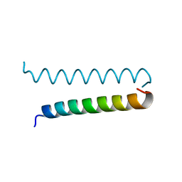
GCN4 Leucine Zipper Peptide Mutant
Descriptor: General control protein GCN4, SODIUM ION
Authors:Du, S, Kettering, R.D, Alvarado, J.J, Tortajada, A, Yeh, J.I.
Deposit date:2010-03-10
Release date:2011-02-16
Last modified:2023-09-06
Method:X-RAY DIFFRACTION (1.451 Å)
Cite:GCN4 Leucine Zipper Peptide Mutant
To be Published
3D42
DownloadVisualize
BU of 3d42 by Molmil
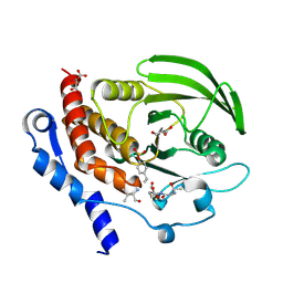
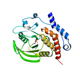
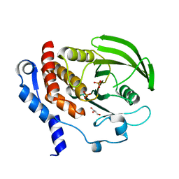
Crystal structure of HePTP in complex with a monophosphorylated Erk2 peptide
Descriptor: D(-)-TARTARIC ACID, GLYCEROL, Mitogen-activated protein kinase 1 peptide, ...
Authors:Critton, D.A, Tortajada, A, Page, R.
Deposit date:2008-05-13
Release date:2009-03-17
Last modified:2024-10-16
Method:X-RAY DIFFRACTION (2.46 Å)
Cite:Structural basis of substrate recognition by hematopoietic tyrosine phosphatase.
Biochemistry, 47, 2008
3D44
DownloadVisualize
BU of 3d44 by Molmil
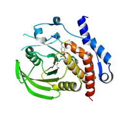
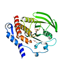
Crystal structure of HePTP in complex with a dually phosphorylated Erk2 peptide mimetic
Descriptor: CHLORIDE ION, GLYCEROL, Mitogen-activated protein kinase 1 peptide, ...
Authors:Critton, D.A, Tortajada, A, Page, R.
Deposit date:2008-05-13
Release date:2009-03-17
Last modified:2023-11-15
Method:X-RAY DIFFRACTION (1.9 Å)
Cite:Structural basis of substrate recognition by hematopoietic tyrosine phosphatase.
Biochemistry, 47, 2008
5FP2
DownloadVisualize
BU of 5fp2 by Molmil
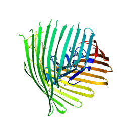
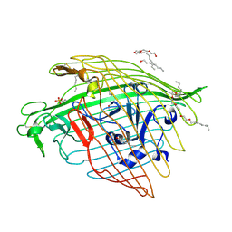
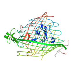
Crystal structure of the siderophore receptor PirA from Pseudomonas aeruginosa
Descriptor: FERRIC ENTEROBACTIN RECEPTOR PIRA, N-OCTANE, SULFATE ION
Authors:Moynie, L, Tortajada, A, Naismith, J.H.
Deposit date:2015-11-27
Release date:2015-12-09
Last modified:2024-11-13
Method:X-RAY DIFFRACTION (2.97 Å)
Cite:Structure and Function of the PiuA and PirA Siderophore-Drug Receptors from Pseudomonas aeruginosa and Acinetobacter baumannii.
Antimicrob. Agents Chemother., 61, 2017
5FR8
DownloadVisualize
BU of 5fr8 by Molmil
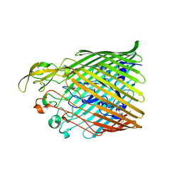
Crystal structure of the siderophore receptor PirA from Acinetobacter baumannii
Descriptor: (HYDROXYETHYLOXY)TRI(ETHYLOXY)OCTANE, TONB-DEPENDENT SIDEROPHORE RECEPTOR
Authors:Moynie, L, Tortajada, A, Naismith, J.H.
Deposit date:2015-12-16
Release date:2016-05-11
Last modified:2024-10-16
Method:X-RAY DIFFRACTION (2.83 Å)
Cite:Structure and Function of the PiuA and PirA Siderophore-Drug Receptors from Pseudomonas aeruginosa and Acinetobacter baumannii.
Antimicrob. Agents Chemother., 61, 2017
5FP1
DownloadVisualize
BU of 5fp1 by Molmil
Crystal structure of the siderophore receptor PiuA from Acinetobacter baumannii
Descriptor: (HYDROXYETHYLOXY)TRI(ETHYLOXY)OCTANE, PHOSPHATE ION, TONB-DEPENDENT SIDEROPHORE RECEPTOR
Authors:Moynie, L, Tortajada, A, Naismith, J.H.
Deposit date:2015-11-26
Release date:2016-05-11
Last modified:2024-11-13
Method:X-RAY DIFFRACTION (1.94 Å)
Cite:Structure and Function of the PiuA and PirA Siderophore-Drug Receptors from Pseudomonas aeruginosa and Acinetobacter baumannii.
Antimicrob. Agents Chemother., 61, 2017
2QDM
DownloadVisualize
BU of 2qdm by Molmil
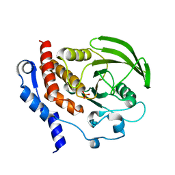
Crystal structure of the HePTP catalytic domain C270S/D236A/Q314A mutant
Descriptor: PHOSPHATE ION, Tyrosine-protein phosphatase non-receptor type 7
Authors:Critton, D.A, Tortajada, A, Page, R.
Deposit date:2007-06-21
Release date:2008-06-24
Last modified:2023-09-20
Method:X-RAY DIFFRACTION (2.05 Å)
Cite:Structural basis of substrate recognition by hematopoietic tyrosine phosphatase.
To be published
2QDP
DownloadVisualize
BU of 2qdp by Molmil
Crystal structure of the HePTP catalytic domain C270S mutant crystallized in ammonium acetate
Descriptor: PHOSPHATE ION, Tyrosine-protein phosphatase non-receptor type 7
Authors:Critton, D.A, Tortajada, A, Page, R.
Deposit date:2007-06-21
Release date:2008-06-24
Last modified:2023-09-20
Method:X-RAY DIFFRACTION (2.72 Å)
Cite:Structural basis of substrate recognition by hematopoietic tyrosine phosphatase.
To be published
2QDC
DownloadVisualize
BU of 2qdc by Molmil
Crystal structure of the HePTP catalytic domain D236A mutant
Descriptor: GLYCEROL, PHOSPHATE ION, Tyrosine-protein phosphatase non-receptor type 7
Authors:Critton, D.A, Tortajada, A, Page, R.
Deposit date:2007-06-20
Release date:2008-06-24
Last modified:2023-09-20
Method:X-RAY DIFFRACTION (2 Å)
Cite:Structural basis of substrate recognition by hematopoietic tyrosine phosphatase.
Biochemistry, 47, 2008
2HVL
DownloadVisualize
BU of 2hvl by Molmil
Crystal structure of the HePTP catalytic domain C270S mutant
Descriptor: GLYCEROL, PHOSPHATE ION, Tyrosine-protein phosphatase non-receptor type 7
Authors:Page, R, Critton, D, Ragusa, M.
Deposit date:2006-07-29
Release date:2007-08-07
Last modified:2024-02-14
Method:X-RAY DIFFRACTION (2.4 Å)
Cite:Structural basis of substrate recognition by hematopoietic tyrosine phosphatase
Biochemistry, 47, 2008
5FOK
DownloadVisualize
BU of 5fok by Molmil
Crystal structure of the siderophore receptor PiuA from Pseudomonas aeruginosa
Descriptor: (HYDROXYETHYLOXY)TRI(ETHYLOXY)OCTANE, IRON TRANSPORT OUTER MEMBRANE RECEPTOR, SODIUM ION, ...
Authors:Moynie, L, Naismith, J.H.
Deposit date:2015-11-23
Release date:2016-05-11
Last modified:2024-11-06
Method:X-RAY DIFFRACTION (1.9 Å)
Cite:Structure and Function of the PiuA and PirA Siderophore-Drug Receptors from Pseudomonas aeruginosa and Acinetobacter baumannii.
Antimicrob. Agents Chemother., 61, 2017
3I1G
DownloadVisualize
BU of 3i1g by Molmil
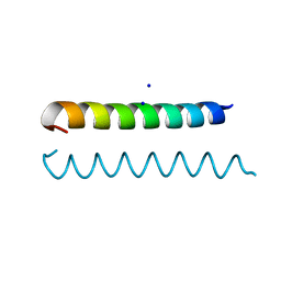
Crystal structure of a GCN4 leucine zipper mutant at 1.6 A resolution
Descriptor: General control protein GCN4
Authors:Diao, J.S, Tortajada, A, Yeh, J.I.
Deposit date:2009-06-26
Release date:2009-07-07
Last modified:2024-04-03
Method:X-RAY DIFFRACTION (1.6 Å)
Cite:Crystal structure of a super leucine zipper, an extended two-stranded super long coiled coil.
Protein Sci., 19, 2010

227344

數據於2024-11-13公開中

PDB statisticsPDBj update infoContact PDBjnumon