Loading
PDBj
MenuPDBj@FacebookPDBj@X(formerly Twitter)PDBj@BlueSkyPDBj@YouTubewwPDB FoundationwwPDB
RCSB PDBPDBeBMRBAdv. SearchSearch help
Search by PDB author
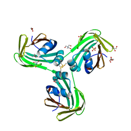
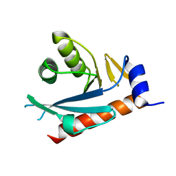
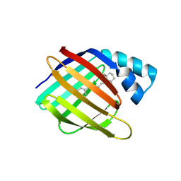
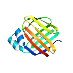
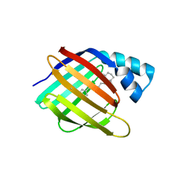
8VZX
DownloadVisualize
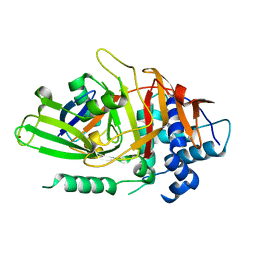
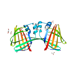
BU of 8vzx by Molmil
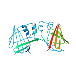
Q108K:K40L:T53A:R58F mutant of hCRBPII bound to synthetic fluorophore TD-1V
Descriptor: (2E)-3-{5-[4-(dimethylamino)phenyl]thiophen-2-yl}but-2-enal, ACETATE ION, Retinol-binding protein 2
Authors:Nossoni, Z, Bingham, C.R, Geiger, J.H.
Deposit date:2024-02-13
Release date:2024-04-03
Last modified:2024-11-13
Method:X-RAY DIFFRACTION (1.47 Å)
Cite:Regulation of Absorption and Emission in a Protein/Fluorophore Complex.
Acs Chem.Biol., 19, 2024
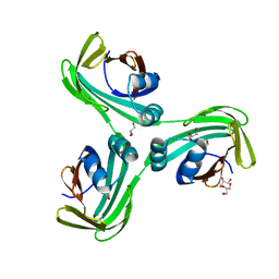
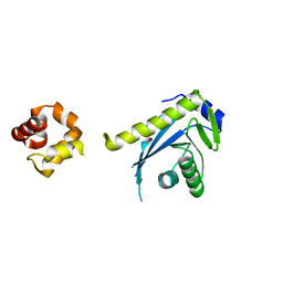
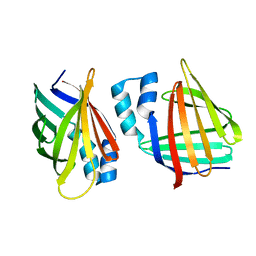
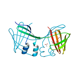
6WNF
DownloadVisualize
BU of 6wnf by Molmil
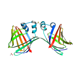
The Crystal Structure of Apo Domain-Swapped Dimer Q108K:K40D:T53A:R58L:Q38F:Q4F:F57H Variant of HCRBPII
Descriptor: ACETATE ION, GLYCEROL, Retinol-binding protein 2
Authors:Ghanbarpour, A, Geiger, J.
Deposit date:2020-04-22
Release date:2020-08-19
Last modified:2023-10-18
Method:X-RAY DIFFRACTION (1.67 Å)
Cite:Human Cellular Retinol Binding Protein II Forms a Domain-Swapped Trimer Representing a Novel Fold and a New Template for Protein Engineering.
Chembiochem, 21, 2020
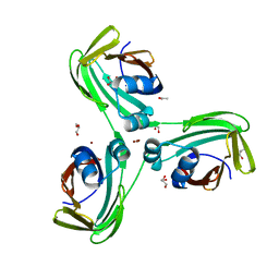
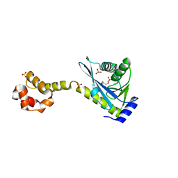
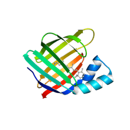
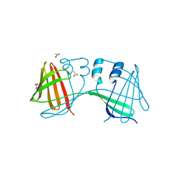
6WNJ
DownloadVisualize
BU of 6wnj by Molmil
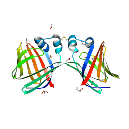
The Crystal Structure of Apo Domain-Swapped Trimer Q108K:T51D:A28C:I32C of HCRBPII
Descriptor: GLYCEROL, Retinol-binding protein 2, SULFATE ION, ...
Authors:Ghanbarpour, A, Geiger, J.
Deposit date:2020-04-22
Release date:2020-08-19
Last modified:2024-11-13
Method:X-RAY DIFFRACTION (2.1 Å)
Cite:Human Cellular Retinol Binding Protein II Forms a Domain-Swapped Trimer Representing a Novel Fold and a New Template for Protein Engineering.
Chembiochem, 21, 2020
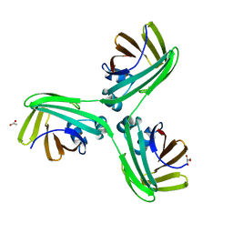
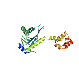
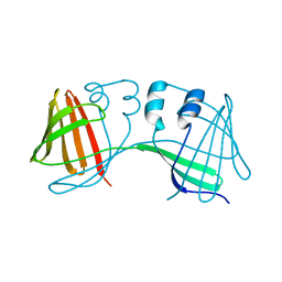
6WP0
DownloadVisualize
BU of 6wp0 by Molmil
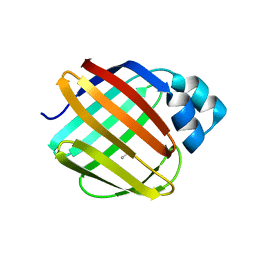
The Crystal Structure of Domain-Swapped Trimer Q108K:T51D variant of HCRBPII
Descriptor: ACETATE ION, GLYCEROL, Retinol-binding protein 2
Authors:Ghanbarpour, A, Geiger, J.
Deposit date:2020-04-26
Release date:2020-08-19
Last modified:2023-10-18
Method:X-RAY DIFFRACTION (2.78 Å)
Cite:Human Cellular Retinol Binding Protein II Forms a Domain-Swapped Trimer Representing a Novel Fold and a New Template for Protein Engineering.
Chembiochem, 21, 2020
6WP2
DownloadVisualize
BU of 6wp2 by Molmil
The Crystal Structure of Apo Zinc-Bound Domain Swapped-Trimer Q108K:K40D:T53A:R58L:Q38F:Q4F:F57H Variant of HCRBPII
Descriptor: ACETATE ION, GLYCEROL, Retinol-binding protein 2, ...
Authors:Ghanbarpour, A, Geiger, J.
Deposit date:2020-04-26
Release date:2020-08-19
Last modified:2023-10-18
Method:X-RAY DIFFRACTION (2.48 Å)
Cite:Human Cellular Retinol Binding Protein II Forms a Domain-Swapped Trimer Representing a Novel Fold and a New Template for Protein Engineering.
Chembiochem, 21, 2020
6WP1
DownloadVisualize
BU of 6wp1 by Molmil
The Crystal Structure of Apo Domain-Swapped Trimer Q108K:K40L:T51K Variant of HCRBPII
Descriptor: ACETATE ION, GLYCEROL, Retinol-binding protein 2
Authors:Ghanbarpour, A, Geiger, J.
Deposit date:2020-04-26
Release date:2020-08-19
Last modified:2023-10-18
Method:X-RAY DIFFRACTION (2.99 Å)
Cite:Human Cellular Retinol Binding Protein II Forms a Domain-Swapped Trimer Representing a Novel Fold and a New Template for Protein Engineering.
Chembiochem, 21, 2020
2HD5
DownloadVisualize
BU of 2hd5 by Molmil
USP2 in complex with ubiquitin
Descriptor: Polyubiquitin, Ubiquitin carboxyl-terminal hydrolase 2, ZINC ION
Authors:Renatus, M, Kroemer, M.
Deposit date:2006-06-20
Release date:2006-08-15
Last modified:2023-08-30
Method:X-RAY DIFFRACTION (1.85 Å)
Cite:Structural Basis of Ubiquitin Recognition by the Deubiquitinating Protease USP2.
Structure, 14, 2006
2FDC
DownloadVisualize
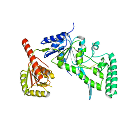
BU of 2fdc by Molmil
Structural Basis of DNA Damage Recognition and Processing by UvrB: crystal structure of a UvrB/DNA complex
Descriptor: 5'-D(P*CP*GP*GP*CP*TP*CP*CP*AP*TP*CP*TP*CP*TP*AP*CP*CP*GP*CP*AP*A)-3', N-[6-(ACETYLAMINO)HEXYL]-3',6'-DIHYDROXY-3-OXO-3H-SPIRO[2-BENZOFURAN-1,9'-XANTHENE]-6-CARBOXAMIDE, UvrABC system protein B
Authors:Truglio, J.J, Kisker, C.
Deposit date:2005-12-13
Release date:2006-03-14
Last modified:2024-02-14
Method:X-RAY DIFFRACTION (3.3 Å)
Cite:Structural basis for DNA recognition and processing by UvrB.
Nat.Struct.Mol.Biol., 13, 2006
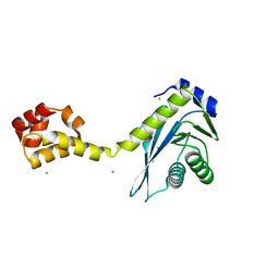
2NRT
DownloadVisualize
BU of 2nrt by Molmil
Crystal structure of the C-terminal half of UvrC
Descriptor: CHLORIDE ION, UvrABC system protein C
Authors:Karakas, E, Truglio, J.J, Kisker, C.
Deposit date:2006-11-02
Release date:2007-02-06
Last modified:2023-08-30
Method:X-RAY DIFFRACTION (1.5 Å)
Cite:Structure of the C-terminal half of UvrC reveals an RNase H endonuclease domain with an Argonaute-like catalytic triad.
Embo J., 26, 2007
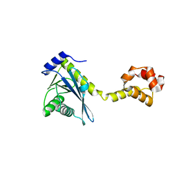
2NRV
DownloadVisualize
BU of 2nrv by Molmil
Crystal structure of the C-terminal half of UvrC
Descriptor: SODIUM ION, UvrABC system protein C
Authors:Karakas, E, Truglio, J.J, Kisker, C.
Deposit date:2006-11-02
Release date:2007-02-06
Last modified:2023-08-30
Method:X-RAY DIFFRACTION (1.8 Å)
Cite:Structure of the C-terminal half of UvrC reveals an RNase H endonuclease domain with an Argonaute-like catalytic triad.
Embo J., 26, 2007
2NRW
DownloadVisualize
BU of 2nrw by Molmil
Crystal structure of the C terminal half of UvrC
Descriptor: UvrABC system protein C
Authors:Karakas, E, Truglio, J.J, Kisker, C.
Deposit date:2006-11-02
Release date:2007-02-06
Last modified:2023-08-30
Method:X-RAY DIFFRACTION (2.3 Å)
Cite:Structure of the C-terminal half of UvrC reveals an RNase H endonuclease domain with an Argonaute-like catalytic triad.
Embo J., 26, 2007
2NRR
DownloadVisualize
BU of 2nrr by Molmil
Crystal structure of the C-terminal RNAseH endonuclase domain of UvrC
Descriptor: UvrABC system protein C
Authors:Karakas, E, Truglio, J.J, Kisker, C.
Deposit date:2006-11-02
Release date:2007-02-06
Last modified:2023-12-27
Method:X-RAY DIFFRACTION (1.2 Å)
Cite:Structure of the C-terminal half of UvrC reveals an RNase H endonuclease domain with an Argonaute-like catalytic triad.
Embo J., 26, 2007
2NRX
DownloadVisualize
BU of 2nrx by Molmil
Crystal structure of the C-terminal half of UvrC, in the presence of sulfate molecules
Descriptor: GLYCEROL, SULFATE ION, UvrABC system protein C
Authors:Karakas, E, Truglio, J.J, Kisker, C.
Deposit date:2006-11-02
Release date:2007-02-06
Last modified:2023-08-30
Method:X-RAY DIFFRACTION (1.9 Å)
Cite:Structure of the C-terminal half of UvrC reveals an RNase H endonuclease domain with an Argonaute-like catalytic triad.
Embo J., 26, 2007
2NRZ
DownloadVisualize
BU of 2nrz by Molmil
Crystal structure of the C-terminal half of UvrC bound to its catalytic divalent cation
Descriptor: MANGANESE (II) ION, UvrABC system protein C
Authors:Karakas, E, Truglio, J.J, Kisker, C.
Deposit date:2006-11-02
Release date:2007-02-06
Last modified:2023-08-30
Method:X-RAY DIFFRACTION (2 Å)
Cite:Structure of the C-terminal half of UvrC reveals an RNase H endonuclease domain with an Argonaute-like catalytic triad.
Embo J., 26, 2007
4YGG
DownloadVisualize
BU of 4ygg by Molmil
CRYSTAL STRUCTURE OF R111K:Y134F:T54V:R132Q:P39Y:R59Y MUTANT OF HUMAN CELLULAR RETINOIC ACID BINDING PROTEIN II WITH RETINAL AT 1.9 ANGSTROM RESOLUTION - VISIBLE LIGHT IRRADIATED CRYSTAL - 2ND CYCLE
Descriptor: Cellular retinoic acid-binding protein 2, RETINAL
Authors:Nosrati, M, Geiger, J.H.
Deposit date:2015-02-26
Release date:2016-03-02
Last modified:2024-11-20
Method:X-RAY DIFFRACTION (1.9 Å)
Cite:A Photoisomerizing Rhodopsin Mimic Observed at Atomic Resolution.
J.Am.Chem.Soc., 138, 2016
6C7Z
DownloadVisualize
BU of 6c7z by Molmil
Crystal structure of the Q108K:K40L:T51V:R58F mutant of human Cellular Retinol Binding Protein II in complex with synthetic Ligand Julolidine
Descriptor: (2E,4E)-3-methyl-5-(2,3,6,7-tetrahydro-1H,5H-pyrido[3,2,1-ij]quinolin-9-yl)penta-2,4-dienal, ACETATE ION, Retinol-binding protein 2
Authors:Nosrati, M, Geiger, J.H.
Deposit date:2018-01-23
Release date:2018-04-25
Last modified:2024-11-13
Method:X-RAY DIFFRACTION (1.42 Å)
Cite:A Genetically Encoded Ratiometric pH Probe: Wavelength Regulation-Inspired Design of pH Indicators.
Chembiochem, 19, 2018
3I17
DownloadVisualize
BU of 3i17 by Molmil
Crystal structure of the apo R132K:L121E mutant of cellular retinoic acid-binding protein II at 1.68 angstrom resolution
Descriptor: Cellular retinoic acid-binding protein 2
Authors:Jia, X, Watson, C.T, Geiger, J.H.
Deposit date:2009-06-25
Release date:2009-07-28
Last modified:2023-09-06
Method:X-RAY DIFFRACTION (1.68 Å)
Cite:Elucidating the exact role of engineered CRABPII residues for the formation of a retinal protonated Schiff base.
Proteins, 77, 2009
4YBU
DownloadVisualize
BU of 4ybu by Molmil
Crystal structure of the R111K:Y134F:T54V:R132Q:P39Q:R59Y mutant of human Cellular Retinoic Acid Binding ProteinII in complex with Retinal after 24 h incubation and 1 hour UV irradiation at 1.92 angstrom - 1st cycle
Descriptor: Cellular retinoic acid-binding protein 2, RETINAL
Authors:Nosrati, M, Geiger, J.H.
Deposit date:2015-02-19
Release date:2016-02-24
Last modified:2024-10-16
Method:X-RAY DIFFRACTION (1.924 Å)
Cite:A Photoisomerizing Rhodopsin Mimic Observed at Atomic Resolution.
J.Am.Chem.Soc., 138, 2016
4YCH
DownloadVisualize
BU of 4ych by Molmil
Crystal Structure of R111K:Y134F:T54V:R132Q:P39Q:R59Y mutant of human Cellular Retinoic Acid Binding ProteinII with Retinal at 1.96 Angstrom - UV irradiated Crystal for 1 hour - 2nd cycle
Descriptor: Cellular retinoic acid-binding protein 2, RETINAL
Authors:Nosrati, M, Geiger, J.H.
Deposit date:2015-02-20
Release date:2016-02-24
Last modified:2024-11-20
Method:X-RAY DIFFRACTION (1.96 Å)
Cite:A Photoisomerizing Rhodopsin Mimic Observed at Atomic Resolution.
J.Am.Chem.Soc., 138, 2016
6E5E
DownloadVisualize
BU of 6e5e by Molmil
Crystal structure of the apo domain-swapped dimer Q108K:T51D mutant of human cellular retinol binding protein II
Descriptor: ACETATE ION, Retinol-binding protein 2
Authors:Ghanbarpour, A, Geiger, J.
Deposit date:2018-07-20
Release date:2019-10-16
Last modified:2023-10-11
Method:X-RAY DIFFRACTION (1.696 Å)
Cite:Engineering the hCRBPII Domain-Swapped Dimer into a New Class of Protein Switches.
J.Am.Chem.Soc., 141, 2019
6E5Q
DownloadVisualize
BU of 6e5q by Molmil
Crystal structure of the apo domain-swapped dimer Q108K:T51D:A28H mutant of human Cellular Retinol Binding Protein II
Descriptor: ACETATE ION, Retinol-binding protein 2
Authors:Ghanbarpour, A, Geiger, J.
Deposit date:2018-07-21
Release date:2019-10-16
Last modified:2023-10-11
Method:X-RAY DIFFRACTION (1.99 Å)
Cite:Engineering the hCRBPII Domain-Swapped Dimer into a New Class of Protein Switches.
J.Am.Chem.Soc., 141, 2019
6E51
DownloadVisualize
BU of 6e51 by Molmil
Crystal structure of the apo domain-swapped dimer Q108K:K40L:T51W mutant of human cellular retinol binding protein II
Descriptor: Retinol-binding protein 2
Authors:Ghanbarpour, A, Geiger, J.
Deposit date:2018-07-19
Release date:2019-10-16
Last modified:2023-10-11
Method:X-RAY DIFFRACTION (2.256 Å)
Cite:Engineering the hCRBPII Domain-Swapped Dimer into a New Class of Protein Switches.
J.Am.Chem.Soc., 141, 2019
6E50
DownloadVisualize
BU of 6e50 by Molmil
Crystal structure of the apo domain-swapped dimer Q108K:K40L:T51F mutant of human Cellular Retinol Binding Protein II
Descriptor: ACETATE ION, Retinol-binding protein 2
Authors:Ghanbarpour, A, Geiger, J.
Deposit date:2018-07-18
Release date:2019-10-16
Last modified:2023-10-11
Method:X-RAY DIFFRACTION (1.965 Å)
Cite:Engineering the hCRBPII Domain-Swapped Dimer into a New Class of Protein Switches.
J.Am.Chem.Soc., 141, 2019
6E5R
DownloadVisualize
BU of 6e5r by Molmil
Crystal structure of the apo domain-swapped dimer Q108K:T51D:A28C mutant of human Cellular Retinol Binding Protein II
Descriptor: ACETATE ION, GLYCEROL, Retinol-binding protein 2
Authors:Ghanbarpour, A, Geiger, J.
Deposit date:2018-07-22
Release date:2019-08-07
Last modified:2024-11-20
Method:X-RAY DIFFRACTION (2.592 Å)
Cite:Engineering the hCRBPII Domain-Swapped Dimer into a New Class of Protein Switches.
J.Am.Chem.Soc., 141, 2019
6E7M
DownloadVisualize
BU of 6e7m by Molmil
Crystal structure of the holo retinal-bound domain-swapped dimer Q108K:T51D:A28C mutant of human Cellular Retinol Binding Protein II
Descriptor: ACETATE ION, GLYCEROL, RETINAL, ...
Authors:Ghanbarpour, A, Geiger, J.
Deposit date:2018-07-26
Release date:2019-10-16
Last modified:2024-11-13
Method:X-RAY DIFFRACTION (2.7 Å)
Cite:Engineering the hCRBPII Domain-Swapped Dimer into a New Class of Protein Switches.
J.Am.Chem.Soc., 141, 2019

227561

數據於2024-11-20公開中

PDB statisticsPDBj update infoContact PDBjnumon