Loading
PDBj
MenuPDBj@FacebookPDBj@TwitterPDBj@YouTubewwPDB FoundationwwPDB
RCSB PDBPDBeBMRBAdv. SearchSearch help
Search by PDB author
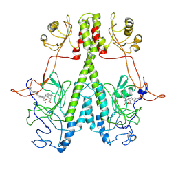
7JRI
DownloadVisualize
BU of 7jri by Molmil
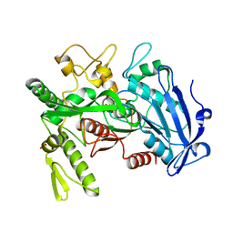
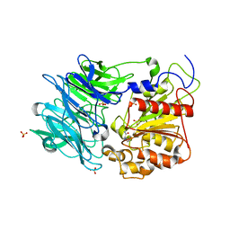
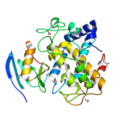
High-resolution Crystal Structures of Transient Intermediates in the Phytochrome Photocycle, 33 ms structure
Descriptor: 3-[2-[[5-[[(3E,4S)-3-ethylidene-4-methyl-5-oxidanylidene-pyrrol-2-yl]methyl]-3-(3-hydroxy-3-oxopropyl)-4-methyl-1H-pyrrol-2-yl]methyl]-5-[(4-ethyl-3-methyl-5-oxidanylidene-pyrrol-2-yl)methyl]-4-methyl-1H-pyrrol-3-yl]propanoic acid, BENZAMIDINE, Photoreceptor-histidine kinase BphP
Authors:Schmidt, M, Stojkovic, E.
Deposit date:2020-08-12
Release date:2021-04-07
Last modified:2023-10-18
Method:X-RAY DIFFRACTION (2.4 Å)
Cite:High-resolution crystal structures of transient intermediates in the phytochrome photocycle.
Structure, 29, 2021
7JR5
DownloadVisualize
BU of 7jr5 by Molmil
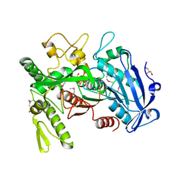
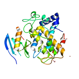
Real Time Reaction Intermediates in Stigmatella Bacteriophytochrome P2
Descriptor: 3-[2-[[5-[(4-ethenyl-3-methyl-5-oxidanylidene-pyrrol-2-yl)methyl]-3-(3-hydroxy-3-oxopropyl)-4-methyl-1~{H}-pyrrol-2-yl]methyl]-5-[[(3~{S})-4-ethyl-3-methyl-2-oxidanylidene-1,3-dihydropyrrol-5-yl]methyl]-4-methyl-1~{H}-pyrrol-3-yl]propanoic acid, BENZAMIDINE, Photoreceptor-histidine kinase BphP
Authors:Schmidt, M.
Deposit date:2020-08-11
Release date:2021-10-06
Last modified:2023-10-18
Method:X-RAY DIFFRACTION (2.4 Å)
Cite:High-resolution crystal structures of transient intermediates in the phytochrome photocycle.
Structure, 29, 2021
2RQP
DownloadVisualize
BU of 2rqp by Molmil
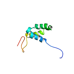
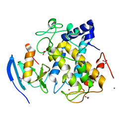
The Solution Structure of Heterochromatin Protein 1-Binding Protein 74 Histone H1 like domain
Descriptor: Heterochromatin protein 1-binding protein 3
Authors:Shimamoto, S, Sugahara, H, Ohkubo, T.
Deposit date:2009-09-04
Release date:2009-12-29
Last modified:2024-05-29
Method:SOLUTION NMR
Cite:The middle region of an HP1-binding protein, HP1-BP74, associates with linker DNA at the entry/exit site of nucleosomal DNA
J.Biol.Chem., 285, 2010
5X7Y
DownloadVisualize
BU of 5x7y by Molmil
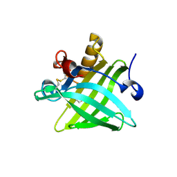
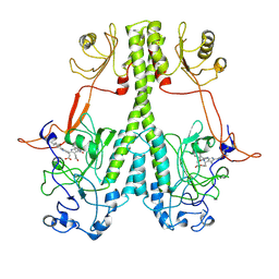
Crystal Structure of the Dog Lipocalin Allergen Can f 6
Descriptor: DI(HYDROXYETHYL)ETHER, Lipocalin-Can f 6 allergen
Authors:Yamamoto, K, Otani, T, Sugiura, K, Nakatsuji, M, Nishimura, S, Inui, T.
Deposit date:2017-02-28
Release date:2018-04-04
Last modified:2023-11-22
Method:X-RAY DIFFRACTION (2.35 Å)
Cite:Crystal structure of the dog allergen Can f 6 and structure-based implications of its cross-reactivity with the cat allergen Fel d 4.
Sci Rep, 9, 2019
4DBG
DownloadVisualize
BU of 4dbg by Molmil
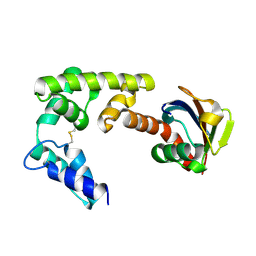
Crystal structure of HOIL-1L-UBL complexed with a HOIP-UBA derivative
Descriptor: RING finger protein 31, RanBP-type and C3HC4-type zinc finger-containing protein 1
Authors:Yagi, H, Hiromoto, T, Mizushima, T, Kurimoto, E, Kato, K.
Deposit date:2012-01-15
Release date:2012-04-04
Last modified:2012-05-16
Method:X-RAY DIFFRACTION (2.71 Å)
Cite:A non-canonical UBA-UBL interaction forms the linear-ubiquitin-chain assembly complex
Embo Rep., 13, 2012
5Z30
DownloadVisualize
BU of 5z30 by Molmil
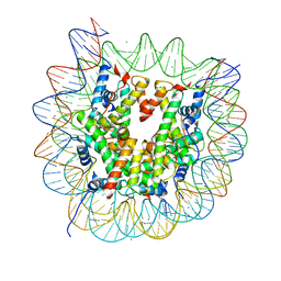
The crystal structure of the nucleosome containing a cancer-associated histone H2A.Z R80C mutant
Descriptor: CHLORIDE ION, DNA (146-MER), Histone H2A.Z, ...
Authors:Horikoshi, N, Arimura, Y, Kurumizaka, H.
Deposit date:2018-01-05
Release date:2018-07-18
Last modified:2023-11-22
Method:X-RAY DIFFRACTION (2.45 Å)
Cite:Cancer-associated mutations of histones H2B, H3.1 and H2A.Z.1 affect the structure and stability of the nucleosome.
Nucleic Acids Res., 46, 2018
7MPB
DownloadVisualize
BU of 7mpb by Molmil
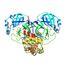
SARS Coronavirus-2 Main Protease 3CL-pro binding Ascorbate
Descriptor: 3C-like proteinase, ASCORBIC ACID, TRIFLUOROETHANOL
Authors:Pandey, S, Malla, T.N, Stojkovic, E.A, Schmidt, M.
Deposit date:2021-05-04
Release date:2021-05-12
Last modified:2023-10-18
Method:X-RAY DIFFRACTION (2.3 Å)
Cite:Vitamin C inhibits SARS coronavirus-2 main protease essential for viral replication
Biorxiv, 2021
6LE4
DownloadVisualize
BU of 6le4 by Molmil
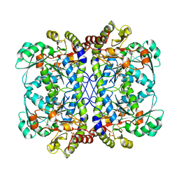
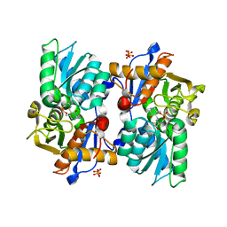
Crystal structure of cystathionine gamma-lyase from Lactobacillus plantarum complexed with cystathionine
Descriptor: (2~{S})-4-[(2~{R})-2-azanyl-3-oxidanyl-3-oxidanylidene-propyl]sulfanyl-2-[(~{E})-[2-methyl-3-oxidanyl-5-(phosphonooxymethyl)pyridin-4-yl]methylideneamino]butanoic acid, Cystathionine gamma-lyase, PHOSPHATE ION
Authors:Oda, K, Matoba, Y.
Deposit date:2019-11-24
Release date:2020-10-07
Last modified:2023-11-22
Method:X-RAY DIFFRACTION (3.1 Å)
Cite:Catalytic specificity of the Lactobacillus plantarum cystathionine gamma-lyase presumed by the crystallographic analysis.
Sci Rep, 10, 2020
6LDO
DownloadVisualize
BU of 6ldo by Molmil
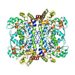
Crystal structure of cystathionine gamma-lyase from Lactobacillus plantarum complexed with L-serine
Descriptor: (E)-N-({3-hydroxy-2-methyl-5-[(phosphonooxy)methyl]pyridin-4-yl}methylidene)-L-serine, Cystathionine gamma-lyase, PHOSPHATE ION
Authors:Oda, K, Matoba, Y.
Deposit date:2019-11-22
Release date:2020-10-07
Last modified:2023-11-22
Method:X-RAY DIFFRACTION (2.75 Å)
Cite:Catalytic specificity of the Lactobacillus plantarum cystathionine gamma-lyase presumed by the crystallographic analysis.
Sci Rep, 10, 2020
5B1H
DownloadVisualize
BU of 5b1h by Molmil
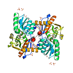
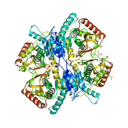
Crystal structure of cystathionine beta-synthase from Lactobacillus plantarum
Descriptor: Cystathionine beta-synthase, GLYCEROL, SULFATE ION
Authors:Matoba, Y, Sugiyama, M.
Deposit date:2015-12-04
Release date:2016-12-07
Last modified:2023-11-15
Method:X-RAY DIFFRACTION (2.4 Å)
Cite:Crystallographic and mutational analyses of cystathionine beta-synthase in the H2 S-synthetic gene cluster in Lactobacillus plantarum
Protein Sci., 26, 2017
5B1I
DownloadVisualize
BU of 5b1i by Molmil
Crystal structure of K42A mutant of cystathionine beta-synthase from Lactobacillus plantarum in a complex with L-methionine
Descriptor: Cystathionine beta-synthase, N-[(3-HYDROXY-2-METHYL-5-{[(TRIHYDROXYPHOSPHORANYL)OXY]METHYL}PYRIDIN-4-YL)METHYLENE]METHIONINE, SULFATE ION
Authors:Matoba, Y, Sugiyama, M.
Deposit date:2015-12-04
Release date:2016-12-07
Last modified:2023-11-08
Method:X-RAY DIFFRACTION (3.3 Å)
Cite:Crystallographic and mutational analyses of cystathionine beta-synthase in the H2 S-synthetic gene cluster in Lactobacillus plantarum
Protein Sci., 26, 2017
5DSV
DownloadVisualize
BU of 5dsv by Molmil
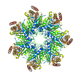
Crystal structure of human proteasome alpha7 tetradecamer
Descriptor: Proteasome subunit alpha type-3
Authors:Satoh, T, Thammaporn, R, Seetaha, S, Kato, K.
Deposit date:2015-09-17
Release date:2015-12-02
Last modified:2023-11-08
Method:X-RAY DIFFRACTION (3.75 Å)
Cite:Disassembly of the self-assembled, double-ring structure of proteasome alpha 7 homo-tetradecamer by alpha 6
Sci Rep, 5, 2015
3WSW
DownloadVisualize
BU of 3wsw by Molmil
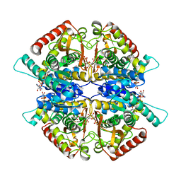
Crystal structure of minor L-lactate dehydrogenase from Enterococcus mundtii in the ligands-bound form
Descriptor: 1,6-di-O-phosphono-beta-D-fructofuranose, GLYCEROL, L-lactate dehydrogenase, ...
Authors:Matoba, Y, Sugiyama, M.
Deposit date:2014-03-27
Release date:2014-09-10
Last modified:2023-11-08
Method:X-RAY DIFFRACTION (2.3 Å)
Cite:An alternative allosteric regulation mechanism of an acidophilic l-lactate dehydrogenase from Enterococcus mundtii 15-1A.
Febs Open Bio, 4, 2014
3WSV
DownloadVisualize
BU of 3wsv by Molmil
Crystal structure of minor L-lactate dehydrogenase from Enterococcus mundtii in the ligands-unbound form
Descriptor: GLYCEROL, L-lactate dehydrogenase
Authors:Matoba, Y, Sugiyama, M.
Deposit date:2014-03-27
Release date:2014-09-10
Last modified:2023-11-08
Method:X-RAY DIFFRACTION (2.38 Å)
Cite:An alternative allosteric regulation mechanism of an acidophilic l-lactate dehydrogenase from Enterococcus mundtii 15-1A.
Febs Open Bio, 4, 2014
3WA6
DownloadVisualize
BU of 3wa6 by Molmil
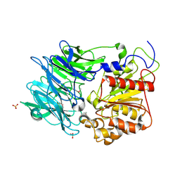
Crystal structure of tannase from Lactobacillus plantarum in the orthorhombic crystal
Descriptor: SULFATE ION, tannase
Authors:Matoba, Y, Tanaka, N, Sugiyama, M.
Deposit date:2013-04-27
Release date:2013-07-24
Last modified:2024-03-20
Method:X-RAY DIFFRACTION (1.8 Å)
Cite:Crystallographic and mutational analyses of tannase from Lactobacillus plantarum
Proteins, 81, 2013
3WA7
DownloadVisualize
BU of 3wa7 by Molmil
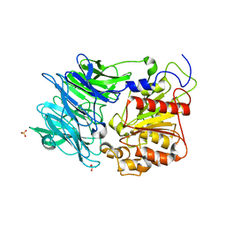
Crystal structure of selenomethionine-labeled tannase from Lactobacillus plantarum in the orthorhombic crystal
Descriptor: ACETATE ION, GLYCEROL, SULFATE ION, ...
Authors:Matoba, Y, Tanaka, N, Sugiyama, M.
Deposit date:2013-04-27
Release date:2013-07-24
Last modified:2013-11-06
Method:X-RAY DIFFRACTION (1.7 Å)
Cite:Crystallographic and mutational analyses of tannase from Lactobacillus plantarum.
Proteins, 81, 2013
3AZO
DownloadVisualize
BU of 3azo by Molmil
Crystal structure of puromycin hydrolase
Descriptor: Aminopeptidase, SULFATE ION
Authors:Matoba, Y, Sugiyama, M.
Deposit date:2011-05-27
Release date:2011-07-27
Last modified:2011-09-28
Method:X-RAY DIFFRACTION (2 Å)
Cite:Structural evidence that puromycin hydrolase is a new type of aminopeptidase with a prolyl oligopeptidase family fold
Proteins, 79, 2011
3AZP
DownloadVisualize
BU of 3azp by Molmil
Crystal structure of puromycin hydrolase S511A mutant
Descriptor: Aminopeptidase, SULFATE ION
Authors:Matoba, Y, Sugiyama, M.
Deposit date:2011-05-27
Release date:2011-07-27
Last modified:2023-11-01
Method:X-RAY DIFFRACTION (2.15 Å)
Cite:Structural evidence that puromycin hydrolase is a new type of aminopeptidase with a prolyl oligopeptidase family fold
Proteins, 79, 2011
3AZQ
DownloadVisualize
BU of 3azq by Molmil
Crystal structure of puromycin hydrolase S511A mutant complexed with PGG
Descriptor: Aminopeptidase, SULFATE ION, tripeptide PGG
Authors:Matoba, Y, Sugiyama, M.
Deposit date:2011-05-27
Release date:2011-07-27
Last modified:2023-11-01
Method:X-RAY DIFFRACTION (2.7 Å)
Cite:Structural evidence that puromycin hydrolase is a new type of aminopeptidase with a prolyl oligopeptidase family fold
Proteins, 79, 2011
3WCK
DownloadVisualize
BU of 3wck by Molmil
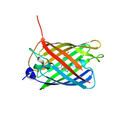
Crystal structure of monomeric photosensitizing fluorescent protein, Supernova
Descriptor: Monomeric photosenitizing fluorescent protein supernova
Authors:Sakai, N, Matsuda, T, Takemoto, K, Nagai, T.
Deposit date:2013-05-27
Release date:2013-10-02
Last modified:2023-11-15
Method:X-RAY DIFFRACTION (2.3 Å)
Cite:SuperNova, a monomeric photosensitizing fluorescent protein for chromophore-assisted light inactivation
Sci Rep, 3, 2013
2ZWF
DownloadVisualize
BU of 2zwf by Molmil
Crystal structure of the copper-bound tyrosinase in complex with a caddie protein from streptomyces castaneoglobisporus obtained by soaking the deoxy-form crystal in dioxygen-saturated solution for 80 minutes
Descriptor: COPPER (II) ION, MelC, NITRATE ION, ...
Authors:Matoba, Y, Sugiyama, M.
Deposit date:2008-12-03
Release date:2009-12-15
Last modified:2023-11-01
Method:X-RAY DIFFRACTION (1.4 Å)
Cite:Crystallographic evidence of drastic movement of a copper ion toward the substrate tyrosine for starting hydroxylation reaction of tyrosinase
To be Published
2ZWE
DownloadVisualize
BU of 2zwe by Molmil
Crystal structure of the copper-bound tyrosinase in complex with a caddie protein from streptomyces castaneoglobisporus obtained by soaking the deoxy-form crystal in dioxygen-saturated solution for 40 minutes
Descriptor: COPPER (II) ION, MelC, NITRATE ION, ...
Authors:Matoba, Y, Sugiyama, M.
Deposit date:2008-12-03
Release date:2009-12-15
Last modified:2023-11-01
Method:X-RAY DIFFRACTION (1.32 Å)
Cite:Crystallographic evidence of drastic movement of a copper ion toward the substrate tyrosine for starting hydroxylation reaction of tyrosinase
To be Published
2ZMZ
DownloadVisualize
BU of 2zmz by Molmil
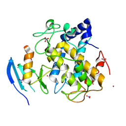
The 1.37-A crystal structure of the hydroxylamine-induced deoxy-form of the copper-bound tyrosinase in complex with a caddie protein from Streptomyces castaneoglobisporus
Descriptor: CADDIE, COPPER (I) ION, NITRATE ION, ...
Authors:Matoba, Y, Sugiyama, M.
Deposit date:2008-04-21
Release date:2009-05-05
Last modified:2023-11-01
Method:X-RAY DIFFRACTION (1.37 Å)
Cite:X-Ray Snapshots of a Hydroxylation Mechanism of Tyrosinase
To be Published
2ZWD
DownloadVisualize
BU of 2zwd by Molmil
Crystal structure of the copper-bound tyrosinase in complex with a caddie protein from streptomyces castaneoglobisporus obtained by soaking the deoxy-form crystal in dioxygen-saturated solution for 5 minutes
Descriptor: COPPER (I) ION, MelC, NITRATE ION, ...
Authors:Matoba, Y, Sugiyama, M.
Deposit date:2008-12-03
Release date:2009-12-15
Last modified:2023-11-01
Method:X-RAY DIFFRACTION (1.35 Å)
Cite:Crystallographic evidence of drastic movement of a copper ion toward the substrate tyrosine for starting hydroxylation reaction of tyrosinase
To be Published
2ZMY
DownloadVisualize
BU of 2zmy by Molmil
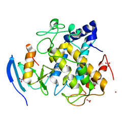
Crystal structure of the met2-form of the copper-bound tyrosinase in complex with a caddie protein from Streptomyces castaneoglobisporus obtained by soaking in cupric sulfate solution for 80 hours
Descriptor: CADDIE, COPPER (II) ION, NITRATE ION, ...
Authors:Matoba, Y, Sugiyama, M.
Deposit date:2008-04-21
Release date:2009-05-05
Last modified:2023-11-01
Method:X-RAY DIFFRACTION (1.45 Å)
Cite:X-Ray Snapshots of a Hydroxylation Mechanism of Tyrosinase
To be Published

220760

數據於2024-06-05公開中

PDB statisticsPDBj update infoContact PDBjnumon