Loading
PDBj
MenuPDBj@FacebookPDBj@TwitterPDBj@YouTubewwPDB FoundationwwPDB
RCSB PDBPDBeBMRBAdv. SearchSearch help
Search by PDB author
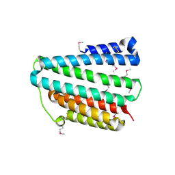
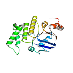
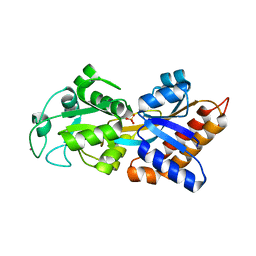
5V77
DownloadVisualize
BU of 5v77 by Molmil
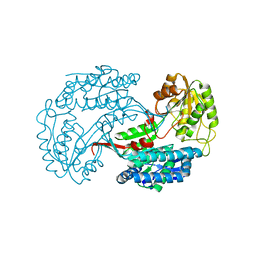
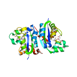
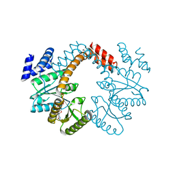
Crystal structure of an uncharacterized protein from Neisseria gonorrhoeae
Descriptor: SULFATE ION, Uncharacterized protein
Authors:Seattle Structural Genomics Center for Infectious Disease (SSGCID)
Deposit date:2017-03-18
Release date:2017-04-12
Last modified:2024-03-06
Method:X-RAY DIFFRACTION (1.85 Å)
Cite:Crystal structure of an uncharacterized protein from Neisseria gonorrhoeae
To be Published
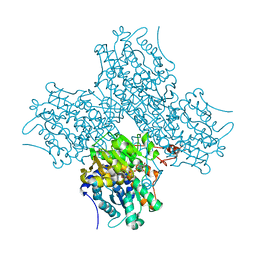
5WNN
DownloadVisualize
BU of 5wnn by Molmil
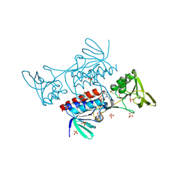
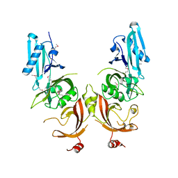
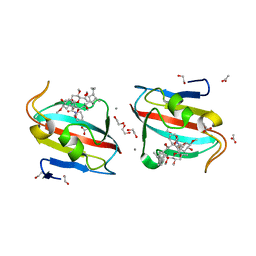
Crystal structure of Phosphate-binding protein PstS protein from Burkholderia pseudomallei
Descriptor: PHOSPHATE ION, POTASSIUM ION, Phosphate-binding protein PstS
Authors:Seattle Structural Genomics Center for Infectious Disease (SSGCID)
Deposit date:2017-08-01
Release date:2017-08-30
Last modified:2023-10-04
Method:X-RAY DIFFRACTION (1.85 Å)
Cite:Crystal structure of Phosphate-binding protein PstS protein from Burkholderia pseudomallei
To Be Published
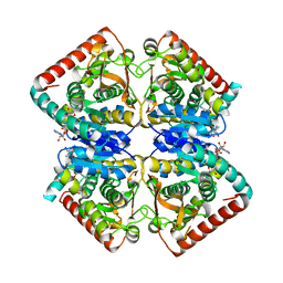
5UJU
DownloadVisualize
BU of 5uju by Molmil
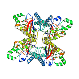
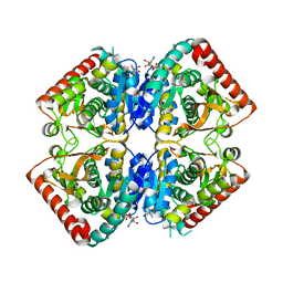
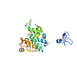
Crystal structure of NAD-dependent aldehyde dehydrogenase from Burkholderia multivorans
Descriptor: 1,2-ETHANEDIOL, IMIDAZOLE, NAD-dependent aldehyde dehydrogenase, ...
Authors:Seattle Structural Genomics Center for Infectious Disease (SSGCID)
Deposit date:2017-01-19
Release date:2017-04-12
Last modified:2023-10-04
Method:X-RAY DIFFRACTION (2.05 Å)
Cite:Crystal structure of NAD-dependent aldehyde dehydrogenase from Burkholderia multivorans
TO BE PUBLISHED
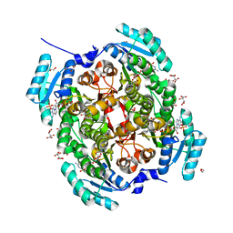
5UM0
DownloadVisualize
BU of 5um0 by Molmil
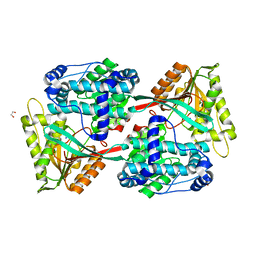
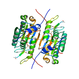
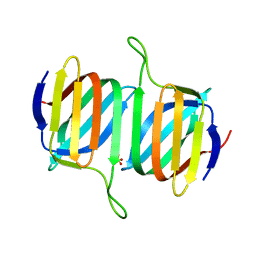
Crystal structure of 2,3-bisphosphoglycerate-dependent phosphoglycerate mutase from Neisseria gonorrhoeae
Descriptor: 2,3-bisphosphoglycerate-dependent phosphoglycerate mutase, L(+)-TARTARIC ACID
Authors:Seattle Structural Genomics Center for Infectious Disease (SSGCID)
Deposit date:2017-01-25
Release date:2017-02-15
Last modified:2023-10-04
Method:X-RAY DIFFRACTION (1.85 Å)
Cite:Crystal structure of 2,3-bisphosphoglycerate-dependent phosphoglycerate mutase from Neisseria gonorrhoeae
TO BE PUBLISHED
3I44
DownloadVisualize
BU of 3i44 by Molmil
Crystal structure of aldehyde dehydrogenase from bartonella henselae at 2.0A resolution
Descriptor: Aldehyde dehydrogenase, SODIUM ION, UNKNOWN ATOM OR ION
Authors:Seattle Structural Genomics Center for Infectious Disease (SSGCID)
Deposit date:2009-07-01
Release date:2009-07-14
Last modified:2023-09-06
Method:X-RAY DIFFRACTION (2 Å)
Cite:Crystal structure of aldehyde dehydrogenase from bartonella henselae at 2.0A resolution
To be Published
5UTH
DownloadVisualize
BU of 5uth by Molmil
Crystal structure of thioredoxin reductase from Mycobacterium smegmatis in complex with FAD
Descriptor: FLAVIN-ADENINE DINUCLEOTIDE, SULFATE ION, Thioredoxin reductase
Authors:Seattle Structural Genomics Center for Infectious Disease (SSGCID)
Deposit date:2017-02-15
Release date:2017-03-08
Last modified:2024-10-23
Method:X-RAY DIFFRACTION (1.95 Å)
Cite:Crystal structure of thioredoxin reductase from Mycobacterium smegmatis in complex with FAD
to be published
6AR7
DownloadVisualize
BU of 6ar7 by Molmil
Crystal structure of a putative uncharacterized protein from Burkholderia thailandensis
Descriptor: Uncharacterized protein
Authors:Seattle Structural Genomics Center for Infectious Disease (SSGCID)
Deposit date:2017-08-21
Release date:2017-09-20
Last modified:2024-11-06
Method:X-RAY DIFFRACTION (2.1 Å)
Cite:Crystal structure of a Putative uncharacterized protein from Burkholderia thailandensis
To be Published
6APH
DownloadVisualize
BU of 6aph by Molmil
Crystal structure of Adenosylhomocysteinase from Elizabethkingia anophelis NUHP1 in complex with NAD and Adenosine
Descriptor: (4S)-2-METHYL-2,4-PENTANEDIOL, ADENOSINE, Adenosylhomocysteinase, ...
Authors:Seattle Structural Genomics Center for Infectious Disease (SSGCID)
Deposit date:2017-08-17
Release date:2017-08-30
Last modified:2024-03-13
Method:X-RAY DIFFRACTION (1.65 Å)
Cite:Crystal structure of Adenosylhomocysteinase from Elizabethkingia anophelis NUHP1 in complex with NAD and Adenosine
To be Published
3GVH
DownloadVisualize
BU of 3gvh by Molmil
Crystal structure of Lactate/malate dehydrogenase from Brucella melitensis
Descriptor: Malate dehydrogenase, NICOTINAMIDE-ADENINE-DINUCLEOTIDE
Authors:Seattle Structural Genomics Center for Infectious Disease (SSGCID)
Deposit date:2009-03-31
Release date:2009-04-14
Last modified:2023-09-06
Method:X-RAY DIFFRACTION (2.3 Å)
Cite:Crystal structure of Lactate/malate dehydrogenase from Brucella melitensis
To be Published
5U9P
DownloadVisualize
BU of 5u9p by Molmil
Crystal structure of a gluconate 5-dehydrogenase from Burkholderia cenocepacia J2315 in complex with NADP and tartrate
Descriptor: 1,2-ETHANEDIOL, Gluconate 5-dehydrogenase, L(+)-TARTARIC ACID, ...
Authors:Seattle Structural Genomics Center for Infectious Disease (SSGCID)
Deposit date:2016-12-17
Release date:2017-01-18
Last modified:2023-10-04
Method:X-RAY DIFFRACTION (1.65 Å)
Cite:Crystal structure of a gluconate 5-dehydrogenase from Burkholderia cenocepacia J2315 in complex with NADP and tartrate
TO BE PUBLISHED
3GVI
DownloadVisualize
BU of 3gvi by Molmil
Crystal structure of Lactate/malate dehydrogenase from Brucella melitensis in complex with ADP
Descriptor: ADENOSINE-5'-DIPHOSPHATE, Malate dehydrogenase
Authors:Seattle Structural Genomics Center for Infectious Disease (SSGCID)
Deposit date:2009-03-31
Release date:2009-04-14
Last modified:2023-09-06
Method:X-RAY DIFFRACTION (2.25 Å)
Cite:Crystal structure of Lactate/malate dehydrogenase from Brucella melitensis in complex with ADP
To be Published
5V6D
DownloadVisualize
BU of 5v6d by Molmil
Crystal structure of nucleoside diphosphate kinase from Neisseria gonorrhoeae in complex with citrate
Descriptor: 1,2-ETHANEDIOL, CITRIC ACID, Nucleoside diphosphate kinase
Authors:Seattle Structural Genomics Center for Infectious Disease (SSGCID)
Deposit date:2017-03-16
Release date:2017-04-19
Last modified:2023-10-04
Method:X-RAY DIFFRACTION (1.85 Å)
Cite:Crystal structure of nucleoside diphosphate kinase from Neisseria gonorrhoeae in complex with citrate
To be Published
4Q6U
DownloadVisualize
BU of 4q6u by Molmil
Crystal structure of a putative uncharacterized protein from Mycobacterium tuberculosis
Descriptor: PENTAETHYLENE GLYCOL, Uncharacterized protein
Authors:Seattle Structural Genomics Center for Infectious Disease (SSGCID)
Deposit date:2014-04-23
Release date:2014-06-18
Last modified:2018-03-07
Method:X-RAY DIFFRACTION (1.95 Å)
Cite:Mycobacterium tuberculosis Rv3651 is a triple sensor-domain protein.
Protein Sci., 27, 2018
3RJM
DownloadVisualize
BU of 3rjm by Molmil
CASPASE2 IN COMPLEX WITH CHDI LIGAND 33c
Descriptor: Caspase-2, Peptide inhibitor (ACE)VDV(3PX)D-CHO
Authors:Abendroth, J, Lorimer, D, Stewart, L, Maillard, M, Kiselyov, A.S.
Deposit date:2011-04-15
Release date:2011-09-21
Last modified:2023-12-06
Method:X-RAY DIFFRACTION (2.55 Å)
Cite:Exploiting differences in caspase-2 and -3 S(2) subsites for selectivity: Structure-based design, solid-phase synthesis and in vitro activity of novel substrate-based caspase-2 inhibitors.
Bioorg.Med.Chem., 19, 2011
5W07
DownloadVisualize
BU of 5w07 by Molmil
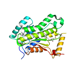
CRYSTAL STRUCTURE OF THE INHA FROM MYCOBACTERIUM TUBERCULOSIS IN COMPLEX WITH AN12855, EBSI 4333.
Descriptor: (~{N}~{E})-~{N}-[[2-[[2-ethylsulfonyl-1,1-bis(oxidanyl)-3,4-dihydro-2,3,1$l^{4}-benzodiazaborinin-7-yl]oxy]-5-(trifluoromethyl)phenyl]methylidene]hydroxylamine, Enoyl-[acyl-carrier-protein] reductase [NADH]
Authors:Abendroth, J, Edwards, T.E, Lorimer, D.
Deposit date:2017-05-30
Release date:2018-06-13
Last modified:2024-03-13
Method:X-RAY DIFFRACTION (2.65 Å)
Cite:Discovery of a cofactor-independent inhibitor of Mycobacterium tuberculosis InhA
To Be Published
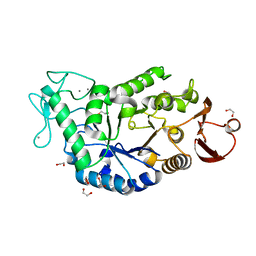
1AMY
DownloadVisualize
BU of 1amy by Molmil
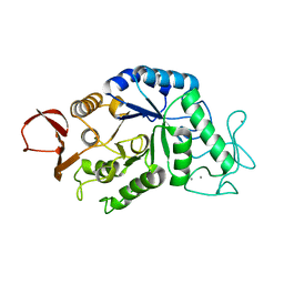
CRYSTAL AND MOLECULAR STRUCTURE OF BARLEY ALPHA-AMYLASE
Descriptor: 1,4-ALPHA-D-GLUCAN GLUCANOHYDROLASE, CALCIUM ION
Authors:Kadziola, A, Haser, R.
Deposit date:1994-03-10
Release date:1995-05-13
Last modified:2024-02-07
Method:X-RAY DIFFRACTION (2.8 Å)
Cite:Crystal and molecular structure of barley alpha-amylase.
J.Mol.Biol., 239, 1994
8IG0
DownloadVisualize
BU of 8ig0 by Molmil
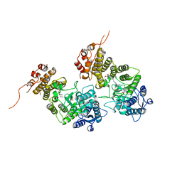
Crystal structure of menin in complex with DS-1594b
Descriptor: (1R,2S,4R)-4-[[4-(5,6-dimethoxypyridazin-3-yl)phenyl]methylamino]-2-[methyl-[6-[2,2,2-tris(fluoranyl)ethyl]thieno[2,3-d]pyrimidin-4-yl]amino]cyclopentan-1-ol, DIMETHYL SULFOXIDE, Menin, ...
Authors:Suzuki, M, Yoneyama, T, Imai, E.
Deposit date:2023-02-20
Release date:2023-03-22
Last modified:2023-09-20
Method:X-RAY DIFFRACTION (2.6 Å)
Cite:A novel Menin-MLL1 inhibitor, DS-1594a, prevents the progression of acute leukemia with rearranged MLL1 or mutated NPM1.
Cancer Cell Int, 23, 2023
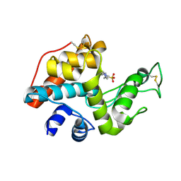
5B59
DownloadVisualize
BU of 5b59 by Molmil
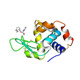
Hen egg-white lysozyme modified with a keto-ABNO.
Descriptor: (1~{S},5~{R})-9-oxidanyl-9-azabicyclo[3.3.1]nonan-3-one, Lysozyme C
Authors:Sasaki, D, Seki, Y, Sohma, Y, Oisaki, K, Kanai, M.
Deposit date:2016-04-28
Release date:2016-09-14
Last modified:2024-11-06
Method:X-RAY DIFFRACTION (2 Å)
Cite:Transition Metal-Free Tryptophan-Selective Bioconjugation of Proteins
J.Am.Chem.Soc., 138, 2016
1HT6
DownloadVisualize
BU of 1ht6 by Molmil
CRYSTAL STRUCTURE AT 1.5A RESOLUTION OF THE BARLEY ALPHA-AMYLASE ISOZYME 1
Descriptor: 1,2-ETHANEDIOL, ALPHA-AMYLASE ISOZYME 1, CALCIUM ION
Authors:Robert, X, Haser, R, Aghajari, N.
Deposit date:2000-12-29
Release date:2003-07-08
Last modified:2024-04-03
Method:X-RAY DIFFRACTION (1.5 Å)
Cite:The structure of barley alpha-amylase isozyme 1 reveals a novel role of domain C in substrate recognition and binding: a pair of sugar tongs
Structure, 11, 2003
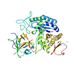
1AVA
DownloadVisualize
BU of 1ava by Molmil
AMY2/BASI PROTEIN-PROTEIN COMPLEX FROM BARLEY SEED
Descriptor: BARLEY ALPHA-AMYLASE 2(CV MENUET), BARLEY ALPHA-AMYLASE/SUBTILISIN INHIBITOR, CALCIUM ION
Authors:Vallee, F, Kadziola, A, Bourne, Y, Juy, M, Svensson, B, Haser, R.
Deposit date:1997-09-15
Release date:1999-03-16
Last modified:2024-10-23
Method:X-RAY DIFFRACTION (1.9 Å)
Cite:Barley alpha-amylase bound to its endogenous protein inhibitor BASI: crystal structure of the complex at 1.9 A resolution.
Structure, 6, 1998
2DBT
DownloadVisualize
BU of 2dbt by Molmil
Crystal structure of chitinase C from Streptomyces griseus HUT6037
Descriptor: 4-(2-HYDROXYETHYL)-1-PIPERAZINE ETHANESULFONIC ACID, chitinase C
Authors:Kezuka, Y, Watanabe, T, Nonaka, T.
Deposit date:2005-12-16
Release date:2006-03-28
Last modified:2023-10-25
Method:X-RAY DIFFRACTION (3.14 Å)
Cite:Structural Studies of a Two-domain Chitinase from Streptomyces griseus HUT6037
J.Mol.Biol., 358, 2006
4WBS
DownloadVisualize
BU of 4wbs by Molmil
Crystal structure of an ABC transporter related protein from Burkholderia phymatum
Descriptor: ABC transporter related, CHLORIDE ION
Authors:Seattle Structural Genomics Center for Infectious Disease (SSGCID)
Deposit date:2014-09-03
Release date:2014-09-24
Last modified:2023-09-27
Method:X-RAY DIFFRACTION (2 Å)
Cite:Crystal structure of an ABC transporter related protein from Burkholderia phymatum
to be published
3NXS
DownloadVisualize
BU of 3nxs by Molmil
Crystal structure of LAO/AO transport system from Mycobacterium smegmatis bound to GDP
Descriptor: CHLORIDE ION, GUANOSINE-5'-DIPHOSPHATE, LAO/AO transport system ATPase
Authors:Seattle Structural Genomics Center for Infectious Disease (SSGCID)
Deposit date:2010-07-14
Release date:2010-07-28
Last modified:2023-09-06
Method:X-RAY DIFFRACTION (2.3 Å)
Cite:Crystal structures of Mycobacterial MeaB and MMAA-like GTPases
J.Struct.Funct.Genom., 16, 2015
4DZ3
DownloadVisualize
BU of 4dz3 by Molmil
Crystal structure of a Peptidyl-prolyl cis-trans isomerase with surface mutation M61H from Burkholderia pseudomallei complexed with FK506
Descriptor: 1,2-ETHANEDIOL, 8-DEETHYL-8-[BUT-3-ENYL]-ASCOMYCIN, ACETATE ION, ...
Authors:Seattle Structural Genomics Center for Infectious Disease (SSGCID)
Deposit date:2012-02-29
Release date:2012-03-28
Last modified:2023-09-13
Method:X-RAY DIFFRACTION (2 Å)
Cite:A structural biology approach enables the development of antimicrobials targeting bacterial immunophilins.
Antimicrob.Agents Chemother., 58, 2014
2DKV
DownloadVisualize
BU of 2dkv by Molmil
Crystal structure of class I chitinase from Oryza sativa L. japonica
Descriptor: (4S)-2-METHYL-2,4-PENTANEDIOL, 2-(N-MORPHOLINO)-ETHANESULFONIC ACID, chitinase
Authors:Kezuka, Y, Nishizawa, Y, Watanabe, T, Nonaka, T.
Deposit date:2006-04-14
Release date:2007-05-01
Last modified:2024-10-16
Method:X-RAY DIFFRACTION (2 Å)
Cite:Structure of full-length class I chitinase from rice revealed by X-ray crystallography and small-angle X-ray scattering.
Proteins, 78, 2010

227111

數據於2024-11-06公開中

PDB statisticsPDBj update infoContact PDBjnumon