Loading
PDBj
MenuPDBj@FacebookPDBj@X(formerly Twitter)PDBj@BlueSkyPDBj@YouTubewwPDB FoundationwwPDB
RCSB PDBPDBeBMRBAdv. SearchSearch help
Search by PDB author
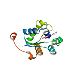
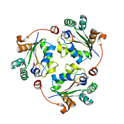
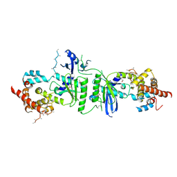
4BXI
DownloadVisualize
BU of 4bxi by Molmil
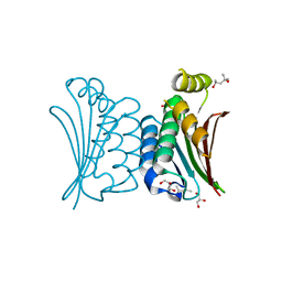
Crystal structure of ATP binding domain of AgrC from Staphylococcus aureus
Descriptor: (4S)-2-METHYL-2,4-PENTANEDIOL, ACCESSORY GENE REGULATOR PROTEIN C, ACETATE ION, ...
Authors:Srivastava, S.K, Rajasree, K, Gopal, B.
Deposit date:2013-07-12
Release date:2014-06-04
Last modified:2023-12-20
Method:X-RAY DIFFRACTION (2.2 Å)
Cite:Influence of the Agrc-Agra Complex in the Response Time of Staphylococcus Aureus Quorum Sensing
J.Bacteriol., 196, 2014
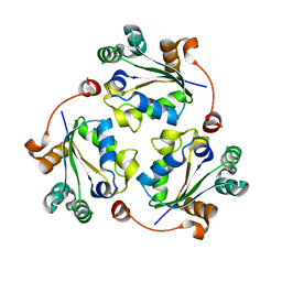
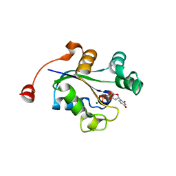
3Q8Y
DownloadVisualize
BU of 3q8y by Molmil
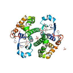
Crystal structure of Staphylococcus aureus nucleoside diphosphate kinase complexed with ADP and Vanadate
Descriptor: ADENOSINE-5'-DIPHOSPHATE, MAGNESIUM ION, Nucleoside diphosphate kinase, ...
Authors:Srivastava, S.K, Rajasree, K, Gopal, B.
Deposit date:2011-01-07
Release date:2011-07-27
Last modified:2023-11-01
Method:X-RAY DIFFRACTION (2.7 Å)
Cite:Conformational basis for substrate recognition and regulation of catalytic activity in Staphylococcus aureus nucleoside di-phosphate kinase.
Biochim.Biophys.Acta, 2011
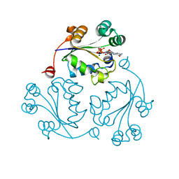
3Q83
DownloadVisualize
BU of 3q83 by Molmil
Crystal structure of Staphylococcus aureus nucleoside diphosphate kinase
Descriptor: Nucleoside diphosphate kinase
Authors:Srivastava, S.K, Rajasree, K, Gopal, B.
Deposit date:2011-01-06
Release date:2011-07-27
Last modified:2023-11-01
Method:X-RAY DIFFRACTION (2.5 Å)
Cite:Conformational basis for substrate recognition and regulation of catalytic activity in Staphylococcus aureus nucleoside di-phosphate kinase.
Biochim.Biophys.Acta, 1814, 2011
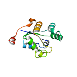
3Q86
DownloadVisualize
BU of 3q86 by Molmil
Crystal structure of Staphylococcus aureus nucleoside diphosphate kinase complexed with GTP
Descriptor: GUANOSINE-5'-TRIPHOSPHATE, MAGNESIUM ION, Nucleoside diphosphate kinase
Authors:Srivastava, S.K, Rajasree, K, Gopal, B.
Deposit date:2011-01-06
Release date:2011-07-27
Last modified:2023-11-01
Method:X-RAY DIFFRACTION (2.38 Å)
Cite:Conformational basis for substrate recognition and regulation of catalytic activity in Staphylococcus aureus nucleoside di-phosphate kinase.
Biochim.Biophys.Acta, 1814, 2011
3Q89
DownloadVisualize
BU of 3q89 by Molmil
Crystal structure of Staphylococcus aureus nucleoside diphosphate kinase complexed with CDP
Descriptor: CYTIDINE-5'-DIPHOSPHATE, MAGNESIUM ION, Nucleoside diphosphate kinase
Authors:Srivastava, S.K, Rajasree, K, Gopal, B.
Deposit date:2011-01-06
Release date:2011-07-27
Last modified:2023-11-01
Method:X-RAY DIFFRACTION (2.9 Å)
Cite:Conformational basis for substrate recognition and regulation of catalytic activity in Staphylococcus aureus nucleoside di-phosphate kinase.
Biochim.Biophys.Acta, 2011
3Q8U
DownloadVisualize
BU of 3q8u by Molmil
Crystal structure of Staphylococcus aureus nucleoside diphosphate kinase complexed with ADP
Descriptor: ADENOSINE-5'-DIPHOSPHATE, MAGNESIUM ION, Nucleoside diphosphate kinase
Authors:Srivastava, S.K, Rajasree, K, Gopal, B.
Deposit date:2011-01-07
Release date:2011-07-27
Last modified:2023-11-01
Method:X-RAY DIFFRACTION (2.22 Å)
Cite:Conformational basis for substrate recognition and regulation of catalytic activity in Staphylococcus aureus nucleoside di-phosphate kinase.
Biochim.Biophys.Acta, 1814, 2011
3Q8V
DownloadVisualize
BU of 3q8v by Molmil
Crystal structure of Staphylococcus aureus nucleoside diphosphate kinase complexed with UDP
Descriptor: MAGNESIUM ION, Nucleoside diphosphate kinase, URIDINE-5'-DIPHOSPHATE
Authors:Srivastava, S.K, Rajasree, K, Gopal, B.
Deposit date:2011-01-07
Release date:2011-07-27
Last modified:2023-11-01
Method:X-RAY DIFFRACTION (2.5 Å)
Cite:Conformational basis for substrate recognition and regulation of catalytic activity in Staphylococcus aureus nucleoside di-phosphate kinase.
Biochim.Biophys.Acta, 2011
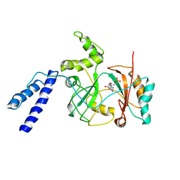
1ZAU
DownloadVisualize
BU of 1zau by Molmil
Adenylation domain of NAD+ dependent DNA ligase from M.tuberculosis
Descriptor: ADENOSINE MONOPHOSPHATE, DNA ligase
Authors:Srivastava, S.K, Ramachandran, R.
Deposit date:2005-04-07
Release date:2005-07-05
Last modified:2024-02-14
Method:X-RAY DIFFRACTION (3.15 Å)
Cite:NAD+-dependent DNA Ligase (Rv3014c) from Mycobacterium tuberculosis: CRYSTAL STRUCTURE OF THE ADENYLATION DOMAIN AND IDENTIFICATION OF NOVEL INHIBITORS
J.Biol.Chem., 280, 2005
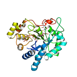
2F2K
DownloadVisualize
BU of 2f2k by Molmil
Aldose reductase tertiary complex with NADPH and DEG
Descriptor: Aldose reductase, GAMMA-GLUTAMYL-S-(1,2-DICARBOXYETHYL)CYSTEINYLGLYCINE, NADPH DIHYDRO-NICOTINAMIDE-ADENINE-DINUCLEOTIDE PHOSPHATE
Authors:Singh, R, White, M.A, Ramana, K.V, Petrash, J.M, Watowich, S.J, Bhatnagar, A, Srivastava, S.K.
Deposit date:2005-11-17
Release date:2006-05-16
Last modified:2023-08-23
Method:X-RAY DIFFRACTION (1.94 Å)
Cite:Structure of a glutathione conjugate bound to the active site of aldose reductase.
Proteins, 64, 2006
3PQ1
DownloadVisualize
BU of 3pq1 by Molmil
Crystal structure of human mitochondrial poly(A) polymerase (PAPD1)
Descriptor: Poly(A) RNA polymerase
Authors:Bai, Y, Srivastava, S.K, Chang, J.H, Tong, L.
Deposit date:2010-11-25
Release date:2011-03-30
Last modified:2024-10-30
Method:X-RAY DIFFRACTION (3.1 Å)
Cite:Structural basis for dimerization and activity of human PAPD1, a noncanonical poly(A) polymerase.
Mol.Cell, 41, 2011
6KV3
DownloadVisualize
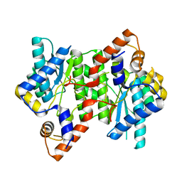
BU of 6kv3 by Molmil
Crystal Structure of NAD+ Synthetase from Staphylococcus aureus
Descriptor: NH(3)-dependent NAD(+) synthetase
Authors:Nasrin, S.K, Sandeep, S.K.
Deposit date:2019-09-03
Release date:2020-09-09
Last modified:2023-11-22
Method:X-RAY DIFFRACTION (2.3 Å)
Cite:Crystallographic and molecular dynamics simulation analysis of NAD synthetase from methicillin resistant Staphylococcus aureus (MRSA).
Int.J.Biol.Macromol., 165, 2020
1PGT
DownloadVisualize
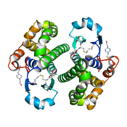
BU of 1pgt by Molmil
CRYSTAL STRUCTURE OF HUMAN GLUTATHIONE S-TRANSFERASE P1-1[V104] COMPLEXED WITH S-HEXYLGLUTATHIONE
Descriptor: 4-(2-HYDROXYETHYL)-1-PIPERAZINE ETHANESULFONIC ACID, GLUTATHIONE S-TRANSFERASE, S-HEXYLGLUTATHIONE
Authors:Ji, X.
Deposit date:1997-02-17
Release date:1997-09-04
Last modified:2023-08-30
Method:X-RAY DIFFRACTION (1.8 Å)
Cite:Structure and function of the xenobiotic substrate-binding site and location of a potential non-substrate-binding site in a class pi glutathione S-transferase.
Biochemistry, 36, 1997
1ABN
DownloadVisualize
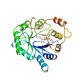
BU of 1abn by Molmil
THE CRYSTAL STRUCTURE OF THE ALDOSE REDUCTASE NADPH BINARY COMPLEX
Descriptor: ALDOSE REDUCTASE, NADPH DIHYDRO-NICOTINAMIDE-ADENINE-DINUCLEOTIDE PHOSPHATE
Authors:Borhani, D.W, Harter, T.M, Petrash, J.M.
Deposit date:1992-09-03
Release date:1994-01-31
Last modified:2024-02-07
Method:X-RAY DIFFRACTION (2.4 Å)
Cite:The crystal structure of the aldose reductase.NADPH binary complex.
J.Biol.Chem., 267, 1992
4PGT
DownloadVisualize
BU of 4pgt by Molmil
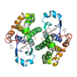
CRYSTAL STRUCTURE OF HGSTP1-1[V104] COMPLEXED WITH THE GSH CONJUGATE OF (+)-ANTI-BPDE
Descriptor: 2-(N-MORPHOLINO)-ETHANESULFONIC ACID, 2-AMINO-4-[1-(CARBOXYMETHYL-CARBAMOYL)-2-(9-HYDROXY-7,8-DIOXO-7,8,9,10-TETRAHYDRO-BENZO[DEF]CHRYSEN-10-YLSULFANYL)-ETHYLCARBAMOYL]-BUTYRIC ACID, PROTEIN (GLUTATHIONE S-TRANSFERASE), ...
Authors:Ji, X, Blaszczyk, J.
Deposit date:1999-03-22
Release date:1999-09-01
Last modified:2024-11-13
Method:X-RAY DIFFRACTION (2.1 Å)
Cite:Structure and function of residue 104 and water molecules in the xenobiotic substrate-binding site in human glutathione S-transferase P1-1.
Biochemistry, 38, 1999
3PGT
DownloadVisualize
BU of 3pgt by Molmil
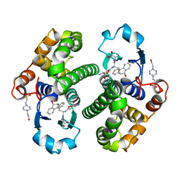
CRYSTAL STRUCTURE OF HGSTP1-1[I104] COMPLEXED WITH THE GSH CONJUGATE OF (+)-ANTI-BPDE
Descriptor: 2-(N-MORPHOLINO)-ETHANESULFONIC ACID, 2-AMINO-4-[1-(CARBOXYMETHYL-CARBAMOYL)-2-(9-HYDROXY-7,8-DIOXO-7,8,9,10-TETRAHYDRO-BENZO[DEF]CHRYSEN-10-YLSULFANYL)-ETHYLCARBAMOYL]-BUTYRIC ACID, PROTEIN (GLUTATHIONE S-TRANSFERASE), ...
Authors:Ji, X, Xiao, B.
Deposit date:1999-03-22
Release date:1999-09-01
Last modified:2023-09-06
Method:X-RAY DIFFRACTION (2.14 Å)
Cite:Structure and function of residue 104 and water molecules in the xenobiotic substrate-binding site in human glutathione S-transferase P1-1.
Biochemistry, 38, 1999
2PGT
DownloadVisualize
BU of 2pgt by Molmil
CRYSTAL STRUCTURE OF HUMAN GLUTATHIONE S-TRANSFERASE P1-1[V104] COMPLEXED WITH (9R,10R)-9-(S-GLUTATHIONYL)-10-HYDROXY-9,10-DIHYDROPHENANTHRENE
Descriptor: (9R,10R)-9-(S-GLUTATHIONYL)-10-HYDROXY-9,10-DIHYDROPHENANTHRENE, 4-(2-HYDROXYETHYL)-1-PIPERAZINE ETHANESULFONIC ACID, GLUTATHIONE S-TRANSFERASE, ...
Authors:Ji, X.
Deposit date:1997-02-17
Release date:1997-09-04
Last modified:2023-08-30
Method:X-RAY DIFFRACTION (1.9 Å)
Cite:Structure and function of the xenobiotic substrate-binding site and location of a potential non-substrate-binding site in a class pi glutathione S-transferase.
Biochemistry, 36, 1997

227344

数据于2024-11-13公开中

PDB statisticsPDBj update infoContact PDBjnumon