Loading
PDBj
MenuPDBj@FacebookPDBj@TwitterPDBj@YouTubewwPDB FoundationwwPDB
RCSB PDBPDBeBMRBAdv. SearchSearch help
Search by PDB author
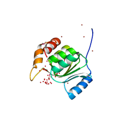
2G36
DownloadVisualize
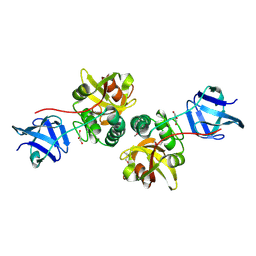
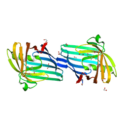
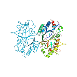
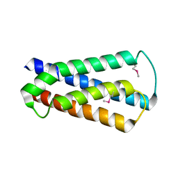
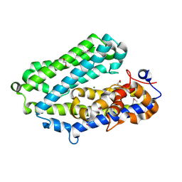
BU of 2g36 by Molmil
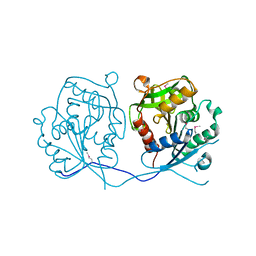
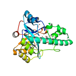
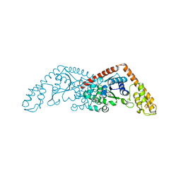
Crystal structure of Tryptophanyl-tRNA synthetase (EC 6.1.1.2) (Tryptophan-tRNA ligase)(TrpRS) (tm0492) from THERMOTOGA MARITIMA at 2.50 A resolution
Descriptor: IRON/SULFUR CLUSTER, TRYPTOPHAN, Tryptophanyl-tRNA synthetase
Authors:Joint Center for Structural Genomics (JCSG)
Deposit date:2006-02-17
Release date:2006-03-28
Last modified:2024-04-03
Method:X-RAY DIFFRACTION (2.5 Å)
Cite:Structure of a tryptophanyl-tRNA synthetase containing an iron-sulfur cluster.
Acta Crystallogr.,Sect.F, 66, 2010
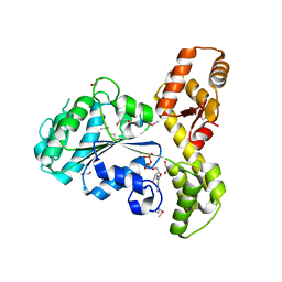
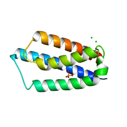
2ETS
DownloadVisualize
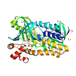
BU of 2ets by Molmil
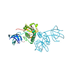
CRYSTAL STRUCTURE OF A BACTERIAL DOMAIN OF UNKNOWN FUNCTION FROM DUF1798 FAMILY (MW1337) FROM STAPHYLOCOCCUS AUREUS SUBSP. AUREUS AT 2.25 A RESOLUTION
Descriptor: CHLORIDE ION, PHOSPHATE ION, hypothetical protein
Authors:Joint Center for Structural Genomics (JCSG)
Deposit date:2005-10-27
Release date:2005-11-08
Last modified:2023-01-25
Method:X-RAY DIFFRACTION (2.25 Å)
Cite:Crystal structures of MW1337R and lin2004: representatives of a novel protein family that adopt a four-helical bundle fold.
Proteins, 71, 2008
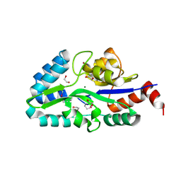
2GHR
DownloadVisualize
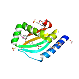
BU of 2ghr by Molmil
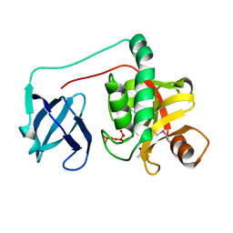
Crystal structure of homoserine o-succinyltransferase (NP_981826.1) from Bacillus cereus ATCC 10987 at 2.40 A resolution
Descriptor: Homoserine O-succinyltransferase, SULFATE ION
Authors:Joint Center for Structural Genomics (JCSG)
Deposit date:2006-03-27
Release date:2006-04-11
Last modified:2023-01-25
Method:X-RAY DIFFRACTION (2.4 Å)
Cite:Crystal structure of homoserine O-succinyltransferase from Bacillus cereus at 2.4 A resolution
Proteins, 68, 2007
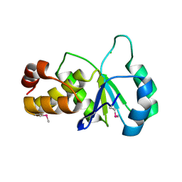
2EVR
DownloadVisualize
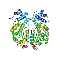
BU of 2evr by Molmil
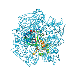
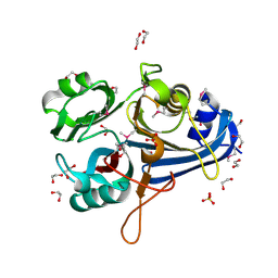
CRYSTAL STRUCTURE OF A PUTATIVE GAMMA-D-GLUTAMYL-L-DIAMINO ACID ENDOPEPTIDASE (NPUN_R0659) FROM NOSTOC PUNCTIFORME PCC 73102 AT 1.60 A RESOLUTION
Descriptor: 1,2-ETHANEDIOL, CHLORIDE ION, COG0791: Cell wall-associated hydrolases (invasion-associated proteins), ...
Authors:Joint Center for Structural Genomics (JCSG)
Deposit date:2005-10-31
Release date:2005-11-22
Last modified:2023-01-25
Method:X-RAY DIFFRACTION (1.6 Å)
Cite:Structural Basis of Murein Peptide Specificity of a gamma-D-Glutamyl-L-Diamino Acid Endopeptidase.
Structure, 17, 2009
2HBW
DownloadVisualize
BU of 2hbw by Molmil
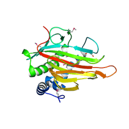
Crystal structure of a putative endopeptidase (ava_3396) from anabaena variabilis atcc 29413 at 1.05 A resolution
Descriptor: ACETATE ION, NLP/P60 protein, UNKNOWN LIGAND
Authors:Joint Center for Structural Genomics (JCSG)
Deposit date:2006-06-14
Release date:2006-08-08
Last modified:2023-01-25
Method:X-RAY DIFFRACTION (1.05 Å)
Cite:Structural Basis of Murein Peptide Specificity of a gamma-D-Glutamyl-L-Diamino Acid Endopeptidase.
Structure, 17, 2009
2GVK
DownloadVisualize
BU of 2gvk by Molmil
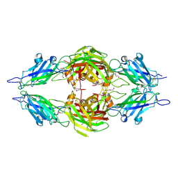
Crystal structure of a dye-decolorizing peroxidase (DyP) from Bacteroides thetaiotaomicron VPI-5482 at 1.6 A resolution
Descriptor: 1,2-ETHANEDIOL, ACETIC ACID, CHLORIDE ION, ...
Authors:Joint Center for Structural Genomics (JCSG)
Deposit date:2006-05-02
Release date:2006-05-16
Last modified:2023-01-25
Method:X-RAY DIFFRACTION (1.6 Å)
Cite:Crystal structures of two novel dye-decolorizing peroxidases reveal a beta-barrel fold with a conserved heme-binding motif.
Proteins, 69, 2007
2FG0
DownloadVisualize
BU of 2fg0 by Molmil
Crystal structure of a putative gamma-d-glutamyl-l-diamino acid endopeptidase (npun_r0659) from nostoc punctiforme pcc 73102 at 1.79 A resolution
Descriptor: COG0791: Cell wall-associated hydrolases (invasion-associated proteins), GLYCEROL
Authors:Joint Center for Structural Genomics (JCSG)
Deposit date:2005-12-20
Release date:2006-01-10
Last modified:2023-01-25
Method:X-RAY DIFFRACTION (1.79 Å)
Cite:Structural Basis of Murein Peptide Specificity of a gamma-D-Glutamyl-L-Diamino Acid Endopeptidase.
Structure, 17, 2009
2H1T
DownloadVisualize
BU of 2h1t by Molmil
Crystal structure of a duf1089 family protein (pa1994) from pseudomonas aeruginosa at 1.80 A resolution
Descriptor: (4S)-2-METHYL-2,4-PENTANEDIOL, 1,2-ETHANEDIOL, Hypothetical protein
Authors:Joint Center for Structural Genomics (JCSG)
Deposit date:2006-05-16
Release date:2006-05-30
Last modified:2023-01-25
Method:X-RAY DIFFRACTION (1.8 Å)
Cite:The structure of the first representative of Pfam family PF06475 reveals a new fold with possible involvement in glycolipid metabolism.
Acta Crystallogr.,Sect.F, 66, 2010
2HAG
DownloadVisualize
BU of 2hag by Molmil
Crystal structure of a putative dyp-type peroxidase protein (so_0740) from shewanella oneidensis at 2.75 A resolution
Descriptor: Melanin biosynthesis protein TyrA, putative
Authors:Joint Center for Structural Genomics (JCSG)
Deposit date:2006-06-12
Release date:2006-08-08
Last modified:2023-01-25
Method:X-RAY DIFFRACTION (2.75 Å)
Cite:Crystal structures of two novel dye-decolorizing peroxidases reveal a beta-barrel fold with a conserved heme-binding motif.
Proteins, 69, 2007
2HUJ
DownloadVisualize
BU of 2huj by Molmil
Crystal structure of a protein of uknown function (NP_471338.1) from Listeria innocua at 1.74 A resolution
Descriptor: Lin2004 protein
Authors:Joint Center for Structural Genomics (JCSG)
Deposit date:2006-07-26
Release date:2006-08-08
Last modified:2023-01-25
Method:X-RAY DIFFRACTION (1.74 Å)
Cite:Crystal structures of MW1337R and lin2004: representatives of a novel protein family that adopt a four-helical bundle fold.
Proteins, 71, 2008
1VK9
DownloadVisualize
BU of 1vk9 by Molmil
CRYSTAL STRUCTURE OF A DUF1893 family protein (TM1506) FROM THERMOTOGA MARITIMA AT 2.70 A RESOLUTION
Descriptor: UNKNOWN LIGAND, ZINC ION, conserved hypothetical protein TM1506
Authors:Joint Center for Structural Genomics (JCSG)
Deposit date:2004-05-06
Release date:2004-05-18
Last modified:2023-01-25
Method:X-RAY DIFFRACTION (2.7 Å)
Cite:Crystal structure of an ADP-ribosylated protein with a cytidine deaminase-like fold, but unknown function (TM1506), from Thermotoga maritima at 2.70 A resolution.
Proteins, 71, 2008
2FNA
DownloadVisualize
BU of 2fna by Molmil
Crystal structure of an archaeal aaa+ atpase (sso1545) from sulfolobus solfataricus p2 at 2.00 A resolution
Descriptor: 1,2-ETHANEDIOL, ADENOSINE-5'-DIPHOSPHATE, Conserved hypothetical protein, ...
Authors:Joint Center for Structural Genomics (JCSG)
Deposit date:2006-01-10
Release date:2006-02-07
Last modified:2023-01-25
Method:X-RAY DIFFRACTION (2 Å)
Cite:Crystal structure of a novel archaeal AAA+ ATPase SSO1545 from Sulfolobus solfataricus.
Proteins, 74, 2009
2FEA
DownloadVisualize
BU of 2fea by Molmil
Crystal structure of MtnX phosphatase from Bacillus Subtilis at 2.00 A resolution
Descriptor: 1,2-ETHANEDIOL, 2-hydroxy-3-keto-5-methylthiopentenyl-1-phosphate phosphatase, MAGNESIUM ION, ...
Authors:Joint Center for Structural Genomics (JCSG)
Deposit date:2005-12-15
Release date:2005-12-27
Last modified:2023-01-25
Method:X-RAY DIFFRACTION (2 Å)
Cite:Crystal structure of MtnX phosphatase from Bacillus subtilis at 2.0 A resolution provides a structural basis for bipartite phosphomonoester hydrolysis of 2-hydroxy-3-keto-5-methylthiopentenyl-1-phosphate.
Proteins, 69, 2007
2F46
DownloadVisualize
BU of 2f46 by Molmil
Crystal structure of a putative phosphatase (nma1982) from neisseria meningitidis z2491 at 1.41 A resolution
Descriptor: CHLORIDE ION, UNKNOWN LIGAND, hypothetical protein
Authors:Joint Center for Structural Genomics (JCSG)
Deposit date:2005-11-22
Release date:2006-02-07
Last modified:2023-01-25
Method:X-RAY DIFFRACTION (1.41 Å)
Cite:Crystal structure of NMA1982 from Neisseria meningitidis at 1.5 A resolution provides a structural scaffold for nonclassical, eukaryotic-like phosphatases.
Proteins, 69, 2007
1K8F
DownloadVisualize
BU of 1k8f by Molmil
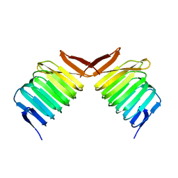
CRYSTAL STRUCTURE OF THE HUMAN C-TERMINAL CAP1-ADENYLYL CYCLASE ASSOCIATED PROTEIN
Descriptor: ADENYLYL CYCLASE-ASSOCIATED PROTEIN
Authors:Patskovsky, Y.V, Chance, M, Almo, S.C, Burley, S.K, New York SGX Research Center for Structural Genomics (NYSGXRC)
Deposit date:2001-10-24
Release date:2003-07-01
Last modified:2023-08-16
Method:X-RAY DIFFRACTION (2.8 Å)
Cite:Crystal structure of the actin binding domain of the cyclase-associated protein.
Biochemistry, 43, 2004
3O0F
DownloadVisualize
BU of 3o0f by Molmil
Crystal structure of a putative metal-dependent phosphoesterase (BAD_1165) from bifidobacterium adolescentis atcc 15703 at 1.94 A resolution
Descriptor: 1,2-ETHANEDIOL, ADENOSINE MONOPHOSPHATE, FE (III) ION, ...
Authors:Joint Center for Structural Genomics (JCSG)
Deposit date:2010-07-19
Release date:2010-08-25
Last modified:2023-02-01
Method:X-RAY DIFFRACTION (1.94 Å)
Cite:Crystal structure of a metal-dependent phosphoesterase (YP_910028.1) from Bifidobacterium adolescentis: Computational prediction and experimental validation of phosphoesterase activity.
Proteins, 79, 2011
3PAY
DownloadVisualize
BU of 3pay by Molmil
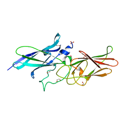
Crystal structure of a putative adhesin (BACOVA_04077) from Bacteroides ovatus at 2.50 A resolution
Descriptor: DI(HYDROXYETHYL)ETHER, putative adhesin
Authors:Joint Center for Structural Genomics (JCSG)
Deposit date:2010-10-19
Release date:2010-11-10
Last modified:2020-04-22
Method:X-RAY DIFFRACTION (2.5 Å)
Cite:A Distinct Type of Pilus from the Human Microbiome.
Cell, 165, 2016
3PL0
DownloadVisualize
BU of 3pl0 by Molmil
Crystal structure of a bsmA homolog (Mpe_A2762) from Methylobium petroleophilum PM1 at 1.91 A resolution
Descriptor: CHLORIDE ION, GLYCEROL, Uncharacterized protein
Authors:Joint Center for Structural Genomics (JCSG)
Deposit date:2010-11-12
Release date:2010-12-08
Last modified:2023-02-01
Method:X-RAY DIFFRACTION (1.91 Å)
Cite:Crystal structure of a member of a novel family of dioxygenases (PF10014) reveals a conserved cupin fold and active site.
Proteins, 82, 2014
3N8U
DownloadVisualize
BU of 3n8u by Molmil
Crystal structure of an imelysin peptidase (BACOVA_03801) from Bacteroides ovatus at 1.44 A resolution
Descriptor: 1,2-ETHANEDIOL, CHLORIDE ION, MAGNESIUM ION, ...
Authors:Joint Center for Structural Genomics (JCSG)
Deposit date:2010-05-28
Release date:2010-07-28
Last modified:2023-02-01
Method:X-RAY DIFFRACTION (1.44 Å)
Cite:Structural and sequence analysis of imelysin-like proteins implicated in bacterial iron uptake.
Plos One, 6, 2011
3OZ2
DownloadVisualize
BU of 3oz2 by Molmil
Crystal structure of a geranylgeranyl bacteriochlorophyll reductase-like (Ta0516) from Thermoplasma acidophilum at 1.60 A resolution
Descriptor: (2R)-3-{[(R)-{[(2S)-2,3-dihydroxypropyl]oxy}(hydroxy)phosphoryl]oxy}-2-[(6Z)-tridec-6-enoyloxy]propyl (9Z)-octadec-9-enoate, 1,2-ETHANEDIOL, Digeranylgeranylglycerophospholipid reductase, ...
Authors:Joint Center for Structural Genomics (JCSG)
Deposit date:2010-09-24
Release date:2010-10-27
Last modified:2023-02-01
Method:X-RAY DIFFRACTION (1.6 Å)
Cite:Insights into substrate specificity of geranylgeranyl reductases revealed by the structure of digeranylgeranylglycerophospholipid reductase, an essential enzyme in the biosynthesis of archaeal membrane lipids.
J.Mol.Biol., 404, 2010
3NPD
DownloadVisualize
BU of 3npd by Molmil
Crystal structure of a putative secreted protein (PA3611) from PSEUDOMONAS AERUGINOSA at 1.60 A resolution
Descriptor: 1,2-ETHANEDIOL, 3-CYCLOHEXYL-1-PROPYLSULFONIC ACID, SULFATE ION, ...
Authors:Joint Center for Structural Genomics (JCSG)
Deposit date:2010-06-28
Release date:2010-07-28
Last modified:2023-02-01
Method:X-RAY DIFFRACTION (1.6 Å)
Cite:Crystal structure of a putative quorum sensing-regulated protein (PA3611) from the Pseudomonas-specific DUF4146 family.
Proteins, 82, 2014
3PXP
DownloadVisualize
BU of 3pxp by Molmil
Crystal structure of a PAS and DNA binding domain containing protein (Caur_2278) from CHLOROFLEXUS AURANTIACUS J-10-FL at 2.30 A resolution
Descriptor: 1,2-ETHANEDIOL, CHLORIDE ION, Helix-turn-helix domain protein, ...
Authors:Joint Center for Structural Genomics (JCSG)
Deposit date:2010-12-10
Release date:2011-01-19
Last modified:2023-02-01
Method:X-RAY DIFFRACTION (2.3 Å)
Cite:Structure of an MmyB-Like Regulator from C. aurantiacus, Member of a New Transcription Factor Family Linked to Antibiotic Metabolism in Actinomycetes.
Plos One, 7, 2012
3R4R
DownloadVisualize
BU of 3r4r by Molmil
Crystal structure of a fimbrial assembly protein (BDI_3522) from Parabacteroides distasonis ATCC 8503 at 2.38 A resolution
Descriptor: 1,2-ETHANEDIOL, SULFATE ION, hypothetical fimbrial assembly protein
Authors:Joint Center for Structural Genomics (JCSG)
Deposit date:2011-03-17
Release date:2011-03-30
Last modified:2023-02-01
Method:X-RAY DIFFRACTION (2.38 Å)
Cite:A Distinct Type of Pilus from the Human Microbiome.
Cell, 165, 2016
3OHG
DownloadVisualize
BU of 3ohg by Molmil
Crystal structure of a protein with unknown function from DUF2233 family (BACOVA_00430) from Bacteroides ovatus at 1.80 A resolution
Descriptor: 1,2-ETHANEDIOL, CHLORIDE ION, SULFATE ION, ...
Authors:Joint Center for Structural Genomics (JCSG)
Deposit date:2010-08-17
Release date:2010-09-08
Last modified:2023-02-01
Method:X-RAY DIFFRACTION (1.8 Å)
Cite:Structure and function of the DUF2233 domain in bacteria and in the human mannose 6-phosphate uncovering enzyme.
J.Biol.Chem., 288, 2013
3OYV
DownloadVisualize
BU of 3oyv by Molmil
Crystal structure of an imelysin peptidase (BACOVA_03801) from Bacteroides ovatus ATCC 8483 at 1.25 A resolution
Descriptor: CHLORIDE ION, GLYCEROL, Imelysin, ...
Authors:Joint Center for Structural Genomics (JCSG)
Deposit date:2010-09-23
Release date:2010-10-20
Last modified:2023-02-01
Method:X-RAY DIFFRACTION (1.25 Å)
Cite:Structural and sequence analysis of imelysin-like proteins implicated in bacterial iron uptake.
Plos One, 6, 2011

222926

数据于2024-07-24公开中

PDB statisticsPDBj update infoContact PDBjnumon