Loading
PDBj
MenuPDBj@FacebookPDBj@TwitterPDBj@YouTubewwPDB FoundationwwPDB
RCSB PDBPDBeBMRBAdv. SearchSearch help
Search by PDB author
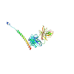
4KHX
DownloadVisualize
BU of 4khx by Molmil
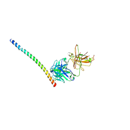
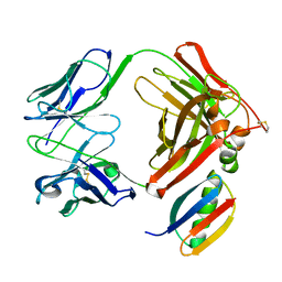
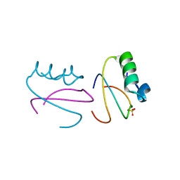
Crystal structure of gp41 helix complexed with antibody 8062
Descriptor: 8062 heavy chain, 8062 light chain, gp41 helix
Authors:Li, M, Gustchina, A, Wlodawer, A.
Deposit date:2013-05-01
Release date:2014-03-12
Last modified:2017-11-15
Method:X-RAY DIFFRACTION (2.921 Å)
Cite:Complexes of neutralizing and non-neutralizing affinity matured Fabs with a mimetic of the internal trimeric coiled-coil of HIV-1 gp41.
Plos One, 8, 2013
3EST
DownloadVisualize
BU of 3est by Molmil
STRUCTURE OF NATIVE PORCINE PANCREATIC ELASTASE AT 1.65 ANGSTROMS RESOLUTION
Descriptor: CALCIUM ION, PORCINE PANCREATIC ELASTASE, SULFATE ION
Authors:Meyer, E.F, Cole, G, Radhakrishnan, R, Epp, O.
Deposit date:1987-09-17
Release date:1988-01-16
Last modified:2024-06-05
Method:X-RAY DIFFRACTION (1.65 Å)
Cite:Structure of native porcine pancreatic elastase at 1.65 A resolutions.
Acta Crystallogr.,Sect.B, 44, 1988
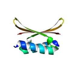
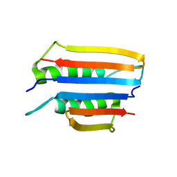
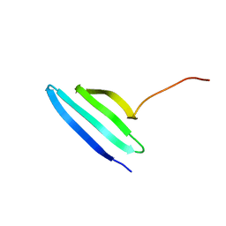
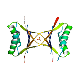
1MVK
DownloadVisualize
BU of 1mvk by Molmil
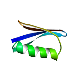
X-ray structure of the tetrameric mutant of the B1 domain of streptococcal protein G
Descriptor: Immunoglobulin G binding protein G, SULFATE ION
Authors:Frank, M.K, Dyda, F, Dobrodumov, A, Gronenborn, A.M.
Deposit date:2002-09-25
Release date:2002-10-30
Last modified:2024-02-14
Method:X-RAY DIFFRACTION (2.5 Å)
Cite:Core mutations switch monomeric protein GB1 into an intertwined tetramer.
Nat.Struct.Biol., 9, 2002
1F5J
DownloadVisualize
BU of 1f5j by Molmil
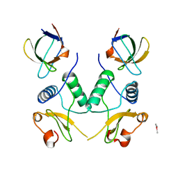
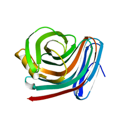
CRYSTAL STRUCTURE OF XYNB, A HIGHLY THERMOSTABLE BETA-1,4-XYLANASE FROM DICTYOGLOMUS THERMOPHILUM RT46B.1, AT 1.8 A RESOLUTION
Descriptor: BETA-1,4-XYLANASE, SULFATE ION
Authors:McCarthy, A.A, Baker, E.N.
Deposit date:2000-07-26
Release date:2000-11-15
Last modified:2024-02-07
Method:X-RAY DIFFRACTION (1.8 Å)
Cite:Structure of XynB, a highly thermostable beta-1,4-xylanase from Dictyoglomus thermophilum Rt46B.1, at 1.8 A resolution.
Acta Crystallogr.,Sect.D, 56, 2000
1FL1
DownloadVisualize
BU of 1fl1 by Molmil
KSHV PROTEASE
Descriptor: POTASSIUM ION, PROTEASE
Authors:Reiling, K.K, Pray, T.R, Craik, C.S, Stroud, R.M.
Deposit date:2000-08-11
Release date:2000-11-22
Last modified:2024-02-07
Method:X-RAY DIFFRACTION (2.2 Å)
Cite:Functional consequences of the Kaposi's sarcoma-associated herpesvirus protease structure: regulation of activity and dimerization by conserved structural elements.
Biochemistry, 39, 2000
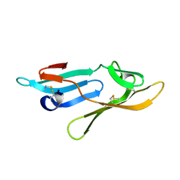
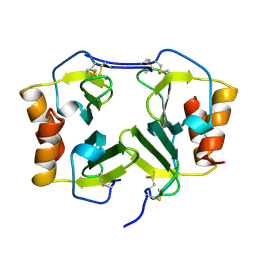
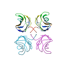
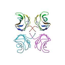
3EZM
DownloadVisualize
BU of 3ezm by Molmil
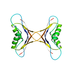
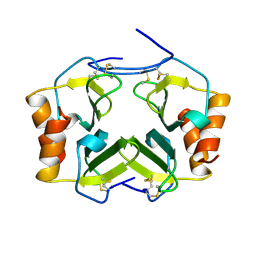
CYANOVIRIN-N
Descriptor: PROTEIN (CYANOVIRIN-N)
Authors:Yang, F, Wlodawer, A.
Deposit date:1998-12-15
Release date:1998-12-23
Last modified:2023-09-06
Method:X-RAY DIFFRACTION (1.5 Å)
Cite:Crystal structure of cyanovirin-N, a potent HIV-inactivating protein, shows unexpected domain swapping.
J.Mol.Biol., 288, 1999
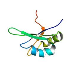
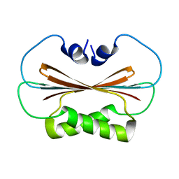
7QBY
DownloadVisualize
BU of 7qby by Molmil
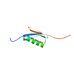
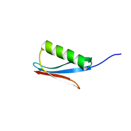
Refined structure of the T193A mutant in the C-terminal domain of DNAJB6b
Descriptor: Isoform B of DnaJ homolog subfamily B member 6
Authors:Karamanos, T.K, Cawood, E.E.
Deposit date:2021-11-21
Release date:2022-03-30
Last modified:2022-05-11
Method:SOLUTION NMR
Cite:Microsecond Backbone Motions Modulate the Oligomerization of the DNAJB6 Chaperone.
Angew.Chem.Int.Ed.Engl., 61, 2022
2Y1S
DownloadVisualize
BU of 2y1s by Molmil
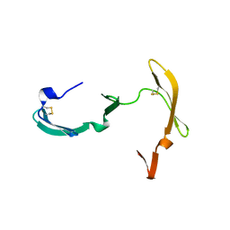
Microvirin lectin
Descriptor: Mannan-binding lectin
Authors:Bewley, C.A, Hussan, S.
Deposit date:2010-12-10
Release date:2011-04-06
Last modified:2023-06-14
Method:SOLUTION NMR
Cite:Solution structure of the monovalent lectin microvirin in complex with Man(alpha)(1-2)Man provides a basis for anti-HIV activity with low toxicity.
J. Biol. Chem., 286, 2011
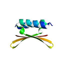
6U6R
DownloadVisualize
BU of 6u6r by Molmil
Solution NMR Structure Of The delta30-ngMinE Protein From Neisseria gonorrheae
Descriptor: Cell division topological specificity factor
Authors:Cai, M, Shen, Y, Clore, M.
Deposit date:2019-08-30
Release date:2020-07-08
Last modified:2024-05-01
Method:SOLID-STATE NMR
Cite:Probing transient excited states of the bacterial cell division regulator MinE by relaxation dispersion NMR spectroscopy.
Proc.Natl.Acad.Sci.USA, 116, 2019
6U6P
DownloadVisualize
BU of 6u6p by Molmil
Solution NMR Structure Of The Full Length Latent Form MinE Protein From Neisseria gonorrheae
Descriptor: Cell division topological specificity factor
Authors:Cai, M, Shen, Y, Clore, M.
Deposit date:2019-08-30
Release date:2020-07-08
Last modified:2024-05-01
Method:SOLUTION NMR
Cite:Probing transient excited states of the bacterial cell division regulator MinE by relaxation dispersion NMR spectroscopy.
Proc.Natl.Acad.Sci.USA, 116, 2019
6U6S
DownloadVisualize
BU of 6u6s by Molmil
Solution NMR Structure Of The I24N-delta10-ngMinE Protein From Neisseria gonorrheae
Descriptor: Cell division topological specificity factor
Authors:Cai, M, Shen, Y, Clore, M.
Deposit date:2019-08-30
Release date:2020-07-08
Last modified:2024-05-01
Method:SOLUTION NMR
Cite:Probing transient excited states of the bacterial cell division regulator MinE by relaxation dispersion NMR spectroscopy.
Proc.Natl.Acad.Sci.USA, 116, 2019
6U6Q
DownloadVisualize
BU of 6u6q by Molmil
Solution NMR Structure Of The Partially Activated MTS Deleted Form MinE Protein (delta10-ngMinE) From Neisseria gonorrheae
Descriptor: Cell division topological specificity factor
Authors:Cai, M, Shen, Y, Clore, M.
Deposit date:2019-08-30
Release date:2020-07-08
Last modified:2024-05-01
Method:SOLUTION NMR
Cite:Probing transient excited states of the bacterial cell division regulator MinE by relaxation dispersion NMR spectroscopy.
Proc.Natl.Acad.Sci.USA, 116, 2019
1N51
DownloadVisualize
BU of 1n51 by Molmil
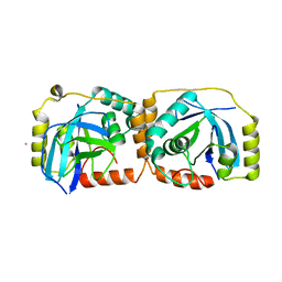
Aminopeptidase P in complex with the inhibitor apstatin
Descriptor: MANGANESE (II) ION, Xaa-Pro aminopeptidase, apstatin
Authors:Graham, S.C, Maher, M.J, Lee, M.H, Simmons, W.H, Freeman, H.C, Guss, J.M.
Deposit date:2002-11-03
Release date:2003-12-16
Last modified:2023-11-15
Method:X-RAY DIFFRACTION (2.3 Å)
Cite:Structure of Escherichia coli aminopeptidase P in complex with the inhibitor apstatin.
Acta Crystallogr.,Sect.D, 60, 2004
1PGX
DownloadVisualize
BU of 1pgx by Molmil
THE 1.66 ANGSTROMS X-RAY STRUCTURE OF THE B2 IMMUNOGLOBULIN-BINDING DOMAIN OF STREPTOCOCCAL PROTEIN G AND COMPARISON TO THE NMR STRUCTURE OF THE B1 DOMAIN
Descriptor: PROTEIN G
Authors:Whitlow, M, Achari, A, Howard, A.J.
Deposit date:1992-04-03
Release date:1992-07-15
Last modified:2024-02-14
Method:X-RAY DIFFRACTION (1.66 Å)
Cite:1.67-A X-ray structure of the B2 immunoglobulin-binding domain of streptococcal protein G and comparison to the NMR structure of the B1 domain.
Biochemistry, 31, 1992
1PGB
DownloadVisualize
BU of 1pgb by Molmil
TWO CRYSTAL STRUCTURES OF THE B1 IMMUNOGLOBULIN-BINDING DOMAIN OF STREPTOCCOCAL PROTEIN G AND COMPARISON WITH NMR
Descriptor: PROTEIN G
Authors:Gallagher, T, Alexander, P, Bryan, P, Gilliland, G.L.
Deposit date:1993-11-23
Release date:1994-04-30
Last modified:2024-02-14
Method:X-RAY DIFFRACTION (1.92 Å)
Cite:Two crystal structures of the B1 immunoglobulin-binding domain of streptococcal protein G and comparison with NMR.
Biochemistry, 33, 1994
1NAP
DownloadVisualize
BU of 1nap by Molmil
THE CRYSTAL STRUCTURE OF RECOMBINANT HUMAN NEUTROPHIL-ACTIVATING PEPTIDE-2 (M6L) AT 1.9-ANGSTROMS RESOLUTION
Descriptor: NEUTROPHIL ACTIVATING PEPTIDE-2
Authors:Malkowski, M.G, Edwards, B.F.P.
Deposit date:1994-12-19
Release date:1995-12-19
Last modified:2019-08-14
Method:X-RAY DIFFRACTION (1.9 Å)
Cite:The crystal structure of recombinant human neutrophil-activating peptide-2 (M6L) at 1.9-A resolution.
J.Biol.Chem., 270, 1995
1PGA
DownloadVisualize
BU of 1pga by Molmil
TWO CRYSTAL STRUCTURES OF THE B1 IMMUNOGLOBULIN-BINDING DOMAIN OF STREPTOCOCCAL PROTEIN G AND COMPARISON WITH NMR
Descriptor: PROTEIN G
Authors:Gallagher, T, Alexander, P, Bryan, P, Gilliland, G.L.
Deposit date:1993-11-23
Release date:1994-04-30
Last modified:2024-02-14
Method:X-RAY DIFFRACTION (2.07 Å)
Cite:Two crystal structures of the B1 immunoglobulin-binding domain of streptococcal protein G and comparison with NMR.
Biochemistry, 33, 1994
2SXL
DownloadVisualize
BU of 2sxl by Molmil
SEX-LETHAL RBD1, NMR, MINIMIZED AVERAGE STRUCTURE
Descriptor: SEX-LETHAL PROTEIN
Authors:Inoue, M, Muto, Y, Sakamoto, H, Kigawa, T, Takio, K, Shimura, Y, Yokoyama, S, RIKEN Structural Genomics/Proteomics Initiative (RSGI)
Deposit date:1997-07-16
Release date:1998-07-22
Last modified:2024-05-22
Method:SOLUTION NMR
Cite:A characteristic arrangement of aromatic amino acid residues in the solution structure of the amino-terminal RNA-binding domain of Drosophila sex-lethal.
J.Mol.Biol., 272, 1997
1IGD
DownloadVisualize
BU of 1igd by Molmil
THE THIRD IGG-BINDING DOMAIN FROM STREPTOCOCCAL PROTEIN G: AN ANALYSIS BY X-RAY CRYSTALLOGRAPHY OF THE STRUCTURE ALONE AND IN A COMPLEX WITH FAB
Descriptor: PROTEIN G
Authors:Derrick, J.P, Wigley, D.B.
Deposit date:1994-08-05
Release date:1994-11-01
Last modified:2024-02-07
Method:X-RAY DIFFRACTION (1.1 Å)
Cite:The third IgG-binding domain from streptococcal protein G. An analysis by X-ray crystallography of the structure alone and in a complex with Fab.
J.Mol.Biol., 243, 1994
1IGC
DownloadVisualize
BU of 1igc by Molmil
IGG1 FAB FRAGMENT (MOPC21) COMPLEX WITH DOMAIN III OF PROTEIN G FROM STREPTOCOCCUS
Descriptor: IGG1-KAPPA MOPC21 FAB (HEAVY CHAIN), IGG1-KAPPA MOPC21 FAB (LIGHT CHAIN), STREPTOCOCCAL PROTEIN G (DOMAIN III)
Authors:Derrick, J.P, Wigley, D.B.
Deposit date:1994-08-05
Release date:1995-06-03
Last modified:2024-06-05
Method:X-RAY DIFFRACTION (2.6 Å)
Cite:The third IgG-binding domain from streptococcal protein G. An analysis by X-ray crystallography of the structure alone and in a complex with Fab.
J.Mol.Biol., 243, 1994
1MPE
DownloadVisualize
BU of 1mpe by Molmil
Ensemble of 20 structures of the tetrameric mutant of the B1 domain of streptococcal protein G
Descriptor: Immunoglobulin G binding protein G
Authors:Frank, M.K, Dyda, F, Dobrodumov, A, Gronenborn, A.M.
Deposit date:2002-09-12
Release date:2002-10-30
Last modified:2024-05-22
Method:SOLUTION NMR
Cite:Core mutations switch monomeric protein GB1 into an intertwined tetramer.
Nat.Struct.Biol., 9, 2002
2L5H
DownloadVisualize
BU of 2l5h by Molmil
Solution Structure of the H189Q mutant of the Enzyme I dimer Using Residual Dipolar Couplings and Small Angle X-Ray Scattering
Descriptor: Phosphoenolpyruvate-protein phosphotransferase
Authors:Takayama, Y.D, Schwieters, C.D, Grishaev, A, Guirlando, R, Clore, G.
Deposit date:2010-11-01
Release date:2011-01-12
Last modified:2024-05-01
Method:SOLUTION NMR, SOLUTION SCATTERING
Cite:Combined Use of Residual Dipolar Couplings and Solution X-ray Scattering To Rapidly Probe Rigid-Body Conformational Transitions in a Non-phosphorylatable Active-Site Mutant of the 128 kDa Enzyme I Dimer.
J.Am.Chem.Soc., 133, 2011
1HA1
DownloadVisualize
BU of 1ha1 by Molmil
HNRNP A1 (RBD1,2) FROM HOMO SAPIENS
Descriptor: HNRNP A1
Authors:Shamoo, Y, Krueger, U, Rice, L, Williams, K.R, Steitz, T.A.
Deposit date:1996-10-30
Release date:1997-05-15
Last modified:2024-02-07
Method:X-RAY DIFFRACTION (1.75 Å)
Cite:Crystal structure of the two RNA binding domains of human hnRNP A1 at 1.75 A resolution.
Nat.Struct.Biol., 4, 1997
2LRK
DownloadVisualize
BU of 2lrk by Molmil
Solution Structures of the IIA(Chitobiose)-HPr complex of the N,N'-Diacetylchitobiose
Descriptor: N,N'-diacetylchitobiose-specific phosphotransferase enzyme IIA component, Phosphocarrier protein HPr
Authors:Cai, M, Jung, Y, Clore, M.
Deposit date:2012-04-06
Release date:2012-05-16
Last modified:2024-05-01
Method:SOLUTION NMR
Cite:Solution Structure of the IIAChitobiose-HPr Complex of the N,N'-Diacetylchitobiose Branch of the Escherichia coli Phosphotransferase System.
J.Biol.Chem., 287, 2012
2LOE
DownloadVisualize
BU of 2loe by Molmil
Structure of the Plasmodium 6-cysteine s48/45 Domain
Descriptor: 6-cysteine protein, putative
Authors:Cai, M, Arredondo, S.A, Clore, M.G, Miller, L.H, Takayama, Y, Macdonald, N.J, Enderson, E.D, Aravind, L.
Deposit date:2012-01-23
Release date:2012-04-18
Last modified:2023-06-14
Method:SOLUTION NMR
Cite:Structure of the Plasmodium 6-cysteine s48/45 domain.
Proc.Natl.Acad.Sci.USA, 109, 2012

221051

数据于2024-06-12公开中

PDB statisticsPDBj update infoContact PDBjnumon