Loading
PDBj
MenuPDBj@FacebookPDBj@TwitterPDBj@YouTubewwPDB FoundationwwPDB
RCSB PDBPDBeBMRBAdv. SearchSearch help
Search by PDB author
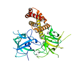
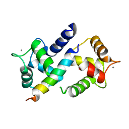
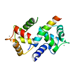
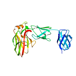
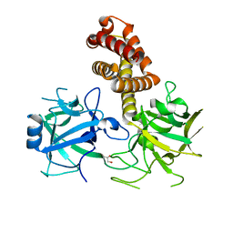
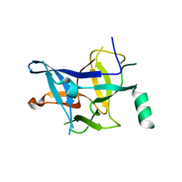
4P9L
DownloadVisualize
BU of 4p9l by Molmil
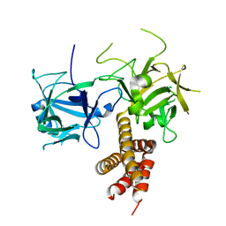
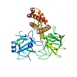
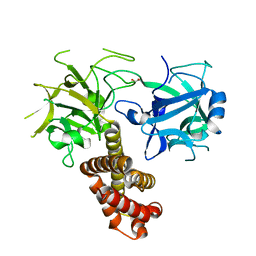
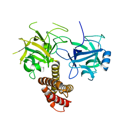
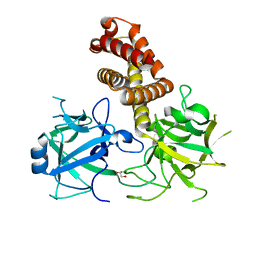
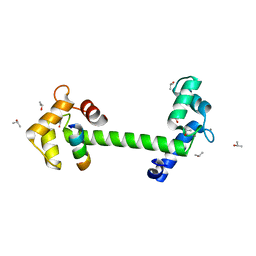
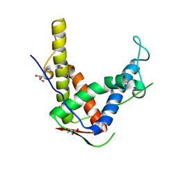
Crystal Structure of mouse Ryanodine Receptor 2 SPRY2 Domain (1080-1253) disease mutant A1107M
Descriptor: Ryanodine receptor 2
Authors:Lau, K, Van Petegem, F.
Deposit date:2014-04-04
Release date:2014-11-05
Last modified:2023-12-27
Method:X-RAY DIFFRACTION (1.4361 Å)
Cite:Crystal structures of wild type and disease mutant forms of the ryanodine receptor SPRY2 domain.
Nat Commun, 5, 2014
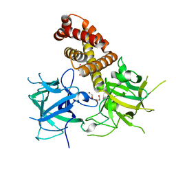
3ILA
DownloadVisualize
BU of 3ila by Molmil
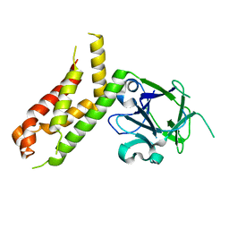
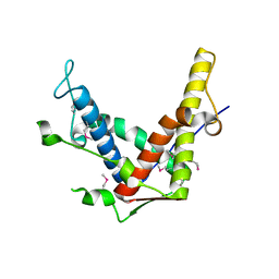
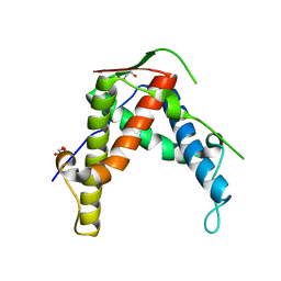
Crystal structure of rabbit ryanodine receptor 1 N-terminal domain (9-205)
Descriptor: Ryanodine receptor 1
Authors:Lobo, P.A, Van Petegem, F.
Deposit date:2009-08-06
Release date:2009-11-17
Last modified:2024-04-03
Method:X-RAY DIFFRACTION (2.9 Å)
Cite:Crystal structures of the N-terminal domains of cardiac and skeletal muscle ryanodine receptors: insights into disease mutations.
Structure, 17, 2009
4L4I
DownloadVisualize
BU of 4l4i by Molmil
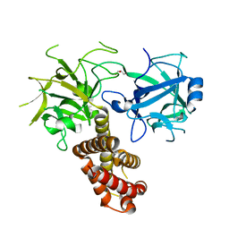
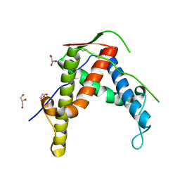
Crystal structure of mouse Ryanodine Receptor isoform 2 (RyR2) 1-547 disease mutant R420Q
Descriptor: GLYCEROL, Ryanodine receptor 2
Authors:Kimlicka, L, Van Petegem, F.
Deposit date:2013-06-07
Release date:2013-07-24
Last modified:2023-09-20
Method:X-RAY DIFFRACTION (2.15 Å)
Cite:Crystal structure of ryanodine receptor disease mutant
To be Published
4L4H
DownloadVisualize
BU of 4l4h by Molmil
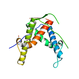
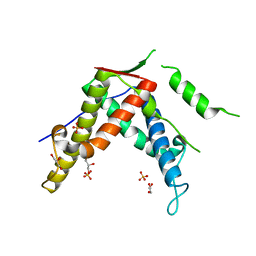
Crystal structure of mouse Ryanodine Receptor isoform 2 (RyR2) 1-547
Descriptor: CHLORIDE ION, DI(HYDROXYETHYL)ETHER, Ryanodine receptor 2, ...
Authors:Kimlicka, L, Tung, C.C, Van Petegem, F.
Deposit date:2013-06-07
Release date:2013-07-24
Last modified:2023-09-20
Method:X-RAY DIFFRACTION (2 Å)
Cite:Crystal structure of a ryanodine receptor domain
To be Published
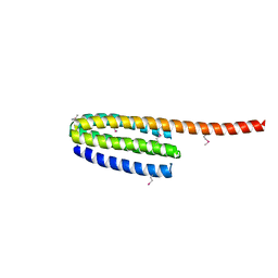
3FYQ
DownloadVisualize
BU of 3fyq by Molmil
Structure of Drosophila melanogaster talin IBS2 domain (residues 1981-2168)
Descriptor: CG6831-PA (Talin)
Authors:Cheung, T.Y.S, Fairchild, M.J, Zarivach, R, Tanentzapf, G, Van Petegem, F.
Deposit date:2009-01-22
Release date:2009-02-03
Last modified:2018-01-24
Method:X-RAY DIFFRACTION (1.95 Å)
Cite:Crystal structure of the talin integrin binding domain 2.
J.Mol.Biol., 387, 2009
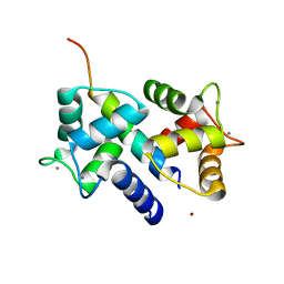
3DVE
DownloadVisualize
BU of 3dve by Molmil
Crystal Structure of Ca2+/CaM-CaV2.2 IQ domain complex
Descriptor: CALCIUM ION, Calmodulin, NICKEL (II) ION, ...
Authors:Kim, E.Y, Rumpf, C.H, Fujiwara, Y, Cooley, E.S, Van Petegem, F, Minor, D.L.
Deposit date:2008-07-18
Release date:2008-11-04
Last modified:2024-02-21
Method:X-RAY DIFFRACTION (2.35 Å)
Cite:Structures of Ca(V)2 Ca(2+)/CaM-IQ Domain Complexes Reveal Binding Modes that Underlie Calcium-Dependent Inactivation and Facilitation.
Structure, 16, 2008
3DVM
DownloadVisualize
BU of 3dvm by Molmil
Crystal Structure of Ca2+/CaM-CaV2.1 IQ domain complex
Descriptor: CALCIUM ION, Calmodulin, Voltage-dependent P/Q-type calcium channel subunit alpha-1A
Authors:Kim, E.Y, Rumpf, C.H, Fujiwara, Y, Cooley, E.S, Van Petegem, F, Minor, D.L.
Deposit date:2008-07-18
Release date:2008-11-04
Last modified:2024-02-21
Method:X-RAY DIFFRACTION (2.6 Å)
Cite:Structures of Ca(V)2 Ca(2+)/CaM-IQ Domain Complexes Reveal Binding Modes that Underlie Calcium-Dependent Inactivation and Facilitation.
Structure, 16, 2008
3DVK
DownloadVisualize
BU of 3dvk by Molmil
Crystal Structure of Ca2+/CaM-CaV2.3 IQ domain complex
Descriptor: CALCIUM ION, Calmodulin, Voltage-dependent R-type calcium channel subunit alpha-1E
Authors:Kim, E.Y, Rumpf, C.H, Fujiwara, Y, Cooley, E.S, Van Petegem, F, Minor, D.L.
Deposit date:2008-07-18
Release date:2008-11-04
Last modified:2024-02-21
Method:X-RAY DIFFRACTION (2.3 Å)
Cite:Structures of Ca(V)2 Ca(2+)/CaM-IQ Domain Complexes Reveal Binding Modes that Underlie Calcium-Dependent Inactivation and Facilitation.
Structure, 16, 2008
7K44
DownloadVisualize
BU of 7k44 by Molmil
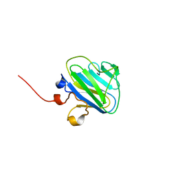
SGBP-B from a complex xyloglucan utilization locus in Bacteroides uniformis
Descriptor: CALCIUM ION, SGBP-B
Authors:Brumer, H, Van Petegem, F, Grondin, J.M.
Deposit date:2020-09-14
Release date:2021-09-29
Last modified:2023-10-18
Method:X-RAY DIFFRACTION (1.45 Å)
Cite:Cell Surface Xyloglucan Recognition and Hydrolysis by the Human Gut Commensal Bacteroides uniformis.
Appl.Environ.Microbiol., 88, 2022
4I8M
DownloadVisualize
BU of 4i8m by Molmil
Crystal structure of rabbit Ryanodine receptor 1 (residues 1-536) disease mutant V219I
Descriptor: GLYCEROL, Ryanodine receptor 1
Authors:Kimlicka, L, Van Petegem, F.
Deposit date:2012-12-03
Release date:2013-02-20
Last modified:2023-09-20
Method:X-RAY DIFFRACTION (2.8 Å)
Cite:Disease mutations in the ryanodine receptor N-terminal region couple to a mobile intersubunit interface.
Nat Commun, 4, 2013
4I6I
DownloadVisualize
BU of 4i6i by Molmil
Crystal structure of rabbit ryanodine receptor 1 (residues 1-559) disease mutant R45C
Descriptor: Ryanodine receptor 1
Authors:Kimlicka, L, Van Petegem, F.
Deposit date:2012-11-29
Release date:2013-02-20
Last modified:2023-09-20
Method:X-RAY DIFFRACTION (2.5 Å)
Cite:Disease mutations in the ryanodine receptor N-terminal region couple to a mobile intersubunit interface.
Nat Commun, 4, 2013
4I1E
DownloadVisualize
BU of 4i1e by Molmil
Crystal structure of Rabbit Ryanodine Receptor 1 (residues 1-536) disease mutant G249R
Descriptor: Ryanodine receptor 1
Authors:Kimlicka, L, Van Petegem, F.
Deposit date:2012-11-20
Release date:2013-02-20
Last modified:2024-02-28
Method:X-RAY DIFFRACTION (2.4 Å)
Cite:Disease mutations in the ryanodine receptor N-terminal region couple to a mobile intersubunit interface.
Nat Commun, 4, 2013
4I3N
DownloadVisualize
BU of 4i3n by Molmil
Crystal structure of rabbit ryanodine receptor 1 (residues 1-536) disease mutant D61N
Descriptor: GLYCEROL, Ryanodine receptor 1
Authors:Kimlicka, L, Van Petegem, F.
Deposit date:2012-11-26
Release date:2013-02-20
Last modified:2023-09-20
Method:X-RAY DIFFRACTION (2.95 Å)
Cite:Disease mutations in the ryanodine receptor N-terminal region couple to a mobile intersubunit interface.
Nat Commun, 4, 2013
4I37
DownloadVisualize
BU of 4i37 by Molmil
Crystal structure of rabbit ryanodine receptor 1 (residues 1-536) disease mutant R402G
Descriptor: Ryanodine receptor 1
Authors:Kimlicka, L, Van Petegem, F.
Deposit date:2012-11-25
Release date:2013-02-20
Last modified:2023-09-20
Method:X-RAY DIFFRACTION (2.95 Å)
Cite:Disease mutations in the ryanodine receptor N-terminal region couple to a mobile intersubunit interface.
Nat Commun, 4, 2013
4I2S
DownloadVisualize
BU of 4i2s by Molmil
Crystal structure of rabbit ryanodine receptor 1 (residues 1-536) disease mutant I404M
Descriptor: GLYCEROL, Ryanodine receptor 1
Authors:Kimlicka, L, Van Petegem, F.
Deposit date:2012-11-22
Release date:2013-02-20
Last modified:2023-09-20
Method:X-RAY DIFFRACTION (2.5 Å)
Cite:Disease mutations in the ryanodine receptor N-terminal region couple to a mobile intersubunit interface.
Nat Commun, 4, 2013
4I96
DownloadVisualize
BU of 4i96 by Molmil
Crystal structure of the N-terminal two domains of the skeletal muscle ryanodine receptor (rabbit RyR1) residues 217-536
Descriptor: Ryanodine receptor 1
Authors:Kimlicka, L, Tung, C.C, Van Petegem, F.
Deposit date:2012-12-04
Release date:2013-02-20
Last modified:2023-09-20
Method:X-RAY DIFFRACTION (2.73 Å)
Cite:Disease mutations in the ryanodine receptor N-terminal region couple to a mobile intersubunit interface.
Nat Commun, 4, 2013
4I7I
DownloadVisualize
BU of 4i7i by Molmil
Crystal structure of rabbit ryanodine receptor 1 (residues 1-536) disease mutant L14R
Descriptor: GLYCEROL, Ryanodine receptor 1
Authors:Kimlicka, L, Van Petegem, F.
Deposit date:2012-11-30
Release date:2013-02-20
Last modified:2023-09-20
Method:X-RAY DIFFRACTION (2.9 Å)
Cite:Disease mutations in the ryanodine receptor N-terminal region couple to a mobile intersubunit interface.
Nat Commun, 4, 2013
4ETU
DownloadVisualize
BU of 4etu by Molmil
Crystal structure of rabbit ryanodine receptor 1 mutant R2939S
Descriptor: GLYCEROL, Ryanodine receptor 1
Authors:Yuchi, Z, Lau, K, Van Petegem, F.
Deposit date:2012-04-24
Release date:2012-06-13
Last modified:2023-09-13
Method:X-RAY DIFFRACTION (2.19 Å)
Cite:Disease mutations in the ryanodine receptor central region: crystal structures of a phosphorylation hot spot domain.
Structure, 20, 2012
4DJC
DownloadVisualize
BU of 4djc by Molmil
1.35 A crystal structure of the NaV1.5 DIII-IV-Ca/CaM complex
Descriptor: CALCIUM ION, Calmodulin, ISOPROPYL ALCOHOL, ...
Authors:Sarhan, M.F, Tung, C.-C, Van Petegem, F, Ahern, C.A.
Deposit date:2012-02-01
Release date:2012-02-22
Last modified:2024-02-28
Method:X-RAY DIFFRACTION (1.35 Å)
Cite:Crystallographic basis for calcium regulation of sodium channels.
Proc.Natl.Acad.Sci.USA, 109, 2012
4ETV
DownloadVisualize
BU of 4etv by Molmil
Crystal structure of mouse ryanodine receptor 2 (2699-2904)
Descriptor: CHLORIDE ION, Ryanodine receptor 2
Authors:Yuchi, Z, Lau, K, Van Petegem, F.
Deposit date:2012-04-24
Release date:2012-06-13
Last modified:2017-11-15
Method:X-RAY DIFFRACTION (1.65 Å)
Cite:Disease mutations in the ryanodine receptor central region: crystal structures of a phosphorylation hot spot domain.
Structure, 20, 2012
4ESU
DownloadVisualize
BU of 4esu by Molmil
Crystal structure of rabbit ryanodine receptor 1 mutant S2776M
Descriptor: GLYCEROL, Ryanodine receptor 1
Authors:Yuchi, Z, Lau, K, Van Petegem, F.
Deposit date:2012-04-23
Release date:2012-06-13
Last modified:2023-09-13
Method:X-RAY DIFFRACTION (1.59 Å)
Cite:Disease mutations in the ryanodine receptor central region: crystal structures of a phosphorylation hot spot domain.
Structure, 20, 2012
4ERV
DownloadVisualize
BU of 4erv by Molmil
Crystal structure of human ryanodine receptor 3 (2597-2800)
Descriptor: GLYCEROL, Ryanodine receptor 3, SULFATE ION
Authors:Yuchi, Z, Lau, K, Van Petegem, F.
Deposit date:2012-04-20
Release date:2012-06-13
Last modified:2024-02-28
Method:X-RAY DIFFRACTION (1.75 Å)
Cite:Disease mutations in the ryanodine receptor central region: crystal structures of a phosphorylation hot spot domain.
Structure, 20, 2012
4ERT
DownloadVisualize
BU of 4ert by Molmil
Crystal structure of rabbit ryanodine receptor 1 (2734-2940)
Descriptor: GLYCEROL, POTASSIUM ION, Ryanodine receptor 1
Authors:Yuchi, Z, Lau, K, Van Petegem, F.
Deposit date:2012-04-20
Release date:2012-06-13
Last modified:2024-02-28
Method:X-RAY DIFFRACTION (1.95 Å)
Cite:Disease mutations in the ryanodine receptor central region: crystal structures of a phosphorylation hot spot domain.
Structure, 20, 2012
4ETT
DownloadVisualize
BU of 4ett by Molmil
Crystal structure of rabbit ryanodine receptor 1 mutant E2764K
Descriptor: GLYCEROL, Ryanodine receptor 1
Authors:Yuchi, Z, Lau, K, Van Petegem, F.
Deposit date:2012-04-24
Release date:2012-06-13
Last modified:2023-09-13
Method:X-RAY DIFFRACTION (2.195 Å)
Cite:Disease mutations in the ryanodine receptor central region: crystal structures of a phosphorylation hot spot domain.
Structure, 20, 2012
2BE9
DownloadVisualize
BU of 2be9 by Molmil
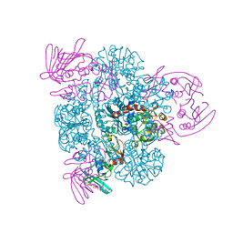
Crystal structure of the CTP-liganded (T-State) aspartate transcarbamoylase from the extremely thermophilic archaeon Sulfolobus acidocaldarius
Descriptor: Aspartate carbamoyltransferase, Aspartate carbamoyltransferase regulatory chain, CYTIDINE-5'-TRIPHOSPHATE, ...
Authors:De Vos, D, Savvides, S.N, Van Beeumen, J.J.
Deposit date:2005-10-23
Release date:2006-10-31
Last modified:2023-08-23
Method:X-RAY DIFFRACTION (2.6 Å)
Cite:Crystal structure of Sulfolobus acidocaldarius aspartate carbamoyltransferase in complex with its allosteric activator CTP.
Biochem.Biophys.Res.Commun., 372, 2008

226262

数据于2024-10-16公开中

PDB statisticsPDBj update infoContact PDBjnumon