Loading
PDBj
MenuPDBj@FacebookPDBj@TwitterPDBj@YouTubewwPDB FoundationwwPDB
RCSB PDBPDBeBMRBAdv. SearchSearch help
Search by PDB author
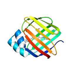
6E5S
DownloadVisualize
BU of 6e5s by Molmil
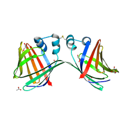
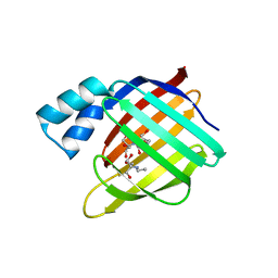
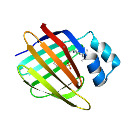
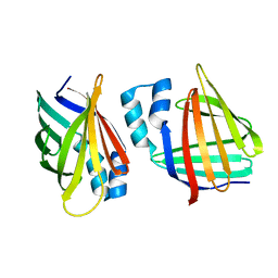
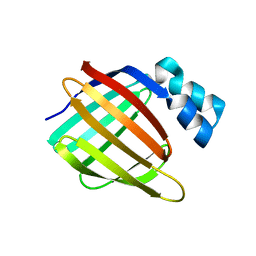
Crystal structure of holo retinal-bound domain-swapped dimer Q108K:T51D mutant of human Cellular Retinol Binding Protein II
Descriptor: RETINAL, Retinol-binding protein 2
Authors:Nosrati, M, Ghanbarpour, A, Geiger, J.
Deposit date:2018-07-22
Release date:2019-07-24
Last modified:2023-10-11
Method:X-RAY DIFFRACTION (2.061 Å)
Cite:Engineering the hCRBPII Domain-Swapped Dimer into a New Class of Protein Switches.
J.Am.Chem.Soc., 141, 2019
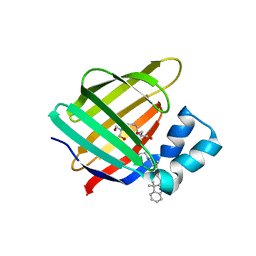
6E7M
DownloadVisualize
BU of 6e7m by Molmil
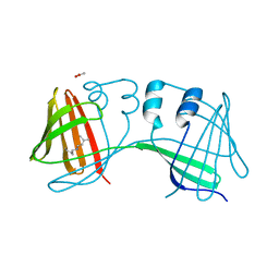
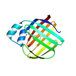
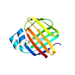
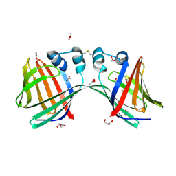
Crystal structure of the holo retinal-bound domain-swapped dimer Q108K:T51D:A28C mutant of human Cellular Retinol Binding Protein II
Descriptor: ACETATE ION, GLYCEROL, RETINAL, ...
Authors:Ghanbarpour, A, Geiger, J.
Deposit date:2018-07-26
Release date:2019-10-16
Last modified:2023-10-11
Method:X-RAY DIFFRACTION (2.7 Å)
Cite:Engineering the hCRBPII Domain-Swapped Dimer into a New Class of Protein Switches.
J.Am.Chem.Soc., 141, 2019
6E5R
DownloadVisualize
BU of 6e5r by Molmil
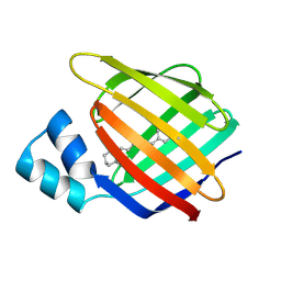
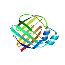
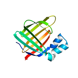
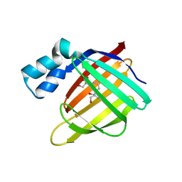
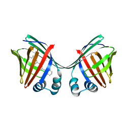
Crystal structure of the apo domain-swapped dimer Q108K:T51D:A28C mutant of human Cellular Retinol Binding Protein II
Descriptor: ACETATE ION, GLYCEROL, Retinol-binding protein 2
Authors:Ghanbarpour, A, Geiger, J.
Deposit date:2018-07-22
Release date:2019-08-07
Last modified:2023-10-11
Method:X-RAY DIFFRACTION (2.592 Å)
Cite:Engineering the hCRBPII Domain-Swapped Dimer into a New Class of Protein Switches.
J.Am.Chem.Soc., 141, 2019
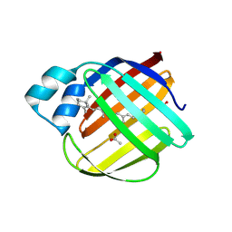
6E6L
DownloadVisualize
BU of 6e6l by Molmil
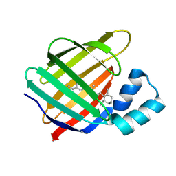
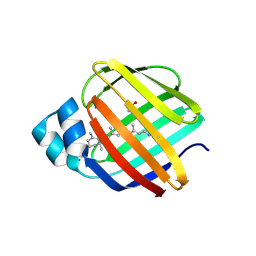
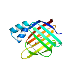
Crystal structure of the holo retinal-bound domain-swapped dimer Q108K:K40L:T51F:Y60A mutant of human cellular retinol binding protein II
Descriptor: ACETATE ION, RETINAL, Retinol-binding protein 2
Authors:Ghanbarpour, A, Geiger, J.
Deposit date:2018-07-25
Release date:2019-10-16
Last modified:2023-10-11
Method:X-RAY DIFFRACTION (2.08 Å)
Cite:Engineering the hCRBPII Domain-Swapped Dimer into a New Class of Protein Switches.
J.Am.Chem.Soc., 141, 2019
4M6S
DownloadVisualize
BU of 4m6s by Molmil
Crystal structure of the R111K:R132Y:Y134F:T54V:R59W:A32W mutant of the Cellular Retinoic Acid Binding Protein Type II in complex with All-Trans Retinal at 2.38 Angstrom Resolution
Descriptor: Cellular retinoic acid-binding protein 2, RETINAL
Authors:Nosrati, M, Geiger, J.H.
Deposit date:2013-08-10
Release date:2013-10-09
Last modified:2023-09-20
Method:X-RAY DIFFRACTION (2.47 Å)
Cite:Rational Design of a Colorimetric pH Sensor from a Soluble Retinoic Acid Chaperone.
J.Am.Chem.Soc., 135, 2013
4M7M
DownloadVisualize
BU of 4m7m by Molmil
Crystal structure of the R111K:R132Q:Y134F:T54V:R59W:A32W mutant of the Cellular Retinoic Acid Binding Protein Type II in complex with All-Trans Retinal at 2.57 Angstrom Resolution
Descriptor: Cellular retinoic acid-binding protein 2, RETINAL
Authors:Nosrati, M, Geiger, J.H.
Deposit date:2013-08-12
Release date:2013-10-09
Last modified:2014-02-19
Method:X-RAY DIFFRACTION (2.571 Å)
Cite:Rational Design of a Colorimetric pH Sensor from a Soluble Retinoic Acid Chaperone.
J.Am.Chem.Soc., 135, 2013
2CEP
DownloadVisualize
BU of 2cep by Molmil
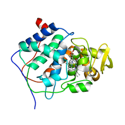
ROLE OF MET-230 IN INTRAMOLECULAR ELECTRON TRANSFER BETWEEN THE OXYFERRYL HEME AND TRP 191 IN CYTOCHROME C PEROXIDASE COMPOUND II
Descriptor: CYTOCHROME C PEROXIDASE, PROTOPORPHYRIN IX CONTAINING FE
Authors:Han, G.W, Miller, M.A, Kraut, J.
Deposit date:1994-05-31
Release date:1994-08-31
Last modified:2024-02-14
Method:X-RAY DIFFRACTION (2.2 Å)
Cite:Role of methionine 230 in intramolecular electron transfer between the oxyferryl heme and tryptophan 191 in cytochrome c peroxidase compound II.
Biochemistry, 33, 1994
3FEL
DownloadVisualize
BU of 3fel by Molmil
Crystal structure of the R132K:R111L:T54E mutant of cellular retinoic acid-binding protein II at 1.85 angstrom resolution
Descriptor: 2-[3-(2-HYDROXY-1,1-DIHYDROXYMETHYL-ETHYLAMINO)-PROPYLAMINO]-2-HYDROXYMETHYL-PROPANE-1,3-DIOL, Cellular retinoic acid-binding protein 2
Authors:Jia, X, Geiger, J.H.
Deposit date:2008-11-30
Release date:2009-11-10
Last modified:2023-09-06
Method:X-RAY DIFFRACTION (1.85 Å)
Cite:Crystal structures of apo cellular retinoic acid-binding protein II mutants: Structural integrity investigated through multiple site mutations
To be Published
4EDE
DownloadVisualize
BU of 4ede by Molmil
Crystal Structure of the Q108K:K40L:T51V:T53C:Y19W:R58W:T29L:A33W Mutant of Cellular Retinol Binding Protein Type II in Complex with All-trans-Retinal at 1.4 Angstrom Resolution
Descriptor: ACETATE ION, RETINAL, Retinol-binding protein 2
Authors:Nossoni, Z, Geiger, J.H.
Deposit date:2012-03-27
Release date:2012-12-26
Last modified:2023-09-13
Method:X-RAY DIFFRACTION (1.4 Å)
Cite:Tuning the electronic absorption of protein-embedded all-trans-retinal.
Science, 338, 2012
4EEJ
DownloadVisualize
BU of 4eej by Molmil
Crystal Structure of the Q108K:K40L:T51V:T53C:Y19W:R58W:T29L:Q4R Mutant of Cellular Retinol Binding Protein Type II in Complex with All-trans-Retinal at 1.5 Angstrom Resolution
Descriptor: ACETATE ION, RETINAL, Retinol-binding protein 2
Authors:Nossoni, Z, Geiger, J.H.
Deposit date:2012-03-28
Release date:2012-12-26
Last modified:2023-09-13
Method:X-RAY DIFFRACTION (1.501 Å)
Cite:Tuning the electronic absorption of protein-embedded all-trans-retinal.
Science, 338, 2012
3FEP
DownloadVisualize
BU of 3fep by Molmil
Crystal structure of the R132K:R111L:L121E:R59W-CRABPII mutant complexed with a synthetic ligand (merocyanin) at 2.60 angstrom resolution.
Descriptor: (2E,4E,6E)-3-methyl-6-(1,3,3-trimethyl-1,3-dihydro-2H-indol-2-ylidene)hexa-2,4-dienal, 2-(N-MORPHOLINO)-ETHANESULFONIC ACID, Cellular retinoic acid-binding protein 2
Authors:Jia, X, Geiger, J.H.
Deposit date:2008-11-30
Release date:2009-11-10
Last modified:2023-09-06
Method:X-RAY DIFFRACTION (2.6 Å)
Cite:"Turn-on" protein fluorescence: in situ formation of cyanine dyes.
J.Am.Chem.Soc., 137, 2015
3FA8
DownloadVisualize
BU of 3fa8 by Molmil
Crystal structure of the apo R132K:Y134F:R111L:L121E mutant of cellular retinoic acid-binding protein II at 1.78 angstrom resolution
Descriptor: Cellular retinoic acid-binding protein 2
Authors:Jia, X, Geiger, J.H.
Deposit date:2008-11-16
Release date:2009-10-27
Last modified:2023-09-06
Method:X-RAY DIFFRACTION (1.78 Å)
Cite:Crystal structures of apo cellular retinoic acid-binding protein II mutants: Structural integrity investigated through multiple site mutations
To be Published
4GKC
DownloadVisualize
BU of 4gkc by Molmil
Crystal structure of Q108K:K40L:T51V:T53C:R58W:T29L:Y19W:Q4A mutant of cellular retinol binding protein II complex with all-trans-retinal at 1.33
Descriptor: RETINAL, Retinol-binding protein 2
Authors:Nossoni, Z, Geiger, J.H.
Deposit date:2012-08-10
Release date:2012-12-26
Last modified:2023-09-13
Method:X-RAY DIFFRACTION (1.3 Å)
Cite:Tuning the electronic absorption of protein-embedded all-trans-retinal.
Science, 338, 2012
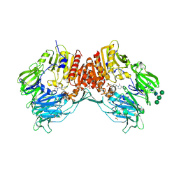
4N8D
DownloadVisualize
BU of 4n8d by Molmil
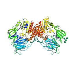
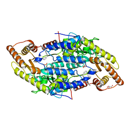
DPP4 complexed with syn-7aa
Descriptor: 1-(cis-1-phenyl-4-{[(2E)-3-phenylprop-2-en-1-yl]oxy}cyclohexyl)methanamine, 2-acetamido-2-deoxy-beta-D-glucopyranose, Dipeptidyl peptidase 4, ...
Authors:Ostermann, N, Zink, F, Kroemer, M.
Deposit date:2013-10-17
Release date:2014-02-12
Last modified:2020-07-29
Method:X-RAY DIFFRACTION (1.65 Å)
Cite:Discovery of C-(1-aryl-cyclohexyl)-methylamines as selective, orally available inhibitors of dipeptidyl peptidase IV.
Bioorg.Med.Chem.Lett., 24, 2014
3I17
DownloadVisualize
BU of 3i17 by Molmil
Crystal structure of the apo R132K:L121E mutant of cellular retinoic acid-binding protein II at 1.68 angstrom resolution
Descriptor: Cellular retinoic acid-binding protein 2
Authors:Jia, X, Watson, C.T, Geiger, J.H.
Deposit date:2009-06-25
Release date:2009-07-28
Last modified:2023-09-06
Method:X-RAY DIFFRACTION (1.68 Å)
Cite:Elucidating the exact role of engineered CRABPII residues for the formation of a retinal protonated Schiff base.
Proteins, 77, 2009
4EFG
DownloadVisualize
BU of 4efg by Molmil
Crystal Structure of the Q108K:K40L:T51V:T53C:Y19W:R58W:T29L Mutant of Cellular Retinol Binding Protein Type II in Complex with All-trans-Retinal at 1.58 Angstrom Resolution
Descriptor: ACETATE ION, RETINAL, Retinol-binding protein 2
Authors:Nossoni, Z, Geiger, J.H.
Deposit date:2012-03-29
Release date:2012-12-26
Last modified:2023-09-13
Method:X-RAY DIFFRACTION (1.58 Å)
Cite:Tuning the electronic absorption of protein-embedded all-trans-retinal.
Science, 338, 2012
3FA7
DownloadVisualize
BU of 3fa7 by Molmil
Crystal structure of the apo R132K:R111L:L121E:R59E mutant of cellular retinoic acid-binding protein II at 1.90 angstrom resolution
Descriptor: 2-[3-(2-HYDROXY-1,1-DIHYDROXYMETHYL-ETHYLAMINO)-PROPYLAMINO]-2-HYDROXYMETHYL-PROPANE-1,3-DIOL, Cellular retinoic acid-binding protein 2
Authors:Jia, X, Geiger, J.H.
Deposit date:2008-11-15
Release date:2009-10-27
Last modified:2023-12-27
Method:X-RAY DIFFRACTION (1.9 Å)
Cite:Crystal structures of apo cellular retinoic acid-binding protein II mutants: Structural integrity investigated through multiple site mutations
To be Published
3FEK
DownloadVisualize
BU of 3fek by Molmil
Crystal structure of the R132K:Y134F:R111L:L121D:T54V mutant of cellular retinoic acid-binding protein II at 1.51 angstrom resolution
Descriptor: ACETATE ION, Cellular retinoic acid-binding protein 2, DI(HYDROXYETHYL)ETHER
Authors:Jia, X, Geiger, J.H.
Deposit date:2008-11-30
Release date:2009-11-10
Last modified:2023-09-06
Method:X-RAY DIFFRACTION (1.51 Å)
Cite:Crystal structures of apo cellular retinoic acid-binding protein II mutants: Structural integrity investigated through multiple site mutations
To be Published
4EXZ
DownloadVisualize
BU of 4exz by Molmil
Crystal Structure of the Q108K:K40L Mutant of Cellular Retinol Binding Protein Type II in Complex with All-trans-Retinal at 1.7 Angstrom Resolution
Descriptor: ACETATE ION, RETINAL, Retinol-binding protein 2
Authors:Nossoni, Z, Geiger, J.H.
Deposit date:2012-05-01
Release date:2012-12-26
Last modified:2023-09-13
Method:X-RAY DIFFRACTION (1.608 Å)
Cite:Tuning the electronic absorption of protein-embedded all-trans-retinal.
Science, 338, 2012
4QGW
DownloadVisualize
BU of 4qgw by Molmil
Crystal sturcture of the R132K:R111L:L121D mutant of Cellular Retinoic Acid Binding ProteinII complexed with a synthetic ligand (Merocyanine) at 1.77 angstrom resolution
Descriptor: (2E,4E,6E)-3-methyl-6-(1,3,3-trimethyl-1,3-dihydro-2H-indol-2-ylidene)hexa-2,4-dienal, 2-[3-(2-HYDROXY-1,1-DIHYDROXYMETHYL-ETHYLAMINO)-PROPYLAMINO]-2-HYDROXYMETHYL-PROPANE-1,3-DIOL, Cellular retinoic acid-binding protein 2
Authors:Nosrati, M, Yapici, I, Geiger, J.H.
Deposit date:2014-05-26
Release date:2015-01-28
Last modified:2015-02-11
Method:X-RAY DIFFRACTION (1.77 Å)
Cite:"Turn-on" protein fluorescence: in situ formation of cyanine dyes.
J.Am.Chem.Soc., 137, 2015
4N8E
DownloadVisualize
BU of 4n8e by Molmil
DPP4 complexed with compound 12a
Descriptor: 1-[cis-4-(aminomethyl)-4-(3-chlorophenyl)cyclohexyl]piperidin-2-one, 2-acetamido-2-deoxy-beta-D-glucopyranose, Dipeptidyl peptidase 4, ...
Authors:Ostermann, N, Zink, F, Kroemer, M.
Deposit date:2013-10-17
Release date:2014-02-12
Last modified:2020-07-29
Method:X-RAY DIFFRACTION (2.3 Å)
Cite:Discovery of C-(1-aryl-cyclohexyl)-methylamines as selective, orally available inhibitors of dipeptidyl peptidase IV.
Bioorg.Med.Chem.Lett., 24, 2014
5F58
DownloadVisualize
BU of 5f58 by Molmil
Crystal structure of the Q108K:K40L:T51V:R58F mutant of human Cellular Retinol Binding Protein II in complex with All-trans-Retinal after 24 hours of incubation at 1.54 Angstrom Resolution
Descriptor: ACETATE ION, RETINAL, Retinol-binding protein 2
Authors:Nosrati, M, Nossoni, Z, Geiger, J.H.
Deposit date:2015-12-04
Release date:2016-12-07
Last modified:2023-09-27
Method:X-RAY DIFFRACTION (1.54 Å)
Cite:Crystal structure of the Q108K:K40L:T51V:R58F mutant of human Cellular Retinol Binding Protein II in complex with All-trans-Retinal after 24 hours of incubation at 1.54 Angstrom Resolution
To Be Published
5F6B
DownloadVisualize
BU of 5f6b by Molmil
Crystal structure of the Q108K:K40L:T51V:R58Y:Y19W mutant of human Cellular Retinol Binding Protein II in complex with All-trans-Retinal at 1.3 Angstrom Resolution
Descriptor: ACETATE ION, RETINAL, Retinol-binding protein 2
Authors:Nosrati, M, Nossoni, Z, Geiger, J.H.
Deposit date:2015-12-05
Release date:2016-12-07
Last modified:2023-09-27
Method:X-RAY DIFFRACTION (1.31 Å)
Cite:Crystal structure of the Q108K:K40L:T51V:R58Y:Y19W mutant of human Cellular Retinol Binding ProteinII in complex with All-trans-Retinal at 1.31 Angstrom Resolution
To Be Published
4Q6R
DownloadVisualize
BU of 4q6r by Molmil
Crystal structure of human sphingosine-1-phosphate lyase in complex with inhibitor 6-[(2R)-4-(4-benzyl-7-chlorophthalazin-1-yl)-2-methylpiperazin-1-yl]pyridine-3-carbonitrile
Descriptor: 6-[(2R)-4-(4-benzyl-7-chlorophthalazin-1-yl)-2-methylpiperazin-1-yl]pyridine-3-carbonitrile, GLYCEROL, PHOSPHATE ION, ...
Authors:Srinivas, H.
Deposit date:2014-04-23
Release date:2014-05-21
Last modified:2023-12-06
Method:X-RAY DIFFRACTION (2.4 Å)
Cite:Orally Active 7-Substituted (4-Benzylphthalazin-1-yl)-2-methylpiperazin-1-yl]nicotinonitriles as Active-Site Inhibitors of Sphingosine 1-Phosphate Lyase for the Treatment of Multiple Sclerosis.
J.Med.Chem., 57, 2014
3FA9
DownloadVisualize
BU of 3fa9 by Molmil
Crystal structure of the apo R132K:Y134F:R111L:L121D mutant of cellular retinoic acid-binding protein II at 1.94 angstrom resolution
Descriptor: ACETATE ION, Cellular retinoic acid-binding protein 2
Authors:Jia, X, Geiger, J.H.
Deposit date:2008-11-16
Release date:2009-10-27
Last modified:2023-12-27
Method:X-RAY DIFFRACTION (1.94 Å)
Cite:Crystal structures of apo cellular retinoic acid-binding protein II mutants: Structural integrity investigated through multiple site mutations
To be Published

223532

数据于2024-08-07公开中

PDB statisticsPDBj update infoContact PDBjnumon