Loading
PDBj
MenuPDBj@FacebookPDBj@TwitterPDBj@YouTubewwPDB FoundationwwPDB
RCSB PDBPDBeBMRBAdv. SearchSearch help
Search by PDB author
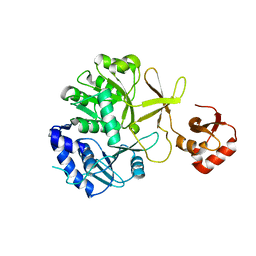
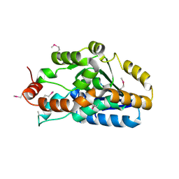
3CLK
DownloadVisualize
BU of 3clk by Molmil
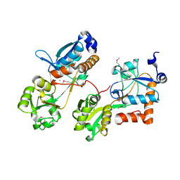
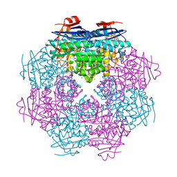
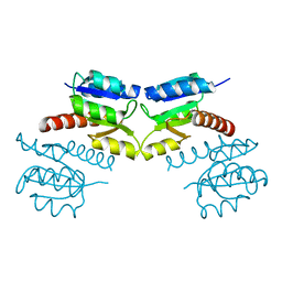
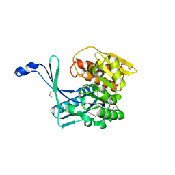
Crystal structure of a transcription regulator from Lactobacillus plantarum
Descriptor: GLYCEROL, Transcription regulator
Authors:Sugadev, R, Burley, S.K, Swaminathan, S, New York SGX Research Center for Structural Genomics (NYSGXRC)
Deposit date:2008-03-19
Release date:2008-04-01
Last modified:2021-02-03
Method:X-RAY DIFFRACTION (2.08 Å)
Cite:Crystal structure of a transcription regulator from lactobacillus plantarum.
To be Published
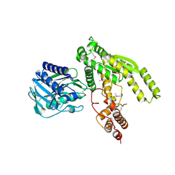
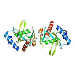
3CO4
DownloadVisualize
BU of 3co4 by Molmil
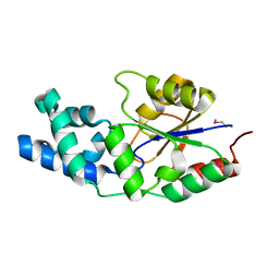
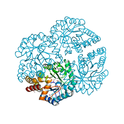
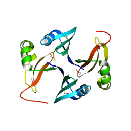
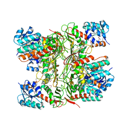
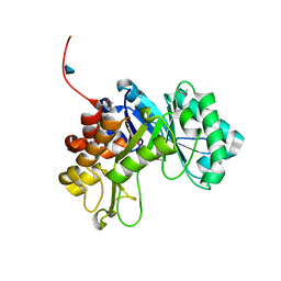
Crystal structure of a chitinase from Bacteroides thetaiotaomicron
Descriptor: 2-amino-2-deoxy-beta-D-glucopyranose, Chitinase
Authors:Damodharan, L, Burley, S.K, Swaminathan, S, New York SGX Research Center for Structural Genomics (NYSGXRC)
Deposit date:2008-03-27
Release date:2008-04-08
Last modified:2021-02-03
Method:X-RAY DIFFRACTION (1.92 Å)
Cite:Crystal structure of a chitinase from Bacteroides thetaiotaomicron.
To be Published
3IPL
DownloadVisualize
BU of 3ipl by Molmil
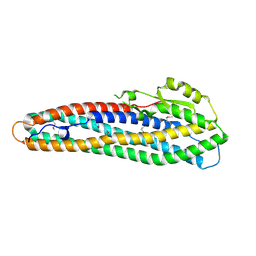
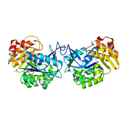
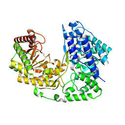
CRYSTAL STRUCTURE OF o-succinylbenzoic acid-CoA ligase FROM Staphylococcus aureus subsp. aureus Mu50
Descriptor: 2-succinylbenzoate--CoA ligase
Authors:Patskovsky, Y, Toro, R, Dickey, M, Chang, S, Sauder, J.M, Burley, S.K, Almo, S.C, New York SGX Research Center for Structural Genomics (NYSGXRC)
Deposit date:2009-08-17
Release date:2009-08-25
Last modified:2024-02-21
Method:X-RAY DIFFRACTION (2.3 Å)
Cite:Crystal structure of o-succinylbenzoic acid-CoA ligase from Staphylococcus aureus
To be Published
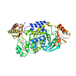
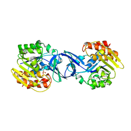
3ILV
DownloadVisualize
BU of 3ilv by Molmil
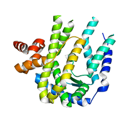
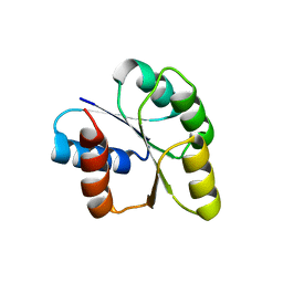
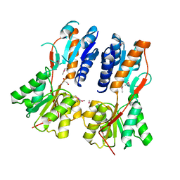
Crystal structure of a glutamine-dependent NAD(+) synthetase from Cytophaga hutchinsonii
Descriptor: Glutamine-dependent NAD(+) synthetase
Authors:Palani, K, Burley, S.K, Swaminathan, S, New York SGX Research Center for Structural Genomics (NYSGXRC)
Deposit date:2009-08-07
Release date:2009-08-18
Last modified:2021-02-10
Method:X-RAY DIFFRACTION (1.79 Å)
Cite:Crystal structure of a glutamine-dependent NAD(+) synthetase from Cytophaga hutchinsonii
To be Published
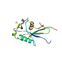
3EXQ
DownloadVisualize
BU of 3exq by Molmil
Crystal structure of a NUDIX family hydrolase from Lactobacillus brevis
Descriptor: NUDIX family hydrolase, SULFATE ION
Authors:Palani, K, Burley, S.K, Swaminathan, S, New York SGX Research Center for Structural Genomics (NYSGXRC)
Deposit date:2008-10-16
Release date:2008-11-04
Last modified:2024-10-09
Method:X-RAY DIFFRACTION (2 Å)
Cite:Crystal structure of a NUDIX family hydrolase from Lactobacillus brevis
To be Published
3FKD
DownloadVisualize
BU of 3fkd by Molmil
The crystal structure of L-threonine-O-3-phosphate decarboxylase from Porphyromonas gingivalis
Descriptor: L-threonine-O-3-phosphate decarboxylase
Authors:Zhang, Z, Eswaramoorthy, S, Burley, S.K, Swaminathan, S, New York SGX Research Center for Structural Genomics (NYSGXRC)
Deposit date:2008-12-16
Release date:2009-02-03
Last modified:2021-02-10
Method:X-RAY DIFFRACTION (2.5 Å)
Cite:The crystal structure of L-threonine-O-3-phosphate decarboxylase from Porphyromonas gingivalis
To be Published
3HP0
DownloadVisualize
BU of 3hp0 by Molmil
Crystal structure of a Putative polyketide biosynthesis enoyl-CoA hydratase (pksH) from Bacillus subtilis
Descriptor: Putative polyketide biosynthesis enoyl-CoA hydratase homolog pksH
Authors:Satyanarayana, L, Eswaramoorthy, S, Burley, S.K, Swaminathan, S, New York SGX Research Center for Structural Genomics (NYSGXRC)
Deposit date:2009-06-03
Release date:2009-06-23
Last modified:2021-02-10
Method:X-RAY DIFFRACTION (2.32 Å)
Cite:Crystal structure of a Putative polyketide biosynthesis enoyl-CoA hydratase (pksH) from Bacillus subtilis
To be Published
3FCM
DownloadVisualize
BU of 3fcm by Molmil
Crystal structure of a NUDIX hydrolase from Clostridium perfringens
Descriptor: Hydrolase, NUDIX family, MANGANESE (II) ION
Authors:Palani, K, Burley, S.K, Swaninathan, S, New York SGX Research Center for Structural Genomics (NYSGXRC)
Deposit date:2008-11-21
Release date:2008-12-09
Last modified:2023-12-27
Method:X-RAY DIFFRACTION (2.2 Å)
Cite:Crystal structure of a NUDIX hydrolase from Clostridium perfringens
To be Published
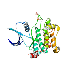
3COM
DownloadVisualize
BU of 3com by Molmil
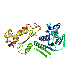
Crystal structure of Mst1 kinase
Descriptor: Serine/threonine-protein kinase 4
Authors:Atwell, S, Burley, S.K, Dickey, M, Leon, B, Sauder, J.M, New York SGX Research Center for Structural Genomics (NYSGXRC)
Deposit date:2008-03-28
Release date:2008-04-15
Last modified:2023-08-30
Method:X-RAY DIFFRACTION (2.2 Å)
Cite:Crystal structure of Mst1 kinase.
To be Published
3I3Y
DownloadVisualize
BU of 3i3y by Molmil
Crystal structure of Ribokinase in Complex with D-Ribose from Klebsiella pneumoniae
Descriptor: Carbohydrate kinase, GLYCEROL, SULFATE ION, ...
Authors:Satyanarayana, L, Burley, S.K, Swaminathan, S, New York SGX Research Center for Structural Genomics (NYSGXRC)
Deposit date:2009-07-01
Release date:2009-07-21
Last modified:2024-10-30
Method:X-RAY DIFFRACTION (2.15 Å)
Cite:Crystal structure of Ribokinase in Complex with D-Ribose from Klebsiella pneumoniae
To be Published
3HV1
DownloadVisualize
BU of 3hv1 by Molmil
Crystal structure of a polar amino acid ABC uptake transporter substrate binding protein from Streptococcus thermophilus
Descriptor: Polar amino acid ABC uptake transporter substrate binding protein
Authors:Palani, K, Burley, S.K, Swaminathan, S, New York SGX Research Center for Structural Genomics (NYSGXRC)
Deposit date:2009-06-15
Release date:2009-06-23
Last modified:2021-02-10
Method:X-RAY DIFFRACTION (1.9 Å)
Cite:Crystal structure of a polar amino acid ABC uptake transporter substrate binding protein from Streptococcus thermophilus
To be Published
2NYV
DownloadVisualize
BU of 2nyv by Molmil
X-ray crystal structure of a phosphoglycolate phosphatase from Aquifex aeolicus
Descriptor: Phosphoglycolate phosphatase
Authors:Ciatto, C, Min, T, Gorman, J, Burley, S.K, Shapiro, L, New York SGX Research Center for Structural Genomics (NYSGXRC)
Deposit date:2006-11-21
Release date:2006-12-05
Last modified:2023-12-27
Method:X-RAY DIFFRACTION (2.103 Å)
Cite:X-ray crystal structure of a phosphoglycolate phosphatase from Aquifex aeolicus
To be Published
2NRJ
DownloadVisualize
BU of 2nrj by Molmil
Crystal Structure of Hemolysin binding component from Bacillus cereus
Descriptor: Hbl B protein
Authors:Madegowda, M, Eswaramoorthy, S, Burley, S.K, Swaminathan, S, New York SGX Research Center for Structural Genomics (NYSGXRC)
Deposit date:2006-11-02
Release date:2006-11-14
Last modified:2024-10-30
Method:X-RAY DIFFRACTION (2.03 Å)
Cite:X-ray crystal structure of the B component of Hemolysin BL from Bacillus cereus
Proteins, 71, 2008
3CMN
DownloadVisualize
BU of 3cmn by Molmil
Crystal structure of a putative hydrolase with a novel fold from Chloroflexus aurantiacus
Descriptor: Putative hydrolase
Authors:Agarwal, R, Burley, S.K, Swaminathan, S, New York SGX Research Center for Structural Genomics (NYSGXRC)
Deposit date:2008-03-24
Release date:2008-04-08
Last modified:2024-02-21
Method:X-RAY DIFFRACTION (2.25 Å)
Cite:Crystal structure of a putative hydrolase with a novel fold from Chloroflexus aurantiacus.
To be Published
2OG9
DownloadVisualize
BU of 2og9 by Molmil
Crystal Structure of mandelate racemase/muconate lactonizing enzyme from Polaromonas sp. JS666
Descriptor: CALCIUM ION, Mandelate racemase/muconate lactonizing enzyme
Authors:Kumaran, D, Burley, S.K, Swaminathan, S, New York SGX Research Center for Structural Genomics (NYSGXRC)
Deposit date:2007-01-05
Release date:2007-02-27
Last modified:2024-10-30
Method:X-RAY DIFFRACTION (1.9 Å)
Cite:Crystal Structure of mandelate racemase/muconate lactonizing enzyme from Polaromonas sp. JS666
To be Published
3DZ1
DownloadVisualize
BU of 3dz1 by Molmil
Crystal structure of Dihydrodipicolinate Synthase from Rhodopseudomonas palustris at 1.87A resolution
Descriptor: Dihydrodipicolinate synthase
Authors:Satyanarayana, L, Eswaramoorthy, S, Burley, S.K, Swaminathan, S, New York SGX Research Center for Structural Genomics (NYSGXRC)
Deposit date:2008-07-29
Release date:2008-08-12
Last modified:2024-10-30
Method:X-RAY DIFFRACTION (1.87 Å)
Cite:Crystal structure of Dihydrodipicolinate Synthase from Rhodopseudomonas palustris at 1.87A resolution
To be Published
3EWM
DownloadVisualize
BU of 3ewm by Molmil
Crystal structure of an uncharacterized sugar kinase PH1459 from pyrococcus horikoshii
Descriptor: Uncharacterized sugar kinase PH1459
Authors:Kumar, G, Eswaramoorthy, S, Burley, S.K, Swaminathan, S, New York SGX Research Center for Structural Genomics (NYSGXRC)
Deposit date:2008-10-15
Release date:2008-11-18
Last modified:2023-12-27
Method:X-RAY DIFFRACTION (1.9 Å)
Cite:Crystal structure of an uncharacterized sugar kinase PH1459 from pyrococcus horikoshii
To be Published
3CNB
DownloadVisualize
BU of 3cnb by Molmil
Crystal structure of signal receiver domain of DNA binding response regulator protein (merR) from Colwellia psychrerythraea 34H
Descriptor: DNA-binding response regulator, merR family
Authors:Patskovsky, Y, Romero, R, Freeman, J, Hu, S, Groshong, C, Wasserman, S.R, Sauder, J.M, Burley, S.K, Almo, S.C, New York SGX Research Center for Structural Genomics (NYSGXRC)
Deposit date:2008-03-25
Release date:2008-04-08
Last modified:2024-02-21
Method:X-RAY DIFFRACTION (2 Å)
Cite:Crystal structure of signal receiver domain of DNA binding response regulator (merR) from Colwellia psychrerythraea 34H.
To be Published
3CWV
DownloadVisualize
BU of 3cwv by Molmil
Crystal structure of B-subunit of the DNA gyrase from Myxococcus xanthus
Descriptor: DNA gyrase, B subunit, truncated
Authors:Ramagopal, U.A, Toro, R, Meyer, A.J, Burley, S.K, Almo, S.C, New York SGX Research Center for Structural Genomics (NYSGXRC)
Deposit date:2008-04-22
Release date:2008-05-06
Last modified:2024-02-21
Method:X-RAY DIFFRACTION (1.95 Å)
Cite:Crystal structure of B-subunit of the DNA gyrase from Myxococcus xanthus.
To be published
3D3A
DownloadVisualize
BU of 3d3a by Molmil
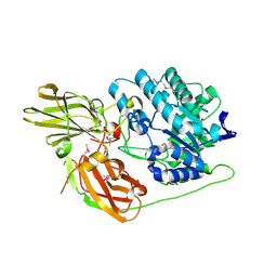
Crystal structure of a beta-galactosidase from Bacteroides thetaiotaomicron
Descriptor: Beta-galactosidase
Authors:Palani, K, Kumaran, D, Burley, S.K, Swaminathan, S, New York SGX Research Center for Structural Genomics (NYSGXRC)
Deposit date:2008-05-09
Release date:2008-05-27
Last modified:2021-02-03
Method:X-RAY DIFFRACTION (2.15 Å)
Cite:Crystal structure of a beta-galactosidase from Bacteroides thetaiotaomicron.
To be Published
3CG0
DownloadVisualize
BU of 3cg0 by Molmil
Crystal structure of signal receiver domain of modulated diguanylate cyclase from Desulfovibrio desulfuricans G20, an example of alternate folding
Descriptor: Response regulator receiver modulated diguanylate cyclase with PAS/PAC sensor
Authors:Patskovsky, Y, Bonanno, J.B, Romero, R, Gilmore, M, Chang, S, Groshong, C, Koss, J, Wasserman, S.R, Sauder, J.M, Burley, S.K, Almo, S.C, New York SGX Research Center for Structural Genomics (NYSGXRC)
Deposit date:2008-03-04
Release date:2008-03-18
Last modified:2024-02-21
Method:X-RAY DIFFRACTION (2.15 Å)
Cite:Crystal Structure of Signal Receiver Domain of Modulated Diguanylate Cyclase from Desulfovibrio desulfuricans.
To be Published
3FCD
DownloadVisualize
BU of 3fcd by Molmil
Crystal Structure of a putative glyoxalase from an environmental bacteria
Descriptor: Lyase
Authors:Silberstein, M, Eswaramoorthy, S, Burley, S.K, Swaminathan, S, New York SGX Research Center for Structural Genomics (NYSGXRC)
Deposit date:2008-11-21
Release date:2008-12-09
Last modified:2023-12-27
Method:X-RAY DIFFRACTION (1.92 Å)
Cite:Crystal Structure of a putative glyoxalase from an environmental bacteria
To be Published
3H49
DownloadVisualize
BU of 3h49 by Molmil
Crystal structure of a putative Ribokinase (Apo Form) from E.coli at 1.8A resolution
Descriptor: Ribokinase
Authors:Satyanarayana, L, Eswaramoorthy, S, Burley, S.K, Swaminathan, S, New York SGX Research Center for Structural Genomics (NYSGXRC)
Deposit date:2009-04-18
Release date:2009-04-28
Last modified:2021-02-10
Method:X-RAY DIFFRACTION (1.8 Å)
Cite:Crystal structure of a putative Ribokinase (Apo Form) from E.coli at 1.8A resolution.
To be Published
3FD8
DownloadVisualize
BU of 3fd8 by Molmil
Crystal Structure of an oxidoreductase from Enterococcus faecalis
Descriptor: Oxidoreductase, Gfo/Idh/MocA family, TRIETHYLENE GLYCOL
Authors:Damodharan, L, Burley, S.K, Swaminathan, S, New York SGX Research Center for Structural Genomics (NYSGXRC)
Deposit date:2008-11-25
Release date:2009-02-03
Last modified:2023-12-27
Method:X-RAY DIFFRACTION (2.45 Å)
Cite:Crystal Structure of an oxidoreductase from Enterococcus faecalis
To be Published
2NQ5
DownloadVisualize
BU of 2nq5 by Molmil
Crystal structure of methyltransferase from Streptococcus mutans
Descriptor: 5-methyltetrahydropteroyltriglutamate--homocysteine methyltransferase
Authors:Fedorov, A.A, Fedorov, E.V, Sauder, J.M, Burley, S.K, Almo, S.C, New York SGX Research Center for Structural Genomics (NYSGXRC)
Deposit date:2006-10-30
Release date:2006-11-14
Last modified:2023-08-30
Method:X-RAY DIFFRACTION (1.9 Å)
Cite:Crystal structure of methyltransferase from Streptococcus mutans
To be Published

226707

数据于2024-10-30公开中

PDB statisticsPDBj update infoContact PDBjnumon