Loading
PDBj
MenuPDBj@FacebookPDBj@TwitterPDBj@YouTubewwPDB FoundationwwPDB
RCSB PDBPDBeBMRBAdv. SearchSearch help
Search by PDB author
3MSW
DownloadVisualize
BU of 3msw by Molmil
Crystal structure of a Protein with unknown function (BF3112) from Bacteroides fragilis NCTC 9343 at 1.90 A resolution
Descriptor: CHLORIDE ION, R-1,2-PROPANEDIOL, uncharacterized protein
Authors:Joint Center for Structural Genomics (JCSG)
Deposit date:2010-04-29
Release date:2010-06-09
Last modified:2023-02-01
Method:X-RAY DIFFRACTION (1.9 Å)
Cite:Structures of single-layer beta-sheet proteins evolved from beta-hairpin repeats.
Protein Sci., 28, 2019
3M81
DownloadVisualize
BU of 3m81 by Molmil
Crystal structure of Acetyl xylan esterase (TM0077) from THERMOTOGA MARITIMA at 2.50 A resolution (native apo structure)
Descriptor: 1,2-ETHANEDIOL, ACETATE ION, Acetyl xylan esterase, ...
Authors:Joint Center for Structural Genomics (JCSG)
Deposit date:2010-03-17
Release date:2010-04-21
Last modified:2023-09-20
Method:X-RAY DIFFRACTION (2.5 Å)
Cite:Functional and structural characterization of a thermostable acetyl esterase from Thermotoga maritima.
Proteins, 80, 2012
3M83
DownloadVisualize
BU of 3m83 by Molmil
Crystal structure of Acetyl xylan esterase (TM0077) from THERMOTOGA MARITIMA at 2.12 A resolution (paraoxon inhibitor complex structure)
Descriptor: 1,2-ETHANEDIOL, ACETATE ION, Acetyl xylan esterase, ...
Authors:Joint Center for Structural Genomics (JCSG)
Deposit date:2010-03-17
Release date:2010-05-12
Last modified:2023-09-20
Method:X-RAY DIFFRACTION (2.12 Å)
Cite:Functional and structural characterization of a thermostable acetyl esterase from Thermotoga maritima.
Proteins, 80, 2012
3M0C
DownloadVisualize
BU of 3m0c by Molmil
The X-ray Crystal Structure of PCSK9 in Complex with the LDL receptor
Descriptor: CALCIUM ION, Low-density lipoprotein receptor, Proprotein convertase subtilisin/kexin type 9
Authors:Spraggon, G, Hampton, E.N.
Deposit date:2010-03-02
Release date:2011-03-16
Last modified:2023-09-06
Method:X-RAY DIFFRACTION (7.01 Å)
Cite:The X-ray Crystal Structure of PCSK9 in Complex with the LDL receptor
To be Published
3BY7
DownloadVisualize
BU of 3by7 by Molmil
CRYSTAL STRUCTURE OF A PROTEIN STRUCTURALLY SIMILAR TO SM/LSM-LIKE RNA-BINDING PROTEINS (JCVI_PEP_1096686650277) FROM UNCULTURED MARINE ORGANISM AT 2.60 A RESOLUTION
Descriptor: uncharacterized protein
Authors:Joint Center for Structural Genomics (JCSG)
Deposit date:2008-01-15
Release date:2008-01-29
Last modified:2023-02-01
Method:X-RAY DIFFRACTION (2.6 Å)
Cite:Crystal structure of a novel Sm-like protein of putative cyanophage origin at 2.60 A resolution.
Proteins, 75, 2009
3B77
DownloadVisualize
BU of 3b77 by Molmil
Crystal structure of a ph domain containing bacterial protein (exig_2160) from exiguobacterium sibiricum 255-15 at 2.42 A resolution
Descriptor: Uncharacterized protein
Authors:Joint Center for Structural Genomics (JCSG)
Deposit date:2007-10-30
Release date:2007-11-13
Last modified:2023-01-25
Method:X-RAY DIFFRACTION (2.42 Å)
Cite:Bacterial pleckstrin homology domains: a prokaryotic origin for the PH domain.
J.Mol.Biol., 396, 2010
3BYQ
DownloadVisualize
BU of 3byq by Molmil
Crystal structure of a duf1185 family protein (bb2672) from bordetella bronchiseptica rb50 at 1.70 A resolution
Descriptor: 1,2-ETHANEDIOL, CHLORIDE ION, TETRAETHYLENE GLYCOL, ...
Authors:Joint Center for Structural Genomics (JCSG)
Deposit date:2008-01-16
Release date:2008-01-29
Last modified:2024-10-16
Method:X-RAY DIFFRACTION (1.7 Å)
Cite:Structures of the first representatives of Pfam family PF06684 (DUF1185) reveal a novel variant of the Bacillus chorismate mutase fold and suggest a role in amino-acid metabolism.
Acta Crystallogr.,Sect.F, 66, 2010
3BOS
DownloadVisualize
BU of 3bos by Molmil
Crystal structure of a putative dna replication regulator HDA (SAMA_1916) from Shewanella amazonensis sb2b at 1.75 A resolution
Descriptor: 1,2-ETHANEDIOL, CHLORIDE ION, CYTIDINE-5'-DIPHOSPHATE, ...
Authors:Joint Center for Structural Genomics (JCSG)
Deposit date:2007-12-17
Release date:2008-01-15
Last modified:2023-01-25
Method:X-RAY DIFFRACTION (1.75 Å)
Cite:A structural basis for the regulatory inactivation of DnaA.
J.Mol.Biol., 385, 2009
3D00
DownloadVisualize
BU of 3d00 by Molmil
Crystal structure of a tungsten formylmethanofuran dehydrogenase subunit e (fmde)-like protein (syn_00638) from syntrophus aciditrophicus at 1.90 A resolution
Descriptor: CHLORIDE ION, Tungsten formylmethanofuran dehydrogenase subunit E, ZINC ION
Authors:Joint Center for Structural Genomics (JCSG)
Deposit date:2008-04-30
Release date:2008-05-20
Last modified:2023-02-01
Method:X-RAY DIFFRACTION (1.9 Å)
Cite:Structures of three members of Pfam PF02663 (FmdE) implicated in microbial methanogenesis reveal a conserved alpha+beta core domain and an auxiliary C-terminal treble-clef zinc finger.
Acta Crystallogr.,Sect.F, 66, 2010
3CGH
DownloadVisualize
BU of 3cgh by Molmil
Crystal structure of a susd homolog (bt_3984) from bacteroides thetaiotaomicron vpi-5482 at 1.70 A resolution
Descriptor: ACETATE ION, SusD homolog, ZINC ION
Authors:Joint Center for Structural Genomics (JCSG)
Deposit date:2008-03-05
Release date:2008-03-18
Last modified:2024-10-09
Method:X-RAY DIFFRACTION (1.7 Å)
Cite:Structure of BT_3984, a member of the SusD/RagB family of nutrient-binding molecules.
Acta Crystallogr.,Sect.F, 66, 2010
1S17
DownloadVisualize
BU of 1s17 by Molmil
Identification of Novel Potent Bicyclic Peptide Deformylase Inhibitors
Descriptor: 2-(3,4-DIHYDRO-3-OXO-2H-BENZO[B][1,4]THIAZIN-2-YL)-N-HYDROXYACETAMIDE, GLYCEROL, NICKEL (II) ION, ...
Authors:Molteni, V, He, X, Nabakka, J, Yang, K, Kreusch, A, Gordon, P, Bursulaya, B, Ryder, N.S, Goldberg, R, He, Y.
Deposit date:2004-01-05
Release date:2004-03-30
Last modified:2023-08-23
Method:X-RAY DIFFRACTION (1.95 Å)
Cite:Identification of novel potent bicyclic peptide deformylase inhibitors
Bioorg.Med.Chem.Lett., 14, 2004
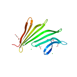
1AJ7
DownloadVisualize
BU of 1aj7 by Molmil
IMMUNOGLOBULIN 48G7 GERMLINE FAB ANTIBODY COMPLEXED WITH HAPTEN 5-(PARA-NITROPHENYL PHOSPHONATE)-PENTANOIC ACID. AFFINITY MATURATION OF AN ESTEROLYTIC ANTIBODY
Descriptor: 5-(PARA-NITROPHENYL PHOSPHONATE)-PENTANOIC ACID, IMMUNOGLOBULIN 48G7 FAB (HEAVY CHAIN), IMMUNOGLOBULIN 48G7 FAB (LIGHT CHAIN)
Authors:Wedemayer, G.J, Wang, L.H, Patten, P.A, Schultz, P.G, Stevens, R.C.
Deposit date:1997-05-15
Release date:1997-11-12
Last modified:2024-10-09
Method:X-RAY DIFFRACTION (2.1 Å)
Cite:Structural insights into the evolution of an antibody combining site.
Science, 276, 1997
2F37
DownloadVisualize
BU of 2f37 by Molmil
Crystal structure of the ankyrin repeat domain of human TRPV2
Descriptor: SULFATE ION, Transient receptor potential cation channel subfamily V member 2
Authors:McCleverty, C.J.
Deposit date:2005-11-18
Release date:2006-10-31
Last modified:2011-07-13
Method:X-RAY DIFFRACTION (1.7 Å)
Cite:Crystal structure of the human TRPV2 channel ankyrin repeat domain.
Protein Sci., 15, 2006
2FNA
DownloadVisualize
BU of 2fna by Molmil
Crystal structure of an archaeal aaa+ atpase (sso1545) from sulfolobus solfataricus p2 at 2.00 A resolution
Descriptor: 1,2-ETHANEDIOL, ADENOSINE-5'-DIPHOSPHATE, Conserved hypothetical protein, ...
Authors:Joint Center for Structural Genomics (JCSG)
Deposit date:2006-01-10
Release date:2006-02-07
Last modified:2024-10-09
Method:X-RAY DIFFRACTION (2 Å)
Cite:Crystal structure of a novel archaeal AAA+ ATPase SSO1545 from Sulfolobus solfataricus.
Proteins, 74, 2009
2G36
DownloadVisualize
BU of 2g36 by Molmil
Crystal structure of Tryptophanyl-tRNA synthetase (EC 6.1.1.2) (Tryptophan-tRNA ligase)(TrpRS) (tm0492) from THERMOTOGA MARITIMA at 2.50 A resolution
Descriptor: IRON/SULFUR CLUSTER, TRYPTOPHAN, Tryptophanyl-tRNA synthetase
Authors:Joint Center for Structural Genomics (JCSG)
Deposit date:2006-02-17
Release date:2006-03-28
Last modified:2024-04-03
Method:X-RAY DIFFRACTION (2.5 Å)
Cite:Structure of a tryptophanyl-tRNA synthetase containing an iron-sulfur cluster.
Acta Crystallogr.,Sect.F, 66, 2010
2FG0
DownloadVisualize
BU of 2fg0 by Molmil
Crystal structure of a putative gamma-d-glutamyl-l-diamino acid endopeptidase (npun_r0659) from nostoc punctiforme pcc 73102 at 1.79 A resolution
Descriptor: COG0791: Cell wall-associated hydrolases (invasion-associated proteins), GLYCEROL
Authors:Joint Center for Structural Genomics (JCSG)
Deposit date:2005-12-20
Release date:2006-01-10
Last modified:2023-01-25
Method:X-RAY DIFFRACTION (1.79 Å)
Cite:Structural Basis of Murein Peptide Specificity of a gamma-D-Glutamyl-L-Diamino Acid Endopeptidase.
Structure, 17, 2009
2EVR
DownloadVisualize
BU of 2evr by Molmil
CRYSTAL STRUCTURE OF A PUTATIVE GAMMA-D-GLUTAMYL-L-DIAMINO ACID ENDOPEPTIDASE (NPUN_R0659) FROM NOSTOC PUNCTIFORME PCC 73102 AT 1.60 A RESOLUTION
Descriptor: 1,2-ETHANEDIOL, CHLORIDE ION, COG0791: Cell wall-associated hydrolases (invasion-associated proteins), ...
Authors:Joint Center for Structural Genomics (JCSG)
Deposit date:2005-10-31
Release date:2005-11-22
Last modified:2024-10-16
Method:X-RAY DIFFRACTION (1.6 Å)
Cite:Structural Basis of Murein Peptide Specificity of a gamma-D-Glutamyl-L-Diamino Acid Endopeptidase.
Structure, 17, 2009
2FNO
DownloadVisualize
BU of 2fno by Molmil
Crystal structure of a glutathione s-transferase (atu5508) from agrobacterium tumefaciens str. c58 at 2.00 A resolution
Descriptor: AGR_pAT_752p, THIOCYANATE ION
Authors:Joint Center for Structural Genomics (JCSG)
Deposit date:2006-01-11
Release date:2006-02-14
Last modified:2023-01-25
Method:X-RAY DIFFRACTION (2 Å)
Cite:Comparative structural analysis of a novel glutathioneS-transferase (ATU5508) from Agrobacterium tumefaciens at 2.0 A resolution.
Proteins, 65, 2006
2ETS
DownloadVisualize
BU of 2ets by Molmil
CRYSTAL STRUCTURE OF A BACTERIAL DOMAIN OF UNKNOWN FUNCTION FROM DUF1798 FAMILY (MW1337) FROM STAPHYLOCOCCUS AUREUS SUBSP. AUREUS AT 2.25 A RESOLUTION
Descriptor: CHLORIDE ION, PHOSPHATE ION, hypothetical protein
Authors:Joint Center for Structural Genomics (JCSG)
Deposit date:2005-10-27
Release date:2005-11-08
Last modified:2023-01-25
Method:X-RAY DIFFRACTION (2.25 Å)
Cite:Crystal structures of MW1337R and lin2004: representatives of a novel protein family that adopt a four-helical bundle fold.
Proteins, 71, 2008
2F46
DownloadVisualize
BU of 2f46 by Molmil
Crystal structure of a putative phosphatase (nma1982) from neisseria meningitidis z2491 at 1.41 A resolution
Descriptor: CHLORIDE ION, UNKNOWN LIGAND, hypothetical protein
Authors:Joint Center for Structural Genomics (JCSG)
Deposit date:2005-11-22
Release date:2006-02-07
Last modified:2023-01-25
Method:X-RAY DIFFRACTION (1.41 Å)
Cite:Crystal structure of NMA1982 from Neisseria meningitidis at 1.5 A resolution provides a structural scaffold for nonclassical, eukaryotic-like phosphatases.
Proteins, 69, 2007
2FEA
DownloadVisualize
BU of 2fea by Molmil
Crystal structure of MtnX phosphatase from Bacillus Subtilis at 2.00 A resolution
Descriptor: 1,2-ETHANEDIOL, 2-hydroxy-3-keto-5-methylthiopentenyl-1-phosphate phosphatase, MAGNESIUM ION, ...
Authors:Joint Center for Structural Genomics (JCSG)
Deposit date:2005-12-15
Release date:2005-12-27
Last modified:2023-01-25
Method:X-RAY DIFFRACTION (2 Å)
Cite:Crystal structure of MtnX phosphatase from Bacillus subtilis at 2.0 A resolution provides a structural basis for bipartite phosphomonoester hydrolysis of 2-hydroxy-3-keto-5-methylthiopentenyl-1-phosphate.
Proteins, 69, 2007
2GHR
DownloadVisualize
BU of 2ghr by Molmil
Crystal structure of homoserine o-succinyltransferase (NP_981826.1) from Bacillus cereus ATCC 10987 at 2.40 A resolution
Descriptor: Homoserine O-succinyltransferase, SULFATE ION
Authors:Joint Center for Structural Genomics (JCSG)
Deposit date:2006-03-27
Release date:2006-04-11
Last modified:2023-01-25
Method:X-RAY DIFFRACTION (2.4 Å)
Cite:Crystal structure of homoserine O-succinyltransferase from Bacillus cereus at 2.4 A resolution
Proteins, 68, 2007
2GLZ
DownloadVisualize
BU of 2glz by Molmil
Crystal structure of a formylmethanofuran dehydrogenase subunit e-like protein (dhaf_2992) from desulfitobacterium hafniense dcb-2 at 1.45 A resolution
Descriptor: 1,2-ETHANEDIOL, NICKEL (II) ION, ZINC ION, ...
Authors:Joint Center for Structural Genomics (JCSG)
Deposit date:2006-04-05
Release date:2006-04-18
Last modified:2023-01-25
Method:X-RAY DIFFRACTION (1.45 Å)
Cite:Structures of three members of Pfam PF02663 (FmdE) implicated in microbial methanogenesis reveal a conserved alpha+beta core domain and an auxiliary C-terminal treble-clef zinc finger.
Acta Crystallogr.,Sect.F, 66, 2010
2HBW
DownloadVisualize
BU of 2hbw by Molmil
Crystal structure of a putative endopeptidase (ava_3396) from anabaena variabilis atcc 29413 at 1.05 A resolution
Descriptor: ACETATE ION, NLP/P60 protein, UNKNOWN LIGAND
Authors:Joint Center for Structural Genomics (JCSG)
Deposit date:2006-06-14
Release date:2006-08-08
Last modified:2023-01-25
Method:X-RAY DIFFRACTION (1.05 Å)
Cite:Structural Basis of Murein Peptide Specificity of a gamma-D-Glutamyl-L-Diamino Acid Endopeptidase.
Structure, 17, 2009
2GVK
DownloadVisualize
BU of 2gvk by Molmil
Crystal structure of a dye-decolorizing peroxidase (DyP) from Bacteroides thetaiotaomicron VPI-5482 at 1.6 A resolution
Descriptor: 1,2-ETHANEDIOL, ACETIC ACID, CHLORIDE ION, ...
Authors:Joint Center for Structural Genomics (JCSG)
Deposit date:2006-05-02
Release date:2006-05-16
Last modified:2023-01-25
Method:X-RAY DIFFRACTION (1.6 Å)
Cite:Crystal structures of two novel dye-decolorizing peroxidases reveal a beta-barrel fold with a conserved heme-binding motif.
Proteins, 69, 2007

226262

数据于2024-10-16公开中

PDB statisticsPDBj update infoContact PDBjnumon