Loading
PDBj
MenuPDBj@FacebookPDBj@TwitterPDBj@YouTubewwPDB FoundationwwPDB
RCSB PDBPDBeBMRBAdv. SearchSearch help
Search by PDB author
1TXN
DownloadVisualize
BU of 1txn by Molmil
Crystal structure of coproporphyrinogen III oxidase
Descriptor: Coproporphyrinogen III oxidase, GLYCEROL
Authors:Kumaran, D, Swaminathan, S, Burley, S.K, New York SGX Research Center for Structural Genomics (NYSGXRC)
Deposit date:2004-07-05
Release date:2004-11-30
Last modified:2021-02-03
Method:X-RAY DIFFRACTION (1.7 Å)
Cite:Crystal structure of coproporphyrinogen III oxidase
To be Published
1U8S
DownloadVisualize
BU of 1u8s by Molmil
Crystal structure of putative glycine cleavage system transcriptional repressor
Descriptor: glycine cleavage system transcriptional repressor, putative
Authors:Kumaran, D, Swaminathan, S.
Deposit date:2004-08-06
Release date:2004-08-17
Last modified:2024-02-14
Method:X-RAY DIFFRACTION (2.45 Å)
Cite:Crystal structure of putative glycine cleavage system transcriptional repressor
To be Published
3NYW
DownloadVisualize
BU of 3nyw by Molmil
Crystal Structure of a betaketoacyl-[ACP] reductase (FabG) from Bacteroides thetaiotaomicron
Descriptor: Putative oxidoreductase
Authors:Satyanarayana, L, Burley, S.K, Swaminathan, S, New York SGX Research Center for Structural Genomics (NYSGXRC)
Deposit date:2010-07-15
Release date:2010-09-15
Last modified:2021-02-10
Method:X-RAY DIFFRACTION (2.16 Å)
Cite:Crystal Structure of a betaketoacyl-[ACP] reductase (FabG) from Bacteroides thetaiotaomicron
To be Published
1U05
DownloadVisualize
BU of 1u05 by Molmil
Crystal structure of protein yfiH from Shigella flexneri, Pfam DUF152
Descriptor: ZINC ION, orf, conserved hypothetical protein
Authors:Seetharaman, J, Swaminathan, S, Burley, S.K, New York SGX Research Center for Structural Genomics (NYSGXRC)
Deposit date:2004-07-12
Release date:2004-08-17
Last modified:2023-08-23
Method:X-RAY DIFFRACTION (2.5 Å)
Cite:Crystal Structure of Conserved Hypothetical Protein
To be Published
3FFZ
DownloadVisualize
BU of 3ffz by Molmil
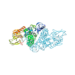
Domain organization in Clostridium butulinum neurotoxin type E is unique: Its implication in faster translocation
Descriptor: ACETATE ION, Botulinum neurotoxin type E, SODIUM ION, ...
Authors:Kumaran, D, Eswaramoorthy, S, Swaminathan, S.
Deposit date:2008-12-04
Release date:2008-12-16
Last modified:2023-09-06
Method:X-RAY DIFFRACTION (2.65 Å)
Cite:Domain organization in Clostridium botulinum neurotoxin type E is unique: its implication in faster translocation.
J.Mol.Biol., 386, 2009
3GPK
DownloadVisualize
BU of 3gpk by Molmil
Crystal Structure of PpiC-type peptidyl-prolyl cis-trans isomerase domain at 1.55A resolution.
Descriptor: PpiC-type peptidyl-prolyl cis-trans isomerase, SULFATE ION
Authors:Satyanarayana, L, Eswaramoorthy, S, Burley, S.K, Swaminathan, S, New York SGX Research Center for Structural Genomics (NYSGXRC)
Deposit date:2009-03-23
Release date:2009-03-31
Last modified:2021-02-10
Method:X-RAY DIFFRACTION (1.55 Å)
Cite:Crystal Structure of PpiC-type peptidyl-prolyl cis-trans isomerase domain at 1.55A resolution.
To be Published
3OVG
DownloadVisualize
BU of 3ovg by Molmil
The crystal structure of an amidohydrolase from Mycoplasma synoviae with Zn ion bound
Descriptor: PHOSPHATE ION, ZINC ION, amidohydrolase
Authors:Zhang, Z, Kumaran, D, Burley, S.K, Swaminathan, S, New York SGX Research Center for Structural Genomics (NYSGXRC)
Deposit date:2010-09-16
Release date:2010-10-13
Last modified:2023-12-06
Method:X-RAY DIFFRACTION (2.059 Å)
Cite:The crystal structure of an amidohydrolase from Mycoplasma synoviae with Zn ion bound
To be Published
3HDG
DownloadVisualize
BU of 3hdg by Molmil
Crystal structure of the N-terminal domain of an uncharacterized protein (WS1339) from Wolinella succinogenes
Descriptor: MAGNESIUM ION, uncharacterized protein
Authors:Eswaramoorthy, S, Burley, S.K, Swaminathan, S, New York SGX Research Center for Structural Genomics (NYSGXRC)
Deposit date:2009-05-07
Release date:2009-05-19
Last modified:2021-02-10
Method:X-RAY DIFFRACTION (2.27 Å)
Cite:Crystal structure of the N-terminal domain of an uncharacterized protein (WS1339) from Wolinella succinogenes
To be Published
3HDP
DownloadVisualize
BU of 3hdp by Molmil
Crystal structure of the NI(II)-bound Glyoxalase-I from Clostridium acetobutylicum
Descriptor: Glyoxalase-I, NICKEL (II) ION
Authors:Satyanarayana, L, Eswaramoorthy, S, Burley, S.K, Swaminathan, S, New York SGX Research Center for Structural Genomics (NYSGXRC)
Deposit date:2009-05-07
Release date:2009-05-26
Last modified:2023-09-06
Method:X-RAY DIFFRACTION (2.06 Å)
Cite:Crystal structure of the NI(II)-bound Glyoxalase-I from Clostridium acetobutylicum.
To be Published
3PAN
DownloadVisualize
BU of 3pan by Molmil
The crystal structure of adenosine deaminase with hypoxanthine bound from Pseudomonas aeruginosa
Descriptor: Adenosine deaminase, HYPOXANTHINE, ZINC ION
Authors:Zhang, Z, Burley, S.K, Swaminathan, S, New York SGX Research Center for Structural Genomics (NYSGXRC)
Deposit date:2010-10-19
Release date:2010-12-22
Last modified:2023-12-06
Method:X-RAY DIFFRACTION (2.627 Å)
Cite:The crystal structure of adenosine deaminase with hypoxanthine bound from Pseudomonas aeruginosa
TO BE PUBLISHED
3G0O
DownloadVisualize
BU of 3g0o by Molmil
Crystal structure of 3-hydroxyisobutyrate dehydrogenase (ygbJ) from Salmonella typhimurium
Descriptor: 3-hydroxyisobutyrate dehydrogenase, CHLORIDE ION, L(+)-TARTARIC ACID
Authors:Eswaramoorthy, S, Burley, S.K, Swaminathan, S, New York SGX Research Center for Structural Genomics (NYSGXRC)
Deposit date:2009-01-28
Release date:2009-02-10
Last modified:2021-02-10
Method:X-RAY DIFFRACTION (1.8 Å)
Cite:Crystal structure of 3-hydroxyisobutyrate dehydrogenase (ygbJ) from Salmonella typhimurium
To be Published
3G12
DownloadVisualize
BU of 3g12 by Molmil
Crystal structure of a putative lactoylglutathione lyase from Bdellovibrio bacteriovorus
Descriptor: Putative lactoylglutathione lyase, SULFATE ION
Authors:Patskovsky, Y, Madegowda, M, Gilmore, M, Chang, S, Maletic, M, Smith, D, Sauder, J.M, Burley, S.K, Swaminathan, S, Almo, S.C, New York SGX Research Center for Structural Genomics (NYSGXRC)
Deposit date:2009-01-29
Release date:2009-02-10
Last modified:2024-02-21
Method:X-RAY DIFFRACTION (2.58 Å)
Cite:Crystal structure of a putative lactoylglutathione lyase from Bdellovibrio bacteriovorus
To be Published
3PAO
DownloadVisualize
BU of 3pao by Molmil
The crystal structure of adenosine deaminase with adenine bound from Pseudomonas aeruginosa
Descriptor: ADENINE, Adenosine deaminase, ZINC ION
Authors:Zhang, Z, Burley, S.K, Swaminathan, S, New York SGX Research Center for Structural Genomics (NYSGXRC)
Deposit date:2010-10-19
Release date:2010-12-22
Last modified:2023-12-06
Method:X-RAY DIFFRACTION (2.493 Å)
Cite:The crystal structure of adenosine deaminase with adenine bound from Pseudomonas aeruginosa
To be Published
3PU6
DownloadVisualize
BU of 3pu6 by Molmil
The crystal structure of an uncharacterized protein from Wolinella succinogenes
Descriptor: Uncharacterized protein
Authors:Zhang, Z, Burley, S.K, Swaminathan, S, New York SGX Research Center for Structural Genomics (NYSGXRC)
Deposit date:2010-12-03
Release date:2011-01-19
Last modified:2021-02-10
Method:X-RAY DIFFRACTION (2.6 Å)
Cite:The crystal structure of an uncharacterized protein from Wolinella succinogenes
TO BE PUBLISHED
3Q1Y
DownloadVisualize
BU of 3q1y by Molmil
Allosteric regulation by Lysine residue: A novel anion-hole formation in the ribokinase family
Descriptor: GLYCEROL, Lin2199 protein, POTASSIUM ION
Authors:Satyanarayana, L, Burley, S.K, Swaminathan, S, New York SGX Research Center for Structural Genomics (NYSGXRC)
Deposit date:2010-12-18
Release date:2011-01-19
Last modified:2023-09-13
Method:X-RAY DIFFRACTION (2.03 Å)
Cite:Allosteric regulation by Lysine residue: A novel anion-hole formation in the ribokinase family
To be Published
3Q34
DownloadVisualize
BU of 3q34 by Molmil
The crystal structure of YceI-like family protein from Pseudomonas syringae
Descriptor: GLYCEROL, Ubiquinone-8, YceI-like family protein
Authors:Zhang, Z, Burley, S.K, Swaminathan, S, New York SGX Research Center for Structural Genomics (NYSGXRC)
Deposit date:2010-12-21
Release date:2011-02-23
Last modified:2021-02-10
Method:X-RAY DIFFRACTION (1.7 Å)
Cite:The crystal structure of YceI-like family protein from Pseudomonas syringae
TO BE PUBLISHED
3PRK
DownloadVisualize
BU of 3prk by Molmil
INHIBITION OF PROTEINASE K BY METHOXYSUCCINYL-ALA-ALA-PRO-ALA-CHLOROMETHYL KETONE. AN X-RAY STUDY AT 2.2-ANGSTROMS RESOLUTION
Descriptor: CALCIUM ION, METHOXYSUCCINYL-ALA-ALA-PRO-ALA-CHLOROMETHYL KETONE, PROTEINASE K
Authors:Wolf, W.M, Bajorath, J, Mueller, A, Raghunathan, S, Singh, T.P, Hinrichs, W, Saenger, W.
Deposit date:1991-08-07
Release date:1994-01-31
Last modified:2017-11-29
Method:X-RAY DIFFRACTION (2.2 Å)
Cite:Inhibition of proteinase K by methoxysuccinyl-Ala-Ala-Pro-Ala-chloromethyl ketone. An x-ray study at 2.2-A resolution.
J.Biol.Chem., 266, 1991
3NAS
DownloadVisualize
BU of 3nas by Molmil
The crystal structure of beta-phosphoglucomutase from Bacillus subtilis
Descriptor: beta-phosphoglucomutase
Authors:Zhang, Z, Burley, S.K, Swaminathan, S, New York SGX Research Center for Structural Genomics (NYSGXRC)
Deposit date:2010-06-02
Release date:2010-07-28
Last modified:2021-02-10
Method:X-RAY DIFFRACTION (3 Å)
Cite:The crystal structure of beta-phosphoglucomutase from Bacillus subtilis
To be Published
3PBK
DownloadVisualize
BU of 3pbk by Molmil
Structural and Functional Studies of Fatty Acyl-Adenylate Ligases from E. coli and L. pneumophila
Descriptor: 5'-O-[(S)-(dodecanoyloxy)(hydroxy)phosphoryl]adenosine, Fatty Acyl-Adenylate Ligase
Authors:Zhang, Z, Burley, S.K, Swaminathan, S, New York SGX Research Center for Structural Genomics (NYSGXRC)
Deposit date:2010-10-20
Release date:2010-12-22
Last modified:2023-12-06
Method:X-RAY DIFFRACTION (3 Å)
Cite:Structural and Functional Studies of Fatty Acyl Adenylate Ligases from E. coli and L. pneumophila.
J.Mol.Biol., 406, 2011
3PBM
DownloadVisualize
BU of 3pbm by Molmil
The crystal structure of adenosine deaminase in complex with chloropurine from Pseudomonas aeruginosa
Descriptor: 6-chloro-9H-purine, Adenosine deaminase, ZINC ION
Authors:Zhang, Z, Burley, S.K, Swaminathan, S, New York SGX Research Center for Structural Genomics (NYSGXRC)
Deposit date:2010-10-20
Release date:2010-12-22
Last modified:2023-12-06
Method:X-RAY DIFFRACTION (2.591 Å)
Cite:The crystal structure of adenosine deaminase in complex with chloropurine from Pseudomonas aeruginosa
To be Published
3OC4
DownloadVisualize
BU of 3oc4 by Molmil
Crystal Structure of a pyridine nucleotide-disulfide family oxidoreductase from the Enterococcus faecalis V583
Descriptor: FLAVIN-ADENINE DINUCLEOTIDE, Oxidoreductase, pyridine nucleotide-disulfide family, ...
Authors:Kumaran, D, Baumann, K, Burley, S.K, Swaminathan, S, New York SGX Research Center for Structural Genomics (NYSGXRC)
Deposit date:2010-08-09
Release date:2010-10-06
Last modified:2021-02-10
Method:X-RAY DIFFRACTION (2.6 Å)
Cite:Crystal Structure of a pyridine nucleotide-disulfide family oxidoreductase from the Enterococcus faecalis V583
To be Published
3NQB
DownloadVisualize
BU of 3nqb by Molmil
Crystal Structure of Adenine Deaminase from Agrobacterium tumefaciens (str. C 58)
Descriptor: Adenine deaminase 2, MANGANESE (II) ION
Authors:Bagaria, A, Kumaran, D, Burley, S.K, Swaminathan, S, New York SGX Research Center for Structural Genomics (NYSGXRC)
Deposit date:2010-06-29
Release date:2010-07-28
Last modified:2023-12-27
Method:X-RAY DIFFRACTION (2.21 Å)
Cite:Crystal Structure of Adenine Deaminase from Agrobacterium tumefaciens (str. C 58)
To be Published
3H49
DownloadVisualize
BU of 3h49 by Molmil
Crystal structure of a putative Ribokinase (Apo Form) from E.coli at 1.8A resolution
Descriptor: Ribokinase
Authors:Satyanarayana, L, Eswaramoorthy, S, Burley, S.K, Swaminathan, S, New York SGX Research Center for Structural Genomics (NYSGXRC)
Deposit date:2009-04-18
Release date:2009-04-28
Last modified:2021-02-10
Method:X-RAY DIFFRACTION (1.8 Å)
Cite:Crystal structure of a putative Ribokinase (Apo Form) from E.coli at 1.8A resolution.
To be Published
3PZL
DownloadVisualize
BU of 3pzl by Molmil
The crystal structure of agmatine ureohydrolase of Thermoplasma volcanium
Descriptor: Agmatine ureohydrolase, MANGANESE (II) ION
Authors:Zhang, Z, Burley, S.K, Swaminathan, S, New York SGX Research Center for Structural Genomics (NYSGXRC)
Deposit date:2010-12-14
Release date:2011-01-19
Last modified:2021-02-10
Method:X-RAY DIFFRACTION (2.7 Å)
Cite:The crystal structure of agmatine ureohydrolase of Thermoplasma volcanium
To be Published
3G1W
DownloadVisualize
BU of 3g1w by Molmil
Crystal structure of sugar ABC transporter (sugar-binding protein) from Bacillus halodurans
Descriptor: Sugar ABC transporter
Authors:Zhang, Z, Eswaramoorthy, S, Swaminathan, S, Burley, S.K, New York SGX Research Center for Structural Genomics (NYSGXRC)
Deposit date:2009-01-30
Release date:2009-02-17
Last modified:2021-02-10
Method:X-RAY DIFFRACTION (2.02 Å)
Cite:The crystal structure of sugar ABC transporter (sugar-binding protein) from Bacillus halodurans.
To be Published

222926

数据于2024-07-24公开中

PDB statisticsPDBj update infoContact PDBjnumon