Loading
PDBj
MenuPDBj@FacebookPDBj@TwitterPDBj@YouTubewwPDB FoundationwwPDB
RCSB PDBPDBeBMRBAdv. SearchSearch help
Search by PDB author
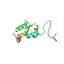
2MYG
DownloadVisualize
BU of 2myg by Molmil
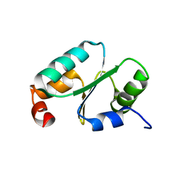
Solution structure of the dithiolic glutaredoxin 2-C-Grx1 from the pathogen Trypanosoma brucei brucei
Descriptor: Dithiol glutaredoxin 1
Authors:Sturlese, M, Stefani, M, Manta, B, Mammi, S, Comini, M, Bellanda, M.
Deposit date:2015-01-22
Release date:2016-02-10
Last modified:2024-05-01
Method:SOLUTION NMR
Cite:Solution structure of the dithiolic glutaredoxin 2-C-Grx1 from the pathogen Trypanosoma brucei brucei
To be Published
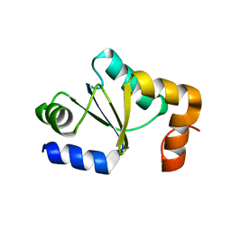
2MXN
DownloadVisualize
BU of 2mxn by Molmil
NMR Structure of the mature form of Trypanosoma brucei 1CGrx1
Descriptor: Mono-cysteine glutaredoxin
Authors:Sturlese, M, Bertarello, A, Manta, B, Lelli, M, Mammi, S, Comini, M, Bellanda, M.
Deposit date:2015-01-08
Release date:2016-01-27
Last modified:2024-05-01
Method:SOLUTION NMR
Cite:The lineage-specific, intrinsically disordered N-terminal extension of monothiol glutaredoxin 1 from trypanosomes contains a regulatory region.
Sci Rep, 8, 2018
2LTK
DownloadVisualize
BU of 2ltk by Molmil
Solution structure of a monomeric truncated mutant of Trypanosoma brucei 1-C-Grx1
Descriptor: Mono-cysteine glutaredoxin
Authors:Pavan, C, Gesiot, L, Sturlese, M, Mammi, S, Manta, B, Comini, M, Bellanda, M.
Deposit date:2012-05-29
Release date:2013-05-08
Last modified:2024-05-01
Method:SOLUTION NMR
Cite:Iron-Sulfur Cluster Binding by Mitochondrial Monothiol Glutaredoxin-1 of Trypanosoma brucei: Molecular Basis of Iron-Sulfur Cluster Coordination and Relevance for Parasite Infectivity.
Antioxid Redox Signal, 19, 2013
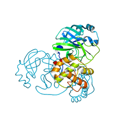
7NIJ
DownloadVisualize
BU of 7nij by Molmil
SARS-CoV-2 main protease (Mpro) in a novel conformational state.
Descriptor: 3C-like proteinase nsp5
Authors:Battistutta, R, Fornasier, E, Giachin, G.
Deposit date:2021-02-12
Release date:2022-03-02
Last modified:2024-01-31
Method:X-RAY DIFFRACTION (1.58 Å)
Cite:A new inactive conformation of SARS-CoV-2 main protease.
Acta Crystallogr D Struct Biol, 78, 2022
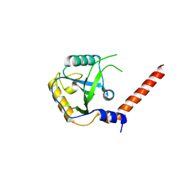
7YYF
DownloadVisualize
BU of 7yyf by Molmil
Orthorombic crystal structure of YTHDF1 YTH domain (G459N mutant) form II
Descriptor: YTH domain-containing family protein 1
Authors:Dalle Vedove, A, Cazzanelli, G, Lolli, G.
Deposit date:2022-02-17
Release date:2023-03-01
Last modified:2024-03-13
Method:X-RAY DIFFRACTION (2.3 Å)
Cite:Pliability in the m 6 A-Binding Region Extends Druggability of YTH Domains.
J.Chem.Inf.Model., 2024
7YYJ
DownloadVisualize
BU of 7yyj by Molmil
Tetragonal crystal structure of YTHDF1 YTH domain (544AVV546 mutant)
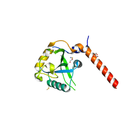
Descriptor: 1,2-ETHANEDIOL, THIOCYANATE ION, YTH domain-containing family protein 1
Authors:Dalle Vedove, A, Cazzanelli, G, Lolli, G.
Deposit date:2022-02-18
Release date:2023-03-01
Last modified:2024-11-06
Method:X-RAY DIFFRACTION (2.1 Å)
Cite:Pliability in the m 6 A-Binding Region Extends Druggability of YTH Domains.
J.Chem.Inf.Model., 2024
7YZ8
DownloadVisualize
BU of 7yz8 by Molmil
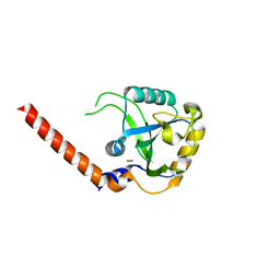
Triclinic crystal structure of YTHDF1 YTH domain (544AVV546 mutant)
Descriptor: THIOCYANATE ION, YTH domain-containing family protein 1
Authors:Dalle Vedove, A, Cazzanelli, G, Lolli, G.
Deposit date:2022-02-19
Release date:2023-03-01
Last modified:2024-03-13
Method:X-RAY DIFFRACTION (2.5 Å)
Cite:Pliability in the m 6 A-Binding Region Extends Druggability of YTH Domains.
J.Chem.Inf.Model., 2024
7YYE
DownloadVisualize
BU of 7yye by Molmil
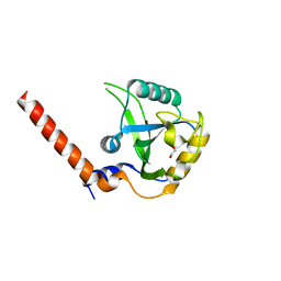
Orthorombic crystal structure of YTHDF1 YTH domain (G459N mutant) form I
Descriptor: 1,2-ETHANEDIOL, MALONIC ACID, YTH domain-containing family protein 1
Authors:Dalle Vedove, A, Cazzanelli, G, Lolli, G.
Deposit date:2022-02-17
Release date:2023-03-01
Last modified:2024-03-13
Method:X-RAY DIFFRACTION (2 Å)
Cite:Pliability in the m 6 A-Binding Region Extends Druggability of YTH Domains.
J.Chem.Inf.Model., 2024

227111

건을2024-11-06부터공개중

PDB statisticsPDBj update infoContact PDBjnumon