Loading
PDBj
MenuPDBj@FacebookPDBj@TwitterPDBj@YouTubewwPDB FoundationwwPDB
RCSB PDBPDBeBMRBAdv. SearchSearch help
Search by PDB author
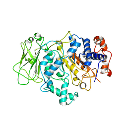
5UQZ
DownloadVisualize
BU of 5uqz by Molmil
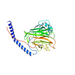
Structural Analysis of the Glucan Binding Protein C of Streptococcus mutans Provides Evidence that it Mediates both Sucrose-Independent and -Dependent Adherence
Descriptor: CALCIUM ION, Glucan-binding protein C, GbpC
Authors:Larson, M.R, Purushotham, S, Mieher, J, Wu, R, Rajashankar, K.R, Wu, H, Deivanayagam, C.
Deposit date:2017-02-08
Release date:2018-03-07
Last modified:2024-03-06
Method:X-RAY DIFFRACTION (1.149 Å)
Cite:Glucan Binding Protein C of Streptococcus mutans Mediates both Sucrose-Independent and Sucrose-Dependent Adherence.
Infect. Immun., 86, 2018
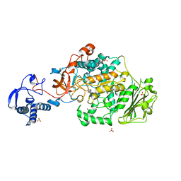
7L0O
DownloadVisualize
BU of 7l0o by Molmil
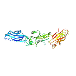
Streptococcus gordonii C123 Domain(s)-Structural and Functional Analysis
Descriptor: Agglutinin receptor, CALCIUM ION
Authors:Schormann, N, Deivanayagam, C.
Deposit date:2020-12-11
Release date:2021-09-08
Last modified:2024-10-09
Method:X-RAY DIFFRACTION (2.7 Å)
Cite:Structural and functional analysis of the C-terminal region of Streptococcus gordonii SspB.
Acta Crystallogr D Struct Biol, 77, 2021
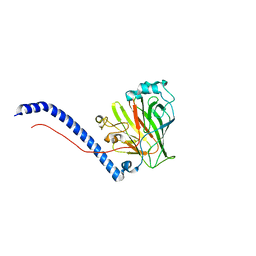
4EIJ
DownloadVisualize
BU of 4eij by Molmil
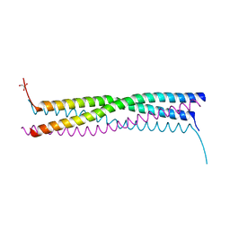
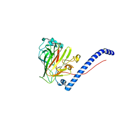
Structure of the Mumps virus phosphoprotein oligomerization domain
Descriptor: GLYCEROL, P protein
Authors:Cox, R, Green, T.J, Luo, M.
Deposit date:2012-04-05
Release date:2013-05-08
Last modified:2024-02-28
Method:X-RAY DIFFRACTION (2.2001 Å)
Cite:Structural and functional characterization of the mumps virus phosphoprotein.
J.Virol., 87, 2013
6E36
DownloadVisualize
BU of 6e36 by Molmil
The structure of the variable domain of Streptococcus intermedius antigen I/II (Pas)
Descriptor: MAGNESIUM ION, Probable cell-surface antigen I/II
Authors:Schormann, N, Deivanayagam, C.
Deposit date:2018-07-13
Release date:2019-07-17
Last modified:2023-10-11
Method:X-RAY DIFFRACTION (1.7 Å)
Cite:Structure-Function Characterization of Streptococcus intermedius Surface Antigen Pas.
J.Bacteriol., 203, 2021
6E3F
DownloadVisualize
BU of 6e3f by Molmil
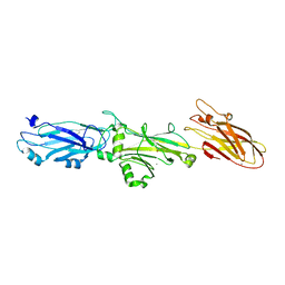
The structure of the C-terminal domains (C123) of Streptococcus intermedius antigen I/II (Pas)
Descriptor: CALCIUM ION, Probable cell-surface antigen I/II
Authors:Schormann, N, Deivanayagam, C.
Deposit date:2018-07-13
Release date:2019-07-17
Last modified:2024-10-30
Method:X-RAY DIFFRACTION (2.7 Å)
Cite:Structure-Function Characterization of Streptococcus intermedius Surface Antigen Pas.
J.Bacteriol., 203, 2021
6CAM
DownloadVisualize
BU of 6cam by Molmil
Glucan Binding Protein C of Streptococcus mutans Mediates both Sucrose-Independent and Sucrose-Dependent Adherence
Descriptor: CALCIUM ION, Glucan-binding protein C, beta-D-glucopyranose
Authors:Schormann, N, Mieher, J.L, Deivanayagam, C.
Deposit date:2018-01-31
Release date:2018-05-09
Last modified:2023-10-04
Method:X-RAY DIFFRACTION (1.75 Å)
Cite:Glucan Binding Protein C of Streptococcus mutans Mediates both Sucrose-Independent and Sucrose-Dependent Adherence.
Infect. Immun., 86, 2018
8FJ9
DownloadVisualize
BU of 8fj9 by Molmil
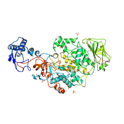
Structure of the catalytic domain of Streptococcus mutans GtfB in tetragonal space group P4322
Descriptor: 1,2-ETHANEDIOL, CALCIUM ION, CHLORIDE ION, ...
Authors:Schormann, N, Deivanayagam, C.
Deposit date:2022-12-19
Release date:2023-05-17
Last modified:2024-05-01
Method:X-RAY DIFFRACTION (2.5 Å)
Cite:The catalytic domains of Streptococcus mutans glucosyltransferases: a structural analysis.
Acta Crystallogr.,Sect.F, 79, 2023
8FJC
DownloadVisualize
BU of 8fjc by Molmil
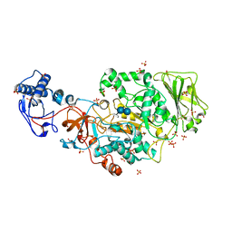
Structure of the catalytic domain of Streptococcus mutans GtfB complexed to acarbose in tetragonal space group P4322
Descriptor: 1,2-ETHANEDIOL, 4,6-dideoxy-4-{[(1S,4R,5S,6S)-4,5,6-trihydroxy-3-(hydroxymethyl)cyclohex-2-en-1-yl]amino}-alpha-D-glucopyranose-(1-4)-alpha-D-glucopyranose-(1-4)-alpha-D-glucopyranose, CALCIUM ION, ...
Authors:Schormann, N, Deivanayagam, C.
Deposit date:2022-12-19
Release date:2023-05-17
Last modified:2024-05-01
Method:X-RAY DIFFRACTION (2.5 Å)
Cite:The catalytic domains of Streptococcus mutans glucosyltransferases: a structural analysis.
Acta Crystallogr.,Sect.F, 79, 2023
8FKL
DownloadVisualize
BU of 8fkl by Molmil
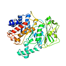
Truncated form of the catalytic domain of Streptococcus mutans GtfB
Descriptor: 1,2-ETHANEDIOL, Glucosyltransferase-I, MAGNESIUM ION
Authors:Schormann, N, Deivanayagam, C.
Deposit date:2022-12-21
Release date:2023-05-17
Last modified:2024-05-01
Method:X-RAY DIFFRACTION (1.48 Å)
Cite:The catalytic domains of Streptococcus mutans glucosyltransferases: a structural analysis.
Acta Crystallogr.,Sect.F, 79, 2023
8FN5
DownloadVisualize
BU of 8fn5 by Molmil
Structure of the truncated catalytic domain of Streptococcus mutans GtfD
Descriptor: Glucosyltransferase-S
Authors:Schormann, N, Deivanayagam, C.
Deposit date:2022-12-27
Release date:2023-05-17
Last modified:2024-05-01
Method:X-RAY DIFFRACTION (1.92 Å)
Cite:The catalytic domains of Streptococcus mutans glucosyltransferases: a structural analysis.
Acta Crystallogr.,Sect.F, 79, 2023
8FK4
DownloadVisualize
BU of 8fk4 by Molmil
Structure of the catalytic domain of Streptococcus mutans GtfB complexed to acarbose in orthorhombic space group P21212
Descriptor: 4,6-dideoxy-4-{[(1S,4R,5S,6S)-4,5,6-trihydroxy-3-(hydroxymethyl)cyclohex-2-en-1-yl]amino}-alpha-D-glucopyranose-(1-4)-alpha-D-glucopyranose-(1-4)-alpha-D-glucopyranose, CALCIUM ION, Glucosyltransferase-I, ...
Authors:Schormann, N, Deivanayagam, C.
Deposit date:2022-12-20
Release date:2023-05-17
Last modified:2024-05-01
Method:X-RAY DIFFRACTION (3.25 Å)
Cite:The catalytic domains of Streptococcus mutans glucosyltransferases: a structural analysis.
Acta Crystallogr.,Sect.F, 79, 2023

227111

건을2024-11-06부터공개중

PDB statisticsPDBj update infoContact PDBjnumon