Loading
PDBj
MenuPDBj@FacebookPDBj@TwitterPDBj@YouTubewwPDB FoundationwwPDB
RCSB PDBPDBeBMRBAdv. SearchSearch help
Search by PDB author
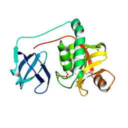
4JG5
DownloadVisualize
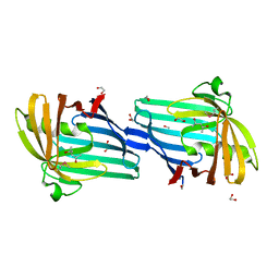
BU of 4jg5 by Molmil
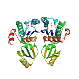
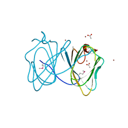
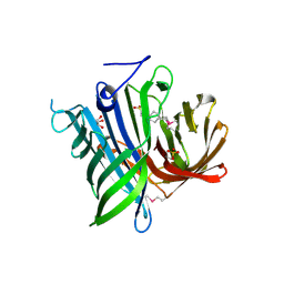
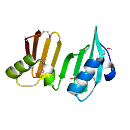
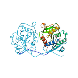
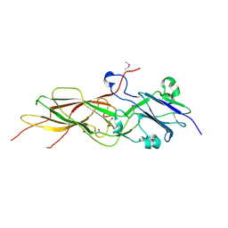
Crystal structure of a putative cell adhesion protein (BDI_3519) from Parabacteroides distasonis ATCC 8503 at 2.34 A resolution (PSI Community Target, Nakayama)
Descriptor: Putative cell adhesion protein
Authors:Joint Center for Structural Genomics (JCSG)
Deposit date:2013-02-28
Release date:2013-03-20
Last modified:2023-02-01
Method:X-RAY DIFFRACTION (2.34 Å)
Cite:A Distinct Type of Pilus from the Human Microbiome.
Cell, 165, 2016
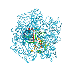
1ZKG
DownloadVisualize
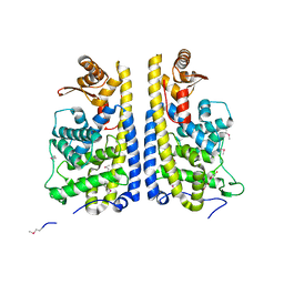
BU of 1zkg by Molmil
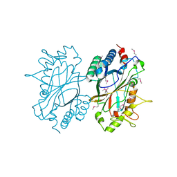
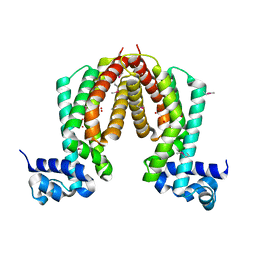
Crystal structure of Transcriptional regulator, TETR family (tm1030) from Thermotoga maritima at 2.30 A resolution
Descriptor: UNKNOWN LIGAND, transcriptional regulator, TetR family
Authors:Joint Center for Structural Genomics (JCSG)
Deposit date:2005-05-02
Release date:2005-07-26
Last modified:2023-01-25
Method:X-RAY DIFFRACTION (2.3 Å)
Cite:Crystal structure of TM1030 from Thermotoga maritima at 2.3 A resolution reveals molecular details of its transcription repressor function.
Proteins, 68, 2007
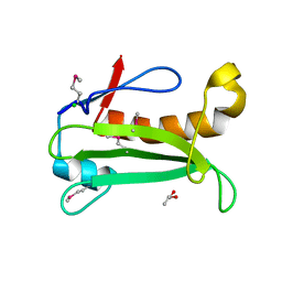
2A6A
DownloadVisualize
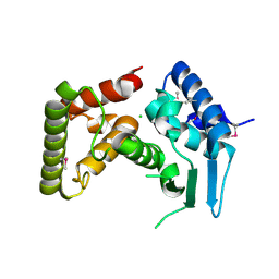
BU of 2a6a by Molmil
Crystal structure of Glycoprotein endopeptidase (tm0874) from THERMOTOGA MARITIMA at 2.50 A resolution
Descriptor: UNKNOWN LIGAND, hypothetical protein TM0874
Authors:Joint Center for Structural Genomics (JCSG)
Deposit date:2005-07-02
Release date:2005-07-19
Last modified:2023-01-25
Method:X-RAY DIFFRACTION (2.5 Å)
Cite:Structure of an essential bacterial protein YeaZ (TM0874) from Thermotoga maritima at 2.5 A resolution.
Acta Crystallogr.,Sect.F, 66, 2010
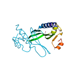
3H50
DownloadVisualize
BU of 3h50 by Molmil
CRYSTAL STRUCTURE OF A TETRACENOMYCIN POLYKETIDE SYNTHESIS PROTEIN (TCMJ) FROM XANTHOMONAS CAMPESTRIS PV. CAMPESTRIS AT 1.60 A RESOLUTION
Descriptor: ACETATE ION, Tetracenomycin polyketide synthesis protein, ZINC ION
Authors:Joint Center for Structural Genomics (JCSG)
Deposit date:2009-04-21
Release date:2009-05-05
Last modified:2023-02-01
Method:X-RAY DIFFRACTION (1.6 Å)
Cite:Conformational changes associated with the binding of zinc acetate at the putative active site of XcTcmJ, a cupin from Xanthomonas campestris pv. campestris.
Acta Crystallogr.,Sect.F, 66, 2010
2ICH
DownloadVisualize
BU of 2ich by Molmil
CRYSTAL STRUCTURE OF A PUTATIVE ATTH (NE1406) FROM NITROSOMONAS EUROPAEA AT 2.00 A RESOLUTION
Descriptor: 2-[N-CYCLOHEXYLAMINO]ETHANE SULFONIC ACID, GLYCEROL, Putative AttH, ...
Authors:Joint Center for Structural Genomics (JCSG)
Deposit date:2006-09-12
Release date:2006-10-03
Last modified:2023-01-25
Method:X-RAY DIFFRACTION (2 Å)
Cite:Structure of the first representative of Pfam family PF09410 (DUF2006) reveals a structural signature of the calycin superfamily that suggests a role in lipid metabolism.
Acta Crystallogr.,Sect.F, 66, 2010
3DUE
DownloadVisualize
BU of 3due by Molmil
CRYSTAL STRUCTURE OF A PUTATIVE PERIPLASMIC PROTEIN FROM DUF2874 FAMILY (BVU_2987) FROM BACTEROIDES VULGATUS ATCC 8482 AT 1.85 A RESOLUTION
Descriptor: CACODYLATE ION, Putative periplasmic protein
Authors:Joint Center for Structural Genomics (JCSG)
Deposit date:2008-07-17
Release date:2008-08-05
Last modified:2023-02-01
Method:X-RAY DIFFRACTION (1.85 Å)
Cite:The structure of BVU2987 from Bacteroides vulgatus reveals a superfamily of bacterial periplasmic proteins with possible inhibitory function.
Acta Crystallogr.,Sect.F, 66, 2010
2H1T
DownloadVisualize
BU of 2h1t by Molmil
Crystal structure of a duf1089 family protein (pa1994) from pseudomonas aeruginosa at 1.80 A resolution
Descriptor: (4S)-2-METHYL-2,4-PENTANEDIOL, 1,2-ETHANEDIOL, Hypothetical protein
Authors:Joint Center for Structural Genomics (JCSG)
Deposit date:2006-05-16
Release date:2006-05-30
Last modified:2023-01-25
Method:X-RAY DIFFRACTION (1.8 Å)
Cite:The structure of the first representative of Pfam family PF06475 reveals a new fold with possible involvement in glycolipid metabolism.
Acta Crystallogr.,Sect.F, 66, 2010
3EQX
DownloadVisualize
BU of 3eqx by Molmil
CRYSTAL STRUCTURE OF A FIC FAMILY PROTEIN (SO_4266) FROM SHEWANELLA ONEIDENSIS AT 1.6 A RESOLUTION
Descriptor: FIC DOMAIN CONTAINING TRANSCRIPTIONAL REGULATOR, TRIETHYLENE GLYCOL
Authors:Joint Center for Structural Genomics (JCSG)
Deposit date:2008-10-01
Release date:2008-10-14
Last modified:2023-02-01
Method:X-RAY DIFFRACTION (1.6 Å)
Cite:Crystal structure of the Fic (Filamentation induced by cAMP) family protein SO4266 (gi|24375750) from Shewanella oneidensis MR-1 at 1.6 A resolution.
Proteins, 75, 2009
3DL1
DownloadVisualize
BU of 3dl1 by Molmil
Crystal structure of a Putative Metal-dependent Hydrolase (YP_001336084.1) from Klebsiella pneumoniae subsp. pneumoniae MGH 78578 at 2.20 A resolution
Descriptor: CHLORIDE ION, Putative Metal-dependent Hydrolase
Authors:Joint Center for Structural Genomics (JCSG)
Deposit date:2008-06-26
Release date:2008-08-26
Last modified:2023-02-01
Method:X-RAY DIFFRACTION (2.2 Å)
Cite:The Structure of Mlc Titration Factor A (MtfA/YeeI) Reveals a Prototypical Zinc Metallopeptidase Related to Anthrax Lethal Factor.
J.Bacteriol., 194, 2012
1Z9F
DownloadVisualize
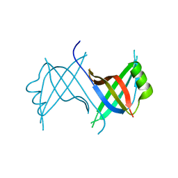
BU of 1z9f by Molmil
Crystal structure of single stranded DNA-binding protein (TM0604) from Thermotoga maritima at 2.60 A resolution
Descriptor: Single-strand binding protein
Authors:Joint Center for Structural Genomics (JCSG)
Deposit date:2005-04-01
Release date:2005-04-12
Last modified:2023-09-20
Method:X-RAY DIFFRACTION (2.3 Å)
Cite:Crystal structure of a single-stranded DNA-binding protein (TM0604) from Thermotoga maritima at 2.60 A resolution.
Proteins, 63, 2006
2HBW
DownloadVisualize
BU of 2hbw by Molmil
Crystal structure of a putative endopeptidase (ava_3396) from anabaena variabilis atcc 29413 at 1.05 A resolution
Descriptor: ACETATE ION, NLP/P60 protein, UNKNOWN LIGAND
Authors:Joint Center for Structural Genomics (JCSG)
Deposit date:2006-06-14
Release date:2006-08-08
Last modified:2023-01-25
Method:X-RAY DIFFRACTION (1.05 Å)
Cite:Structural Basis of Murein Peptide Specificity of a gamma-D-Glutamyl-L-Diamino Acid Endopeptidase.
Structure, 17, 2009
2GVK
DownloadVisualize
BU of 2gvk by Molmil
Crystal structure of a dye-decolorizing peroxidase (DyP) from Bacteroides thetaiotaomicron VPI-5482 at 1.6 A resolution
Descriptor: 1,2-ETHANEDIOL, ACETIC ACID, CHLORIDE ION, ...
Authors:Joint Center for Structural Genomics (JCSG)
Deposit date:2006-05-02
Release date:2006-05-16
Last modified:2023-01-25
Method:X-RAY DIFFRACTION (1.6 Å)
Cite:Crystal structures of two novel dye-decolorizing peroxidases reveal a beta-barrel fold with a conserved heme-binding motif.
Proteins, 69, 2007
2IAY
DownloadVisualize
BU of 2iay by Molmil
Crystal structure of a duf1831 family protein (lp2179) from lactobacillus plantarum at 1.20 A resolution
Descriptor: CHLORIDE ION, GLYCEROL, Hypothetical protein
Authors:Joint Center for Structural Genomics (JCSG)
Deposit date:2006-09-08
Release date:2006-10-10
Last modified:2023-01-25
Method:X-RAY DIFFRACTION (1.2 Å)
Cite:Structure of LP2179, the first representative of Pfam family PF08866, suggests a new fold with a role in amino-acid metabolism.
Acta Crystallogr.,Sect.F, 66, 2010
3D7Q
DownloadVisualize
BU of 3d7q by Molmil
Crystal structure of a xisi-like protein (npun_ar114) from nostoc punctiforme pcc 73102 at 2.30 A resolution
Descriptor: XisI protein-like
Authors:Joint Center for Structural Genomics (JCSG)
Deposit date:2008-05-21
Release date:2008-06-17
Last modified:2019-07-24
Method:X-RAY DIFFRACTION (2.3 Å)
Cite:Site-specific recombination of nitrogen-fixation genes in cyanobacteria by XisF-XisH-XisI complex: Structures and models.
Proteins, 2014
2GHR
DownloadVisualize
BU of 2ghr by Molmil
Crystal structure of homoserine o-succinyltransferase (NP_981826.1) from Bacillus cereus ATCC 10987 at 2.40 A resolution
Descriptor: Homoserine O-succinyltransferase, SULFATE ION
Authors:Joint Center for Structural Genomics (JCSG)
Deposit date:2006-03-27
Release date:2006-04-11
Last modified:2023-01-25
Method:X-RAY DIFFRACTION (2.4 Å)
Cite:Crystal structure of homoserine O-succinyltransferase from Bacillus cereus at 2.4 A resolution
Proteins, 68, 2007
2HAG
DownloadVisualize
BU of 2hag by Molmil
Crystal structure of a putative dyp-type peroxidase protein (so_0740) from shewanella oneidensis at 2.75 A resolution
Descriptor: Melanin biosynthesis protein TyrA, putative
Authors:Joint Center for Structural Genomics (JCSG)
Deposit date:2006-06-12
Release date:2006-08-08
Last modified:2023-01-25
Method:X-RAY DIFFRACTION (2.75 Å)
Cite:Crystal structures of two novel dye-decolorizing peroxidases reveal a beta-barrel fold with a conserved heme-binding motif.
Proteins, 69, 2007
2HUJ
DownloadVisualize
BU of 2huj by Molmil
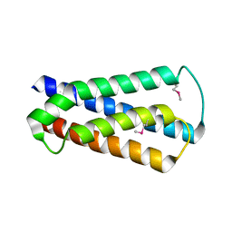
Crystal structure of a protein of uknown function (NP_471338.1) from Listeria innocua at 1.74 A resolution
Descriptor: Lin2004 protein
Authors:Joint Center for Structural Genomics (JCSG)
Deposit date:2006-07-26
Release date:2006-08-08
Last modified:2023-01-25
Method:X-RAY DIFFRACTION (1.74 Å)
Cite:Crystal structures of MW1337R and lin2004: representatives of a novel protein family that adopt a four-helical bundle fold.
Proteins, 71, 2008
2MKS
DownloadVisualize
BU of 2mks by Molmil
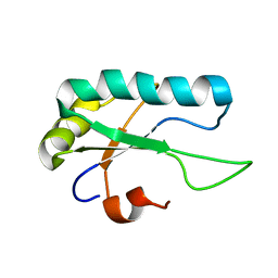
NMR structure of the RRM domain of RBMX from homo sapiens
Descriptor: RNA-binding motif protein, X chromosome
Authors:Serrano, P, Geralt, M, Wuthrich, K, Joint Center for Structural Genomics (JCSG), Partnership for T-Cell Biology (TCELL)
Deposit date:2014-02-14
Release date:2014-03-12
Last modified:2024-05-01
Method:SOLUTION NMR
Cite:NMR structure of the first RRM domain of the protein RBM39 from Homo sapiens
To be Published
2ME6
DownloadVisualize
BU of 2me6 by Molmil
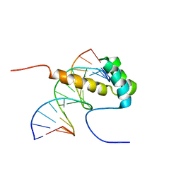
NMR Structure of the homeodomain transcription factor Gbx1 from Homo sapiens in complex with the DNA sequence CGACTAATTAGTCG
Descriptor: DNA (5'-D(*CP*GP*AP*CP*TP*AP*AP*TP*TP*AP*GP*TP*CP*G)-3'), Homeobox protein GBX-1
Authors:Proudfoot, A, Serrano, P, Geralt, M, Wuthrich, K, Joint Center for Structural Genomics (JCSG), Partnership for Stem Cell Biology (STEMCELL)
Deposit date:2013-09-23
Release date:2013-10-30
Last modified:2024-05-01
Method:SOLUTION NMR
Cite:NMR Structure of the homeodomain transcription factor Gbx1 from Homo sapiens in complex with the DNA sequence CGACTAATTAGTCG
To be Published
2M51
DownloadVisualize
BU of 2m51 by Molmil
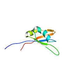
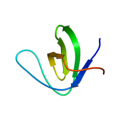
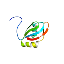
NMR structure of the SH3 domain of human RAS p21 protein activator (GTPase activating protein) 1
Descriptor: Ras GTPase-activating protein 1
Authors:Dutta, S.K, Serrano, P, Geralt, M, Wuthrich, K, Joint Center for Structural Genomics (JCSG), Partnership for T-Cell Biology (TCELL)
Deposit date:2013-02-12
Release date:2013-03-27
Last modified:2024-05-15
Method:SOLUTION NMR
Cite:NMR structure of the SH3 domain of human RAS p21 protein activator (GTPase activating protein) 1
To be Published
2MHN
DownloadVisualize
BU of 2mhn by Molmil
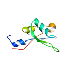
NMR structure of the first RRM domain of the protein RBM39 from Homo sapiens
Descriptor: RNA-binding protein 39
Authors:Serrano, P, Geralt, M, Dutta, S.K, Wuthrich, K, Joint Center for Structural Genomics (JCSG), Partnership for T-Cell Biology (TCELL)
Deposit date:2013-12-02
Release date:2014-01-01
Last modified:2024-05-15
Method:SOLUTION NMR
Cite:NMR structure of the first RRM domain of the protein RBM39 from Homo sapiens
To be Published
2M2B
DownloadVisualize
BU of 2m2b by Molmil
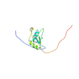
NMR structure of the RRM2 domain of the protein RBM10 from Homo sapiens
Descriptor: RNA-binding protein 10
Authors:Serrano, P, Geralt, M, Dutta, S.K, Wuthrich, K, Wrobel, R.L, Makino, S, Misenhiemer, T.M, Markley, J.L, Fox, B.G, Joint Center for Structural Genomics (JCSG), Partnership for T-Cell Biology (TCELL), Mitochondrial Protein Partnership (MPP)
Deposit date:2012-12-17
Release date:2013-01-16
Last modified:2024-05-15
Method:SOLUTION NMR
Cite:NMR structure of the RRM2 domain of the protein RBM10 from Homo sapiens
To be Published
2M34
DownloadVisualize
BU of 2m34 by Molmil
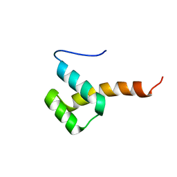
NMR Structure of the homeodomain transcription factor Gbx1 from Homo sapiens
Descriptor: Homeobox protein GBX-1
Authors:Proudfoot, A, Serrano, P, Geralt, M, Wuthrich, K, Partnership for Stem Cell Biology (STEMCELL), Joint Center for Structural Genomics (JCSG)
Deposit date:2013-01-09
Release date:2013-01-23
Last modified:2024-05-15
Method:SOLUTION NMR
Cite:NMR Structure of the homeodomain transcription factor Gbx1 from Homo sapiens
To be Published
2M52
DownloadVisualize
BU of 2m52 by Molmil
NMR Structure of the third RNA Recognition Motif (RRM) of U2 small nuclear ribonucleoprotein auxiliary factor (U2AF) 2
Descriptor: Splicing factor U2AF 65 kDa subunit
Authors:Dutta, S.K, Serrano, P, Geralt, M, Wuthrich, K, Joint Center for Structural Genomics (JCSG), Partnership for T-Cell Biology (TCELL)
Deposit date:2013-02-12
Release date:2013-03-27
Last modified:2024-05-15
Method:SOLUTION NMR
Cite:NMR Structure of the third RNA Recognition Motif (RRM) of U2 small nuclear ribonucleoprotein auxiliary factor (U2AF) 2
To be Published
2M7S
DownloadVisualize
BU of 2m7s by Molmil
NMR structure of RNA recognition motif 2 (RRM2) of Homo sapiens splicing factor, arginine/serine-rich 1
Descriptor: Serine/arginine-rich splicing factor 1
Authors:Dutta, S.K, Serrano, P, Geralt, M, Wuthrich, K, Joint Center for Structural Genomics (JCSG), Partnership for T-Cell Biology (TCELL)
Deposit date:2013-04-29
Release date:2013-05-15
Last modified:2024-05-15
Method:SOLUTION NMR
Cite:NMR structure of RNA recognition motif 2 (RRM2) of Homo sapiens splicing factor, arginine/serine-rich 1
To be Published

223790

건을2024-08-14부터공개중

PDB statisticsPDBj update infoContact PDBjnumon