Loading
PDBj
MenuPDBj@FacebookPDBj@TwitterPDBj@YouTubewwPDB FoundationwwPDB
RCSB PDBPDBeBMRBAdv. SearchSearch help
Search by PDB author
6QK2
DownloadVisualize
BU of 6qk2 by Molmil
R2-like ligand-binding oxidase Y715F mutant with anaerobically reconstituted Mn/Fe cofactor
Descriptor: FE (II) ION, MANGANESE (II) ION, PALMITIC ACID, ...
Authors:Griese, J.J, Hogbom, M.
Deposit date:2019-01-28
Release date:2020-02-19
Last modified:2024-01-24
Method:X-RAY DIFFRACTION (1.596 Å)
Cite:Key Structural Motifs Balance Metal Binding and Oxidative Reactivity in a Heterobimetallic Mn/Fe Protein.
J.Am.Chem.Soc., 142, 2020
6QK0
DownloadVisualize
BU of 6qk0 by Molmil
R2-like ligand-binding oxidase E69D mutant with anaerobically reconstituted Mn/Fe cofactor
Descriptor: FE (II) ION, MANGANESE (II) ION, PALMITIC ACID, ...
Authors:Griese, J.J, Hogbom, M.
Deposit date:2019-01-28
Release date:2020-02-19
Last modified:2024-01-24
Method:X-RAY DIFFRACTION (2.091 Å)
Cite:Key Structural Motifs Balance Metal Binding and Oxidative Reactivity in a Heterobimetallic Mn/Fe Protein.
J.Am.Chem.Soc., 142, 2020
6QJV
DownloadVisualize
BU of 6qjv by Molmil
R2-like ligand-binding oxidase E69D mutant with aerobically reconstituted Mn/Fe cofactor
Descriptor: FE (III) ION, MANGANESE (II) ION, MANGANESE (III) ION, ...
Authors:Griese, J.J, Hogbom, M.
Deposit date:2019-01-25
Release date:2020-02-12
Last modified:2024-01-24
Method:X-RAY DIFFRACTION (1.905 Å)
Cite:Key Structural Motifs Balance Metal Binding and Oxidative Reactivity in a Heterobimetallic Mn/Fe Protein.
J.Am.Chem.Soc., 142, 2020
6QK1
DownloadVisualize
BU of 6qk1 by Molmil
R2-like ligand-binding oxidase Y175F mutant with aerobically reconstituted Mn/Fe cofactor
Descriptor: FE (III) ION, MANGANESE (II) ION, MANGANESE (III) ION, ...
Authors:Griese, J.J, Hogbom, M.
Deposit date:2019-01-28
Release date:2020-02-19
Last modified:2024-01-24
Method:X-RAY DIFFRACTION (1.754 Å)
Cite:Key Structural Motifs Balance Metal Binding and Oxidative Reactivity in a Heterobimetallic Mn/Fe Protein.
J.Am.Chem.Soc., 142, 2020
6QO8
DownloadVisualize
BU of 6qo8 by Molmil
Crystal structure of ribonucleotide reductase NrdF from Bacillus anthracis anaerobically soaked with ferrous ions
Descriptor: 2-[BIS-(2-HYDROXY-ETHYL)-AMINO]-2-HYDROXYMETHYL-PROPANE-1,3-DIOL, FE (II) ION, Ribonucleoside-diphosphate reductase subunit beta, ...
Authors:Grave, K, Hogbom, M.
Deposit date:2019-02-12
Release date:2019-08-21
Last modified:2024-01-24
Method:X-RAY DIFFRACTION (1.31921768 Å)
Cite:Redox-induced structural changes in the di-iron and di-manganese forms of Bacillus anthracis ribonucleotide reductase subunit NrdF suggest a mechanism for gating of radical access.
J.Biol.Inorg.Chem., 24, 2019
6QO9
DownloadVisualize
BU of 6qo9 by Molmil
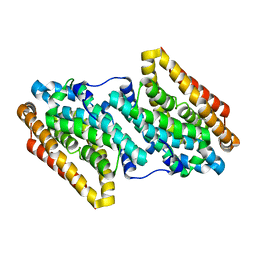
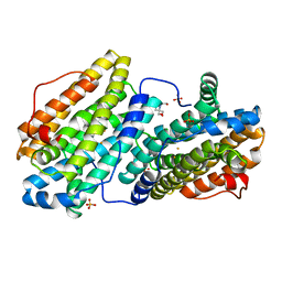
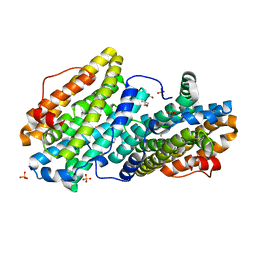
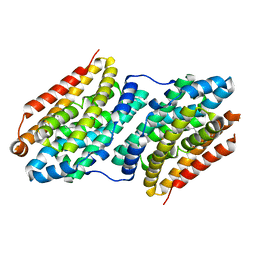
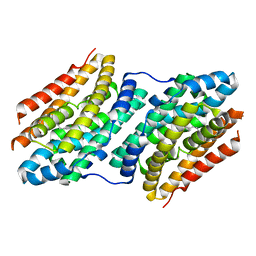
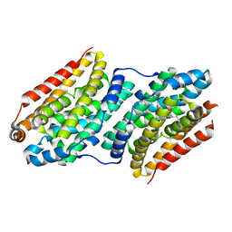
Crystal structure of ribonucleotide reductase NrdF from Bacillus anthracis soaked with manganese ions
Descriptor: 2-[BIS-(2-HYDROXY-ETHYL)-AMINO]-2-HYDROXYMETHYL-PROPANE-1,3-DIOL, MANGANESE (II) ION, Ribonucleoside-diphosphate reductase subunit beta, ...
Authors:Grave, K, Hogbom, M.
Deposit date:2019-02-12
Release date:2019-08-21
Last modified:2024-01-24
Method:X-RAY DIFFRACTION (1.299 Å)
Cite:Redox-induced structural changes in the di-iron and di-manganese forms of Bacillus anthracis ribonucleotide reductase subunit NrdF suggest a mechanism for gating of radical access.
J.Biol.Inorg.Chem., 24, 2019
6QO7
DownloadVisualize
BU of 6qo7 by Molmil
Crystal structure of ribonucleotide reductase NrdF from Bacillus anthracis aerobically soaked with ferrous ions (photo-reduced)
Descriptor: CHLORIDE ION, FE (II) ION, Ribonucleoside-diphosphate reductase subunit beta
Authors:Grave, K, Hogbom, M.
Deposit date:2019-02-12
Release date:2019-08-21
Last modified:2024-05-15
Method:X-RAY DIFFRACTION (1.628 Å)
Cite:Redox-induced structural changes in the di-iron and di-manganese forms of Bacillus anthracis ribonucleotide reductase subunit NrdF suggest a mechanism for gating of radical access.
J.Biol.Inorg.Chem., 24, 2019
6QO5
DownloadVisualize
BU of 6qo5 by Molmil
Crystal structure of apo (metal-free) ribonucleotide reductase NrdF from Bacillus anthracis
Descriptor: CHLORIDE ION, Ribonucleoside-diphosphate reductase subunit beta, SULFATE ION
Authors:Grave, K, Hogbom, M.
Deposit date:2019-02-12
Release date:2019-08-21
Last modified:2024-01-24
Method:X-RAY DIFFRACTION (1.511 Å)
Cite:Redox-induced structural changes in the di-iron and di-manganese forms of Bacillus anthracis ribonucleotide reductase subunit NrdF suggest a mechanism for gating of radical access.
J.Biol.Inorg.Chem., 24, 2019
6QOB
DownloadVisualize
BU of 6qob by Molmil
Crystal structure of ribonucleotide reductase NrdF from Bacillus anthracis with partially oxidised di-iron metallocofactor
Descriptor: 2-[BIS-(2-HYDROXY-ETHYL)-AMINO]-2-HYDROXYMETHYL-PROPANE-1,3-DIOL, FE (III) ION, Ribonucleoside-diphosphate reductase subunit beta, ...
Authors:Grave, K, Hogbom, M.
Deposit date:2019-02-12
Release date:2019-08-21
Last modified:2024-01-24
Method:X-RAY DIFFRACTION (1.457 Å)
Cite:Redox-induced structural changes in the di-iron and di-manganese forms of Bacillus anthracis ribonucleotide reductase subunit NrdF suggest a mechanism for gating of radical access.
J.Biol.Inorg.Chem., 24, 2019
5OCV
DownloadVisualize
BU of 5ocv by Molmil
A Rare Lysozyme Crystal Form Solved Using High-Redundancy 3D Electron Diffraction Data from Micron-Sized Needle Shaped Crystals
Descriptor: Lysozyme C, SODIUM ION
Authors:Xu, H, Lebrette, H, Yang, T, Srinivas, V, Hovmoller, S, Hogbom, M, Zou, X.
Deposit date:2017-07-03
Release date:2018-03-28
Last modified:2024-01-17
Method:ELECTRON CRYSTALLOGRAPHY (2.2 Å)
Cite:A Rare Lysozyme Crystal Form Solved Using Highly Redundant Multiple Electron Diffraction Datasets from Micron-Sized Crystals.
Structure, 26, 2018
5NF8
DownloadVisualize
BU of 5nf8 by Molmil
Solution structure of detergent-solubilized Rcf1, a yeast mitochondrial inner membrane protein involved in respiratory Complex III/IV supercomplex formation
Descriptor: Respiratory supercomplex factor 1, mitochondrial
Authors:Zhou, S, Pettersson, P, Sjoholm, J, Sjostrand, D, Hogbom, M, Brzezinski, P, Maler, L, Adelroth, P.
Deposit date:2017-03-13
Release date:2018-02-28
Last modified:2023-06-14
Method:SOLUTION NMR
Cite:Solution NMR structure of yeast Rcf1, a protein involved in respiratory supercomplex formation.
Proc. Natl. Acad. Sci. U.S.A., 115, 2018
5OMJ
DownloadVisualize
BU of 5omj by Molmil
R2-like ligand-binding oxidase with aerobically reconstituted metal cofactor after photoconversion
Descriptor: FE (III) ION, PALMITIC ACID, Ribonucleotide reductase small subunit
Authors:Griese, J.J, Hogbom, M.
Deposit date:2017-07-31
Release date:2018-01-03
Last modified:2024-01-17
Method:X-RAY DIFFRACTION (2.005 Å)
Cite:Driving Protein Conformational Changes with Light: Photoinduced Structural Rearrangement in a Heterobimetallic Oxidase.
J. Am. Chem. Soc., 140, 2018
2C8T
DownloadVisualize
BU of 2c8t by Molmil
The 3.0 A Resolution Structure of Caseinolytic Clp Protease 1 from Mycobacterium tuberculosis
Descriptor: ATP-DEPENDENT CLP PROTEASE PROTEOLYTIC SUBUNIT 1
Authors:Ingvarsson, H, Hogbom, M, Jones, T.A, Unge, T.
Deposit date:2005-12-07
Release date:2007-02-06
Last modified:2023-12-13
Method:X-RAY DIFFRACTION (3 Å)
Cite:Insights Into the Inter-Ring Plasticity of Caseinolytic Proteases from the X-Ray Structure of Mycobacterium Tuberculosis Clpp1.
Acta Crystallogr.,Sect.D, 63, 2007
6H53
DownloadVisualize
BU of 6h53 by Molmil
Crystal structure of Mycobacterium tuberculosis phosphatidylinositol phosphate synthase (PgsA1) in apo form
Descriptor: CDP-diacylglycerol--inositol 3-phosphatidyltransferase, SULFATE ION, UNKNOWN BRANCHED FRAGMENT OF PHOSPHOLIPID
Authors:Grave, K, Hogbom, M.
Deposit date:2018-07-23
Release date:2019-05-15
Last modified:2024-01-17
Method:X-RAY DIFFRACTION (2.9 Å)
Cite:Structure ofMycobacterium tuberculosisphosphatidylinositol phosphate synthase reveals mechanism of substrate binding and metal catalysis.
Commun Biol, 2, 2019
6H5A
DownloadVisualize
BU of 6h5a by Molmil
Crystal structure of Mycobacterium tuberculosis phosphatidylinositol phosphate synthase (PgsA1) in complex with manganese and citrate
Descriptor: (2R)-2,3-dihydroxypropyl (9Z)-octadec-9-enoate, CDP-diacylglycerol--inositol 3-phosphatidyltransferase, CITRATE ANION, ...
Authors:Grave, K, Hogbom, M.
Deposit date:2018-07-24
Release date:2019-05-15
Last modified:2024-01-17
Method:X-RAY DIFFRACTION (1.88 Å)
Cite:Structure ofMycobacterium tuberculosisphosphatidylinositol phosphate synthase reveals mechanism of substrate binding and metal catalysis.
Commun Biol, 2, 2019
6HWH
DownloadVisualize
BU of 6hwh by Molmil
Structure of a functional obligate respiratory supercomplex from Mycobacterium smegmatis
Descriptor: CARDIOLIPIN, COPPER (II) ION, Co-purified unknown peptide built as polyALA, ...
Authors:Wiseman, B, Nitharwal, R.G, Fedotovskaya, O, Schafer, J, Guo, H, Kuang, Q, Benlekbir, S, Sjostrand, D, Adelroth, P, Rubinstein, J.L, Brzezinski, P, Hogbom, M.
Deposit date:2018-10-12
Release date:2018-11-07
Last modified:2019-11-06
Method:ELECTRON MICROSCOPY (3.3 Å)
Cite:Structure of a functional obligate complex III2IV2respiratory supercomplex from Mycobacterium smegmatis.
Nat. Struct. Mol. Biol., 25, 2018
6H59
DownloadVisualize
BU of 6h59 by Molmil
Crystal structure of Mycobacterium tuberculosis phosphatidylinositol phosphate synthase (PgsA1) with CDP-DAG bound
Descriptor: (2S)-2,3-dihydroxypropyl (9Z)-octadec-9-enoate, CDP-diacylglycerol--inositol 3-phosphatidyltransferase, MAGNESIUM ION, ...
Authors:Grave, K, Hogbom, M.
Deposit date:2018-07-24
Release date:2019-05-15
Last modified:2024-01-17
Method:X-RAY DIFFRACTION (1.8 Å)
Cite:Structure ofMycobacterium tuberculosisphosphatidylinositol phosphate synthase reveals mechanism of substrate binding and metal catalysis.
Commun Biol, 2, 2019
2D1F
DownloadVisualize
BU of 2d1f by Molmil
Structure of Mycobacterium tuberculosis threonine synthase
Descriptor: PYRIDOXAL-5'-PHOSPHATE, Threonine synthase
Authors:Covarrubias, A.S, Bergfors, T, Mannerstedt, K, Oscarson, S, Jones, T.A, Mowbray, S.L, Hogbom, M.
Deposit date:2005-08-20
Release date:2006-09-05
Last modified:2011-07-13
Method:X-RAY DIFFRACTION (2.5 Å)
Cite:Structural, biochemical, and in vivo investigations of the threonine synthase from Mycobacterium tuberculosis.
J.Mol.Biol., 381, 2008
6F6C
DownloadVisualize
BU of 6f6c by Molmil
R2-like ligand-binding oxidase V72A mutant with aerobically reconstituted Mn/Fe cofactor
Descriptor: FE (III) ION, MANGANESE (II) ION, MANGANESE (III) ION, ...
Authors:Griese, J.J, Hogbom, M.
Deposit date:2017-12-05
Release date:2018-07-04
Last modified:2024-01-17
Method:X-RAY DIFFRACTION (1.766 Å)
Cite:Ether cross-link formation in the R2-like ligand-binding oxidase.
J. Biol. Inorg. Chem., 23, 2018
6F6K
DownloadVisualize
BU of 6f6k by Molmil
R2-like ligand-binding oxidase V72L mutant with anaerobically reconstituted Mn/Fe cofactor
Descriptor: FE (II) ION, LAURIC ACID, MANGANESE (II) ION, ...
Authors:Griese, J.J, Hogbom, M.
Deposit date:2017-12-05
Release date:2018-07-04
Last modified:2024-01-17
Method:X-RAY DIFFRACTION (1.982 Å)
Cite:Ether cross-link formation in the R2-like ligand-binding oxidase.
J. Biol. Inorg. Chem., 23, 2018
6F6H
DownloadVisualize
BU of 6f6h by Molmil
R2-like ligand-binding oxidase V72L mutant with aerobically reconstituted Mn/Fe cofactor
Descriptor: FE (III) ION, MANGANESE (II) ION, MANGANESE (III) ION, ...
Authors:Griese, J.J, Hogbom, M.
Deposit date:2017-12-05
Release date:2018-07-04
Last modified:2024-01-17
Method:X-RAY DIFFRACTION (1.764 Å)
Cite:Ether cross-link formation in the R2-like ligand-binding oxidase.
J. Biol. Inorg. Chem., 23, 2018
6F6G
DownloadVisualize
BU of 6f6g by Molmil
R2-like ligand-binding oxidase V72I mutant with anaerobically reconstituted Mn/Fe cofactor
Descriptor: FE (II) ION, MANGANESE (II) ION, PALMITIC ACID, ...
Authors:Griese, J.J, Hogbom, M.
Deposit date:2017-12-05
Release date:2018-07-04
Last modified:2024-01-17
Method:X-RAY DIFFRACTION (1.991 Å)
Cite:Ether cross-link formation in the R2-like ligand-binding oxidase.
J. Biol. Inorg. Chem., 23, 2018
6F6E
DownloadVisualize
BU of 6f6e by Molmil
R2-like ligand-binding oxidase V72A mutant with anaerobically reconstituted Mn/Fe cofactor
Descriptor: FE (II) ION, MANGANESE (II) ION, PALMITIC ACID, ...
Authors:Griese, J.J, Hogbom, M.
Deposit date:2017-12-05
Release date:2018-07-04
Last modified:2024-01-17
Method:X-RAY DIFFRACTION (1.627 Å)
Cite:Ether cross-link formation in the R2-like ligand-binding oxidase.
J. Biol. Inorg. Chem., 23, 2018
6F6F
DownloadVisualize
BU of 6f6f by Molmil
R2-like ligand-binding oxidase V72I mutant with aerobically reconstituted Mn/Fe cofactor
Descriptor: FE (III) ION, MANGANESE (II) ION, MANGANESE (III) ION, ...
Authors:Griese, J.J, Hogbom, M.
Deposit date:2017-12-05
Release date:2018-07-04
Last modified:2024-01-17
Method:X-RAY DIFFRACTION (1.788 Å)
Cite:Ether cross-link formation in the R2-like ligand-binding oxidase.
J. Biol. Inorg. Chem., 23, 2018
3BOR
DownloadVisualize
BU of 3bor by Molmil
Crystal structure of the DEADc domain of human translation initiation factor 4A-2
Descriptor: Human initiation factor 4A-II
Authors:Dimov, S, Hong, B, Tempel, W, MacKenzie, F, Karlberg, T, Arrowsmith, C.H, Edwards, A.M, Weigelt, J, Bochkarev, A, Park, H, Structural Genomics Consortium (SGC)
Deposit date:2007-12-17
Release date:2008-01-01
Last modified:2023-08-30
Method:X-RAY DIFFRACTION (1.85 Å)
Cite:Comparative Structural Analysis of Human DEAD-Box RNA Helicases.
Plos One, 5, 2010

221051

건을2024-06-12부터공개중

PDB statisticsPDBj update infoContact PDBjnumon