Loading
PDBj
MenuPDBj@FacebookPDBj@TwitterPDBj@YouTubewwPDB FoundationwwPDB
RCSB PDBPDBeBMRBAdv. SearchSearch help
Search by PDB author
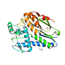
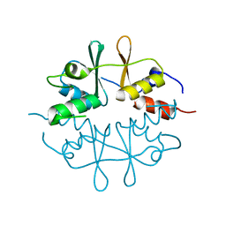
1VKH
DownloadVisualize
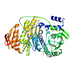
BU of 1vkh by Molmil
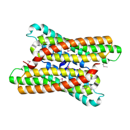
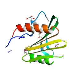
CRYSTAL STRUCTURE OF A PUTATIVE SERINE HYDROLASE (YDR428C) FROM SACCHAROMYCES CEREVISIAE AT 1.85 A RESOLUTION
Descriptor: CHLORIDE ION, GLYCEROL, putative serine hydrolase
Authors:Joint Center for Structural Genomics (JCSG)
Deposit date:2004-05-20
Release date:2004-06-08
Last modified:2024-10-09
Method:X-RAY DIFFRACTION (1.85 Å)
Cite:Crystal structure of an alpha/beta serine hydrolase (YDR428C) from Saccharomyces cerevisiae at 1.85 A resolution
Proteins, 58, 2005
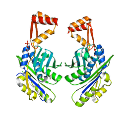
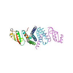
1VJ2
DownloadVisualize
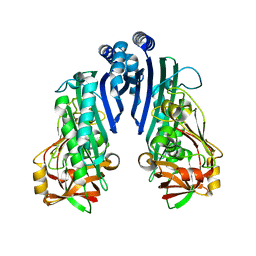
BU of 1vj2 by Molmil
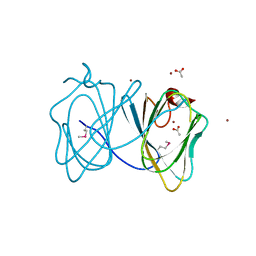
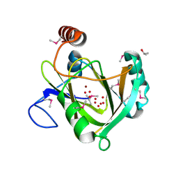
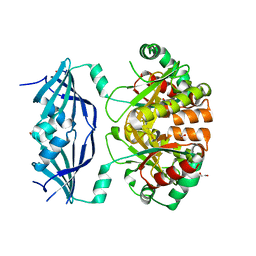
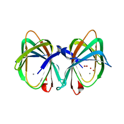
Crystal structure of a novel family of manganese-containing cupin (tm1459) from thermotoga maritima at 1.65 A resolution
Descriptor: MANGANESE (II) ION, UNKNOWN LIGAND, novel manganese-containing cupin TM1459
Authors:Joint Center for Structural Genomics (JCSG)
Deposit date:2003-12-03
Release date:2003-12-09
Last modified:2023-09-20
Method:X-RAY DIFFRACTION (1.65 Å)
Cite:Crystal structure of a novel manganese-containing cupin (TM1459) from Thermotoga maritima at 1.65 A resolution.
Proteins, 56, 2004
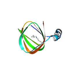
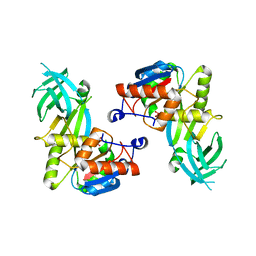
1O5H
DownloadVisualize
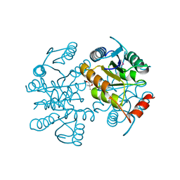
BU of 1o5h by Molmil
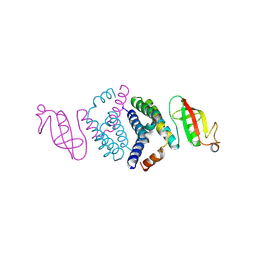
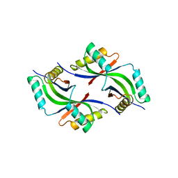
Crystal structure of formiminotetrahydrofolate cyclodeaminase (TM1560) from Thermotoga maritima at 2.80 A resolution
Descriptor: Formiminotetrahydrofolate cyclodeaminase
Authors:Joint Center for Structural Genomics (JCSG)
Deposit date:2003-09-17
Release date:2003-09-30
Last modified:2024-10-30
Method:X-RAY DIFFRACTION (2.8 Å)
Cite:Crystal structure of a formiminotetrahydrofolate cyclodeaminase (TM1560) from Thermotoga maritima at 2.80 A resolution reveals a new fold
Proteins, 58, 2005
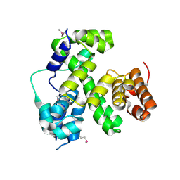
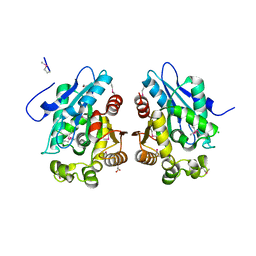
3H50
DownloadVisualize
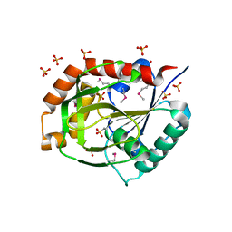
BU of 3h50 by Molmil
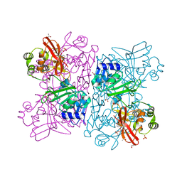
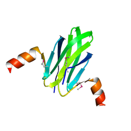
CRYSTAL STRUCTURE OF A TETRACENOMYCIN POLYKETIDE SYNTHESIS PROTEIN (TCMJ) FROM XANTHOMONAS CAMPESTRIS PV. CAMPESTRIS AT 1.60 A RESOLUTION
Descriptor: ACETATE ION, Tetracenomycin polyketide synthesis protein, ZINC ION
Authors:Joint Center for Structural Genomics (JCSG)
Deposit date:2009-04-21
Release date:2009-05-05
Last modified:2024-10-30
Method:X-RAY DIFFRACTION (1.6 Å)
Cite:Conformational changes associated with the binding of zinc acetate at the putative active site of XcTcmJ, a cupin from Xanthomonas campestris pv. campestris.
Acta Crystallogr.,Sect.F, 66, 2010
6WIQ
DownloadVisualize
BU of 6wiq by Molmil
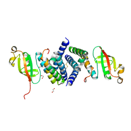
Crystal structure of the co-factor complex of NSP7 and the C-terminal domain of NSP8 from SARS CoV-2
Descriptor: Non-structural protein 7, Non-structural protein 8
Authors:Wilamowski, M, Kim, Y, Jedrzejczak, R, Maltseva, N, Endres, M, Godzik, A, Michalska, K, Joachimiak, A, Center for Structural Genomics of Infectious Diseases (CSGID)
Deposit date:2020-04-10
Release date:2020-04-22
Last modified:2024-10-23
Method:X-RAY DIFFRACTION (2.85 Å)
Cite:Transient and stabilized complexes of Nsp7, Nsp8, and Nsp12 in SARS-CoV-2 replication.
Biophys.J., 120, 2021
6WLC
DownloadVisualize
BU of 6wlc by Molmil
Crystal Structure of NSP15 Endoribonuclease from SARS CoV-2 in the Complex with Uridine-5'-Monophosphate
Descriptor: 1,2-ETHANEDIOL, 2-AMINO-2-HYDROXYMETHYL-PROPANE-1,3-DIOL, ACETATE ION, ...
Authors:Kim, Y, Maltseva, N, Jedrzejczak, R, Endres, M, Chang, C, Godzik, A, Michalska, K, Joachimiak, A, Center for Structural Genomics of Infectious Diseases (CSGID)
Deposit date:2020-04-19
Release date:2020-04-29
Last modified:2023-10-18
Method:X-RAY DIFFRACTION (1.82 Å)
Cite:Tipiracil binds to uridine site and inhibits Nsp15 endoribonuclease NendoU from SARS-CoV-2.
Commun Biol, 4, 2021
6WQD
DownloadVisualize
BU of 6wqd by Molmil
The 1.95 A Crystal Structure of the Co-factor Complex of NSP7 and the C-terminal Domain of NSP8 from SARS-CoV-2
Descriptor: 1,2-ETHANEDIOL, Non-structural protein 7, Non-structural protein 8
Authors:Kim, Y, Wilamowski, M, Jedrzejczak, R, Maltseva, N, Endres, M, Godzik, A, Michalska, K, Joachimiak, A, Center for Structural Genomics of Infectious Diseases (CSGID)
Deposit date:2020-04-28
Release date:2020-05-06
Last modified:2024-10-30
Method:X-RAY DIFFRACTION (1.95 Å)
Cite:Transient and stabilized complexes of Nsp7, Nsp8, and Nsp12 in SARS-CoV-2 replication.
Biophys.J., 120, 2021
3D7Q
DownloadVisualize
BU of 3d7q by Molmil
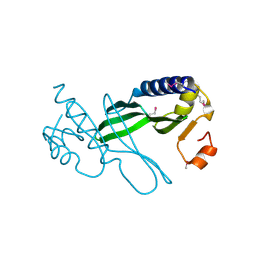
Crystal structure of a xisi-like protein (npun_ar114) from nostoc punctiforme pcc 73102 at 2.30 A resolution
Descriptor: XisI protein-like
Authors:Joint Center for Structural Genomics (JCSG)
Deposit date:2008-05-21
Release date:2008-06-17
Last modified:2019-07-24
Method:X-RAY DIFFRACTION (2.3 Å)
Cite:Site-specific recombination of nitrogen-fixation genes in cyanobacteria by XisF-XisH-XisI complex: Structures and models.
Proteins, 2014
3DL1
DownloadVisualize
BU of 3dl1 by Molmil
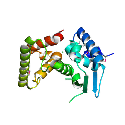
Crystal structure of a Putative Metal-dependent Hydrolase (YP_001336084.1) from Klebsiella pneumoniae subsp. pneumoniae MGH 78578 at 2.20 A resolution
Descriptor: CHLORIDE ION, Putative Metal-dependent Hydrolase
Authors:Joint Center for Structural Genomics (JCSG)
Deposit date:2008-06-26
Release date:2008-08-26
Last modified:2024-10-30
Method:X-RAY DIFFRACTION (2.2 Å)
Cite:The Structure of Mlc Titration Factor A (MtfA/YeeI) Reveals a Prototypical Zinc Metallopeptidase Related to Anthrax Lethal Factor.
J.Bacteriol., 194, 2012
1VR3
DownloadVisualize
BU of 1vr3 by Molmil
Crystal structure of Acireductone dioxygenase (13543033) from Mus musculus at 2.06 A resolution
Descriptor: Acireductone dioxygenase, ISOPROPYL ALCOHOL, NICKEL (II) ION, ...
Authors:Joint Center for Structural Genomics (JCSG)
Deposit date:2005-01-28
Release date:2005-02-15
Last modified:2023-01-25
Method:X-RAY DIFFRACTION (2.06 Å)
Cite:Crystal structure of acireductone dioxygenase (ARD) from Mus musculus at 2.06 angstrom resolution.
Proteins, 64, 2006
1VRM
DownloadVisualize
BU of 1vrm by Molmil
Crystal structure of the apbe protein (tm1553) from thermotoga maritima msb8 at 1.58 A resolution
Descriptor: (4R)-2-METHYLPENTANE-2,4-DIOL, (4S)-2-METHYL-2,4-PENTANEDIOL, UNKNOWN LIGAND, ...
Authors:Joint Center for Structural Genomics (JCSG)
Deposit date:2005-03-10
Release date:2005-03-22
Last modified:2023-01-25
Method:X-RAY DIFFRACTION (1.58 Å)
Cite:Crystal structure of the ApbE protein (TM1553) from Thermotoga maritima at 1.58 A resolution.
Proteins, 64, 2006
1VQ0
DownloadVisualize
BU of 1vq0 by Molmil
Crystal structure of 33 kDa chaperonin (Heat shock protein 33 homolog) (HSP33) (TM1394) from Thermotoga maritima at 2.20 A resolution
Descriptor: 1,2-ETHANEDIOL, 33 kDa chaperonin, CHLORIDE ION, ...
Authors:Joint Center for Structural Genomics (JCSG)
Deposit date:2004-11-30
Release date:2004-12-14
Last modified:2023-09-20
Method:X-RAY DIFFRACTION (2.2 Å)
Cite:Crystal structure of Hsp33 chaperone (TM1394) from Thermotoga maritima at 2.20 A resolution.
Proteins, 61, 2005
1O5U
DownloadVisualize
BU of 1o5u by Molmil
Crystal structure of a duf861 family protein (tm1112) from thermotoga maritima at 1.83 A resolution
Descriptor: UNKNOWN LIGAND, novel Thermotoga maritima enzyme TM1112
Authors:Joint Center for Structural Genomics (JCSG)
Deposit date:2003-10-06
Release date:2003-11-11
Last modified:2023-09-20
Method:X-RAY DIFFRACTION (1.83 Å)
Cite:Crystal structure of a novel Thermotoga maritima enzyme (TM1112) from the cupin family at 1.83 A resolution
Proteins, 56, 2004
1VQR
DownloadVisualize
BU of 1vqr by Molmil
Crystal structure of a virulence factor (cj0248) from campylobacter jejuni subsp. jejuni at 2.25 A resolution
Descriptor: hypothetical protein Cj0248
Authors:Joint Center for Structural Genomics (JCSG)
Deposit date:2004-12-17
Release date:2004-12-28
Last modified:2024-10-09
Method:X-RAY DIFFRACTION (2.25 Å)
Cite:Crystal structure of virulence factor CJ0248 from Campylobacter jejuni at 2.25 A resolution reveals a new fold.
Proteins, 62, 2006
1VR8
DownloadVisualize
BU of 1vr8 by Molmil
Crystal structure of GTP binding regulator (TM1622) from Thermotoga Maritima at 1.75 A resolution
Descriptor: AZIDE ION, GLYCEROL, GTP binding regulator
Authors:Joint Center for Structural Genomics (JCSG)
Deposit date:2005-02-16
Release date:2005-03-15
Last modified:2023-01-25
Method:X-RAY DIFFRACTION (1.75 Å)
Cite:Crystal structure of an ORFan protein (TM1622) from Thermotoga maritima at 1.75 A resolution reveals a fold similar to the Ran-binding protein Mog1p.
Proteins, 65, 2006
1VLR
DownloadVisualize
BU of 1vlr by Molmil
Crystal structure of mRNA decapping enzyme (DcpS) from Mus musculus at 1.83 A resolution
Descriptor: 1,2-ETHANEDIOL, mRNA decapping enzyme
Authors:Joint Center for Structural Genomics (JCSG)
Deposit date:2004-08-10
Release date:2004-08-24
Last modified:2023-09-20
Method:X-RAY DIFFRACTION (1.83 Å)
Cite:Crystal structure of an Apo mRNA decapping enzyme (DcpS) from Mouse at 1.83 A resolution.
Proteins, 60, 2005
1VQ3
DownloadVisualize
BU of 1vq3 by Molmil
Crystal structure of Phosphoribosylformylglycinamidine synthase, purS subunit (EC 6.3.5.3) (TM1244) from Thermotoga maritima at 1.90 A resolution
Descriptor: Phosphoribosylformylglycinamidine synthase, purS subunit
Authors:Joint Center for Structural Genomics (JCSG)
Deposit date:2004-12-15
Release date:2004-12-28
Last modified:2023-09-20
Method:X-RAY DIFFRACTION (1.9 Å)
Cite:Crystal structure of phosphoribosylformyl-glycinamidine synthase II, PurS subunit (TM1244) from Thermotoga maritima at 1.90 A resolution.
Proteins, 65, 2006
1VPZ
DownloadVisualize
BU of 1vpz by Molmil
Crystal structure of a putative carbon storage regulator protein (csra, pa0905) from pseudomonas aeruginosa at 2.05 A resolution
Descriptor: Carbon storage regulator homolog
Authors:Joint Center for Structural Genomics (JCSG)
Deposit date:2004-11-24
Release date:2004-12-14
Last modified:2023-01-25
Method:X-RAY DIFFRACTION (2.05 Å)
Cite:Crystal structure of the global regulatory protein CsrA from Pseudomonas putida at 2.05 A resolution reveals a new fold.
Proteins, 61, 2005
1VK3
DownloadVisualize
BU of 1vk3 by Molmil
Crystal structure of Phosphoribosylformylglycinamidine synthase II (TM1246) from Thermotoga maritima at 2.15 A resolution
Descriptor: CHLORIDE ION, Phosphoribosylformylglycinamidine synthase II
Authors:Joint Center for Structural Genomics (JCSG)
Deposit date:2004-04-23
Release date:2004-05-11
Last modified:2023-01-25
Method:X-RAY DIFFRACTION (2.15 Å)
Cite:Crystal structure of phosphoribosylformylglycinamidine synthase II (smPurL) from Thermotoga maritima at 2.15 A resolution.
Proteins, 63, 2006
1VL4
DownloadVisualize
BU of 1vl4 by Molmil
CRYSTAL STRUCTURE OF A PUTATIVE MODULATOR OF A DNA GYRASE (TM0727) FROM THERMOTOGA MARITIMA MSB8 AT 1.95 A RESOLUTION
Descriptor: pmbA-related protein
Authors:Joint Center for Structural Genomics (JCSG)
Deposit date:2004-07-09
Release date:2004-09-21
Last modified:2024-05-22
Method:X-RAY DIFFRACTION (1.95 Å)
Cite:Crystal structure of a putative modulator of DNA gyrase (pmbA) from Thermotoga maritima at 1.95 A resolution reveals a new fold.
Proteins, 61, 2005
1O1X
DownloadVisualize
BU of 1o1x by Molmil
Crystal structure of a ribose 5-phosphate isomerase rpib (tm1080) from thermotoga maritima at 1.90 A resolution
Descriptor: (4S)-2-METHYL-2,4-PENTANEDIOL, ribose-5-phosphate isomerase RpiB
Authors:Joint Center for Structural Genomics (JCSG)
Deposit date:2003-02-12
Release date:2003-04-01
Last modified:2023-01-25
Method:X-RAY DIFFRACTION (1.9 Å)
Cite:Crystal structure of a ribose-5-phosphate isomerase RpiB (TM1080) from Thermotoga maritima at 1.90 A resolution.
Proteins, 56, 2004
1O1Y
DownloadVisualize
BU of 1o1y by Molmil
Crystal structure of a glutamine amidotransferase (tm1158) from thermotoga maritima at 1.70 A resolution
Descriptor: SULFATE ION, conserved hypothetical protein TM1158
Authors:Joint Center for Structural Genomics (JCSG)
Deposit date:2003-02-12
Release date:2003-03-11
Last modified:2023-01-25
Method:X-RAY DIFFRACTION (1.7 Å)
Cite:Crystal structure of a putative glutamine amido transferase (TM1158) from Thermotoga maritima at 1.7 A resolution.
PROTEINS, 54, 2004
1O50
DownloadVisualize
BU of 1o50 by Molmil
Crystal structure of a cbs domain-containing protein (tm0935) from thermotoga maritima at 1.87 A resolution
Descriptor: CBS domain-containing predicted protein TM0935
Authors:Joint Center for Structural Genomics (JCSG)
Deposit date:2003-07-31
Release date:2003-08-12
Last modified:2024-05-22
Method:X-RAY DIFFRACTION (1.87 Å)
Cite:Crystal structure of a tandem cystathionine-beta-synthase (CBS) domain protein (TM0935) from Thermotoga maritima at 1.87 A resolution
Proteins, 57, 2004
6XIP
DownloadVisualize
BU of 6xip by Molmil
The 1.5 A Crystal Structure of the Co-factor Complex of NSP7 and the C-terminal Domain of NSP8 from SARS CoV-2
Descriptor: 1,2-ETHANEDIOL, Non-structural protein 7, Non-structural protein 8
Authors:Wilamowski, M, Kim, Y, Jedrzejczak, R, Maltseva, N, Endres, M, Godzik, A, Michalska, K, Joachimiak, A, Center for Structural Genomics of Infectious Diseases (CSGID)
Deposit date:2020-06-20
Release date:2020-07-01
Last modified:2024-10-09
Method:X-RAY DIFFRACTION (1.5 Å)
Cite:Transient and stabilized complexes of Nsp7, Nsp8, and Nsp12 in SARS-CoV-2 replication.
Biophys.J., 120, 2021
1VKY
DownloadVisualize
BU of 1vky by Molmil
Crystal structure of S-adenosylmethionine tRNA ribosyltransferase (TM0574) from Thermotoga maritima at 2.00 A resolution
Descriptor: S-adenosylmethionine:tRNA ribosyltransferase-isomerase, UNKNOWN LIGAND
Authors:Joint Center for Structural Genomics (JCSG)
Deposit date:2004-06-28
Release date:2004-08-17
Last modified:2024-05-22
Method:X-RAY DIFFRACTION (2 Å)
Cite:Crystal structure of S-adenosylmethionine:tRNA ribosyltransferase-isomerase (QueA) from Thermotoga maritima at 2.0 A resolution reveals a new fold.
Proteins, 59, 2005

227111

건을2024-11-06부터공개중

PDB statisticsPDBj update infoContact PDBjnumon