Loading
PDBj
MenuPDBj@FacebookPDBj@TwitterPDBj@YouTubewwPDB FoundationwwPDB
RCSB PDBPDBeBMRBAdv. SearchSearch help
Search by PDB author
3GF8
DownloadVisualize
BU of 3gf8 by Molmil
Crystal structure of putative polysaccharide binding proteins (DUF1812) (NP_809975.1) from BACTEROIDES THETAIOTAOMICRON VPI-5482 at 2.20 A resolution
Descriptor: 1,2-ETHANEDIOL, putative polysaccharide binding proteins (DUF1812)
Authors:Joint Center for Structural Genomics (JCSG)
Deposit date:2009-02-26
Release date:2009-03-24
Last modified:2023-02-01
Method:X-RAY DIFFRACTION (2.2 Å)
Cite:A conserved fold for fimbrial components revealed by the crystal structure of a putative fimbrial assembly protein (BT1062) from Bacteroides thetaiotaomicron at 2.2 A resolution
Acta Crystallogr.,Sect.F, 66, 2010
3H0N
DownloadVisualize
BU of 3h0n by Molmil
Crystal structure of a duf1470 family protein (jann_2411) from jannaschia sp. ccs1 at 1.45 A resolution
Descriptor: ACETATE ION, GLYCEROL, NICKEL (II) ION, ...
Authors:Joint Center for Structural Genomics (JCSG)
Deposit date:2009-04-09
Release date:2009-04-21
Last modified:2023-02-01
Method:X-RAY DIFFRACTION (1.45 Å)
Cite:The structure of Jann_2411 (DUF1470) from Jannaschia sp. at 1.45  resolution reveals a new fold (the ABATE domain) and suggests its possible role as a transcription regulator.
Acta Crystallogr.,Sect.F, 66, 2010
3HBZ
DownloadVisualize
BU of 3hbz by Molmil
Crystal structure of a putative glycoside hydrolase (bt_2081) from bacteroides thetaiotaomicron vpi-5482 at 2.05 A resolution
Descriptor: ACETATE ION, CACODYLATE ION, CALCIUM ION, ...
Authors:Joint Center for Structural Genomics (JCSG)
Deposit date:2009-05-05
Release date:2009-05-26
Last modified:2023-02-01
Method:X-RAY DIFFRACTION (2.05 Å)
Cite:Structure of Bacteroides thetaiotaomicron BT2081 at 2.05 A resolution: the first structural representative of a new protein family that may play a role in carbohydrate metabolism.
Acta Crystallogr.,Sect.F, 66, 2010
3DCX
DownloadVisualize
BU of 3dcx by Molmil
Crystal structure of a duf1696 family protein with a pleckstrin-homology domain (shew_0819) from shewanella loihica pv-4 at 2.00 A resolution
Descriptor: (4S)-2-METHYL-2,4-PENTANEDIOL, CHLORIDE ION, Protein of Unknown Function (DUF1696) with Pleckstrin-homology Domains
Authors:Joint Center for Structural Genomics (JCSG)
Deposit date:2008-06-04
Release date:2008-07-29
Last modified:2023-02-01
Method:X-RAY DIFFRACTION (2 Å)
Cite:Bacterial pleckstrin homology domains: a prokaryotic origin for the PH domain.
J.Mol.Biol., 396, 2010
2GVI
DownloadVisualize
BU of 2gvi by Molmil
Crystal structure of a putative formylmethanofuran dehydrogenase subunit e (ta1109) from thermoplasma acidophilum at 1.87 A resolution
Descriptor: 1,2-ETHANEDIOL, ACETIC ACID, UNKNOWN LIGAND, ...
Authors:Joint Center for Structural Genomics (JCSG)
Deposit date:2006-05-02
Release date:2006-06-13
Last modified:2023-01-25
Method:X-RAY DIFFRACTION (1.87 Å)
Cite:Structures of three members of Pfam PF02663 (FmdE) implicated in microbial methanogenesis reveal a conserved alpha+beta core domain and an auxiliary C-terminal treble-clef zinc finger.
Acta Crystallogr.,Sect.F, 66, 2010
3H41
DownloadVisualize
BU of 3h41 by Molmil
CRYSTAL STRUCTURE OF A NLPC/P60 FAMILY PROTEIN (BCE_2878) FROM BACILLUS CEREUS ATCC 10987 AT 1.79 A RESOLUTION
Descriptor: ALANINE, D-GLUTAMIC ACID, NLP/P60 family protein, ...
Authors:Joint Center for Structural Genomics (JCSG)
Deposit date:2009-04-17
Release date:2009-05-05
Last modified:2023-02-01
Method:X-RAY DIFFRACTION (1.79 Å)
Cite:Structure of the gamma-D-glutamyl-L-diamino acid endopeptidase YkfC from Bacillus cereus in complex with L-Ala-[gamma]-D-Glu: insights into substrate recognition by NlpC/P60 cysteine peptidases
Acta Crystallogr.,Sect.F, 66, 2010
3THF
DownloadVisualize
BU of 3thf by Molmil
Crystal structure of the SD2 domain from Drosophila Shroom
Descriptor: Protein Shroom
Authors:Mohan, S, VanDemark, A.P.
Deposit date:2011-08-18
Release date:2012-06-06
Last modified:2024-02-28
Method:X-RAY DIFFRACTION (2.6951 Å)
Cite:Structure of Shroom domain 2 reveals a three-segmented coiled-coil required for dimerization, Rock binding, and apical constriction.
Mol Biol Cell, 23, 2012
3TX8
DownloadVisualize
BU of 3tx8 by Molmil
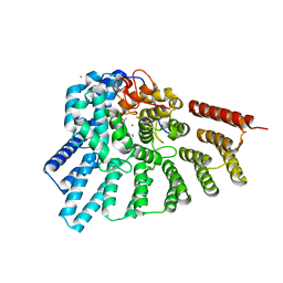
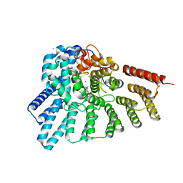
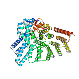
Crystal structure of a succinyl-diaminopimelate desuccinylase (ArgE) from Corynebacterium glutamicum ATCC 13032 at 2.97 A resolution
Descriptor: CHLORIDE ION, PHOSPHATE ION, Succinyl-diaminopimelate desuccinylase
Authors:Joint Center for Structural Genomics (JCSG), Brunger, A.T, Terwilliger, T.C, Read, R.J, Adams, P.D, Levitt, M, Schroder, G.F.
Deposit date:2011-09-22
Release date:2011-10-26
Last modified:2023-12-06
Method:X-RAY DIFFRACTION (2.972 Å)
Cite:Application of DEN refinement and automated model building to a difficult case of molecular-replacement phasing: the structure of a putative succinyl-diaminopimelate desuccinylase from Corynebacterium glutamicum.
Acta Crystallogr.,Sect.D, 68, 2012
2GLZ
DownloadVisualize
BU of 2glz by Molmil
Crystal structure of a formylmethanofuran dehydrogenase subunit e-like protein (dhaf_2992) from desulfitobacterium hafniense dcb-2 at 1.45 A resolution
Descriptor: 1,2-ETHANEDIOL, NICKEL (II) ION, ZINC ION, ...
Authors:Joint Center for Structural Genomics (JCSG)
Deposit date:2006-04-05
Release date:2006-04-18
Last modified:2023-01-25
Method:X-RAY DIFFRACTION (1.45 Å)
Cite:Structures of three members of Pfam PF02663 (FmdE) implicated in microbial methanogenesis reveal a conserved alpha+beta core domain and an auxiliary C-terminal treble-clef zinc finger.
Acta Crystallogr.,Sect.F, 66, 2010
6MCS
DownloadVisualize
BU of 6mcs by Molmil
X-ray crystal structure of wild type HIV-1 protease in complex with GRL-003
Descriptor: (3S,3aR,5R,7aS,8S)-hexahydro-4H-3,5-methanofuro[2,3-b]pyran-8-yl [(2S,3R)-4-[{[2-(cyclopropylamino)-1,3-benzothiazol-6-yl]sulfonyl}(2-methylpropyl)amino]-1-(4-fluorophenyl)-3-hydroxybutan-2-yl]carbamate, Protease
Authors:Bulut, H, Hayashi, H, Hattori, S.I, Aoki, M, Das, D, Ghosh, A.K, Mitsuya, H.
Deposit date:2018-09-02
Release date:2019-04-24
Last modified:2023-10-11
Method:X-RAY DIFFRACTION (1.52 Å)
Cite:Halogen Bond Interactions of Novel HIV-1 Protease Inhibitors (PI) (GRL-001-15 and GRL-003-15) with the Flap of Protease Are Critical for Their Potent Activity against Wild-Type HIV-1 and Multi-PI-Resistant Variants.
Antimicrob.Agents Chemother., 63, 2019
6MCR
DownloadVisualize
BU of 6mcr by Molmil
X-ray crystal structure of wild type HIV-1 protease in complex with GRL-001
Descriptor: (3S,3aR,5R,7aS,8S)-hexahydro-4H-3,5-methanofuro[2,3-b]pyran-8-yl [(2S,3R)-4-[{[2-(cyclopropylamino)-1,3-benzothiazol-6-yl]sulfonyl}(2-methylpropyl)amino]-1-(3-fluorophenyl)-3-hydroxybutan-2-yl]carbamate, 1,2-ETHANEDIOL, Protease
Authors:Bulut, H, Hayashi, H, Hattori, S.I, Aoki, M, Das, D, Ghosh, A.K, Mitsuya, H.
Deposit date:2018-09-02
Release date:2019-04-24
Last modified:2023-10-11
Method:X-RAY DIFFRACTION (1.48 Å)
Cite:Halogen Bond Interactions of Novel HIV-1 Protease Inhibitors (PI) (GRL-001-15 and GRL-003-15) with the Flap of Protease Are Critical for Their Potent Activity against Wild-Type HIV-1 and Multi-PI-Resistant Variants.
Antimicrob.Agents Chemother., 63, 2019
5V4Y
DownloadVisualize
BU of 5v4y by Molmil
X-ray crystal structure of wild type HIV-1 protease in complex with GRL-09510
Descriptor: (3S,3aR,5R,7aS,8S)-hexahydro-4H-3,5-methanofuro[2,3-b]pyran-8-yl [(2S,3R)-3-hydroxy-4-{[(4-methoxyphenyl)sulfonyl](2-methylpropyl)amino}-1-phenylbutan-2-yl]carbamate, Protease
Authors:Yedidi, R.S, Delino, N.S, Das, D, Kaufman, J.D, Wingfield, P.T, Ghosh, A.K, Mitsuya, H.
Deposit date:2017-03-11
Release date:2017-09-13
Last modified:2023-10-04
Method:X-RAY DIFFRACTION (1.9 Å)
Cite:GRL-09510, a Unique P2-Crown-Tetrahydrofuranylurethane -Containing HIV-1 Protease Inhibitor, Maintains Its Favorable Antiviral Activity against Highly-Drug-Resistant HIV-1 Variants in vitro.
Sci Rep, 7, 2017
3BYQ
DownloadVisualize
BU of 3byq by Molmil
Crystal structure of a duf1185 family protein (bb2672) from bordetella bronchiseptica rb50 at 1.70 A resolution
Descriptor: 1,2-ETHANEDIOL, CHLORIDE ION, TETRAETHYLENE GLYCOL, ...
Authors:Joint Center for Structural Genomics (JCSG)
Deposit date:2008-01-16
Release date:2008-01-29
Last modified:2023-02-01
Method:X-RAY DIFFRACTION (1.7 Å)
Cite:Structures of the first representatives of Pfam family PF06684 (DUF1185) reveal a novel variant of the Bacillus chorismate mutase fold and suggest a role in amino-acid metabolism.
Acta Crystallogr.,Sect.F, 66, 2010
3DSW
DownloadVisualize
BU of 3dsw by Molmil
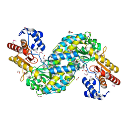
Crystal structure of RabGGTase(DELTA LRR; DELTA IG)in complex with mono-prenylated peptide Ser-Cys(GG)-Ser-Cys derivated from Rab7
Descriptor: CALCIUM ION, GERAN-8-YL GERAN, Geranylgeranyl transferase type-2 subunit alpha, ...
Authors:Guo, Z, Yu, S, Goody, R.S, Alexandrov, K, Blankenfeldt, W.
Deposit date:2008-07-14
Release date:2008-09-09
Last modified:2023-11-01
Method:X-RAY DIFFRACTION (2.15 Å)
Cite:Structures of RabGGTase-substrate/product complexes provide insights into the evolution of protein prenylation
Embo J., 27, 2008
3G0T
DownloadVisualize
BU of 3g0t by Molmil
Crystal structure of putative aspartate aminotransferase (NP_905498.1) from Porphyromonas gingivalis W83 at 1.75 A resolution
Descriptor: 1,2-ETHANEDIOL, 2-{2-[2-(2-{2-[2-(2-ETHOXY-ETHOXY)-ETHOXY]-ETHOXY}-ETHOXY)-ETHOXY]-ETHOXY}-ETHANOL, CHLORIDE ION, ...
Authors:Joint Center for Structural Genomics (JCSG)
Deposit date:2009-01-28
Release date:2009-02-10
Last modified:2023-02-01
Method:X-RAY DIFFRACTION (1.75 Å)
Cite:Molecular characterization of novel pyridoxal-5'-phosphate-dependent enzymes from the human microbiome.
Protein Sci., 23, 2014
3DST
DownloadVisualize
BU of 3dst by Molmil
Crystal structure of RabGGTase(DELTA LRR; DELTA IG)in complex with geranylgeranyl pyrophosphate
Descriptor: CALCIUM ION, GERANYLGERANYL DIPHOSPHATE, Geranylgeranyl transferase type-2 subunit alpha, ...
Authors:Guo, Z, Yu, S, Goody, R.S, Alexandrov, K, Blankenfeldt, W.
Deposit date:2008-07-14
Release date:2008-09-09
Last modified:2023-11-01
Method:X-RAY DIFFRACTION (1.9 Å)
Cite:Structures of RabGGTase-substrate/product complexes provide insights into the evolution of protein prenylation
Embo J., 27, 2008
3DSV
DownloadVisualize
BU of 3dsv by Molmil
Crystal structure of RabGGTase(DELTA LRR; DELTA IG)in complex with mono-prenylated peptide Ser-Cys-Ser-Cys(GG) derivated from Rab7
Descriptor: CALCIUM ION, GERAN-8-YL GERAN, Geranylgeranyl transferase type-2 subunit alpha, ...
Authors:Guo, Z, Yu, S, Goody, R.S, Alexandrov, K, Blankenfeldt, W.
Deposit date:2008-07-14
Release date:2008-09-09
Last modified:2023-11-01
Method:X-RAY DIFFRACTION (2.1 Å)
Cite:Structures of RabGGTase-substrate/product complexes provide insights into the evolution of protein prenylation
Embo J., 27, 2008
3DSU
DownloadVisualize
BU of 3dsu by Molmil
Crystal structure of RabGGTase(DELTA LRR; DELTA IG)in complex with farnesyl pyrophosphate
Descriptor: CALCIUM ION, FARNESYL DIPHOSPHATE, Geranylgeranyl transferase type-2 subunit alpha, ...
Authors:Guo, Z, Yu, S, Goody, R.S, Alexandrov, K, Blankenfeldt, W.
Deposit date:2008-07-14
Release date:2008-09-09
Last modified:2023-11-01
Method:X-RAY DIFFRACTION (1.9 Å)
Cite:Structures of RabGGTase-substrate/product complexes provide insights into the evolution of protein prenylation
Embo J., 27, 2008
2HB3
DownloadVisualize
BU of 2hb3 by Molmil
Wild-type HIV-1 Protease in complex with potent inhibitor GRL06579
Descriptor: (3AS,5R,6AR)-HEXAHYDRO-2H-CYCLOPENTA[B]FURAN-5-YL (2S,3S)-3-HYDROXY-4-(4-(HYDROXYMETHYL)-N-ISOBUTYLPHENYLSULFONAMIDO)-1-PHENYLBUTAN-2-YLCARBAMATE, CHLORIDE ION, GLYCEROL, ...
Authors:Kovalevsky, A.Y, Weber, I.T.
Deposit date:2006-06-13
Release date:2006-08-29
Last modified:2024-02-14
Method:X-RAY DIFFRACTION (1.35 Å)
Cite:Structure-Based Design of Novel HIV-1 Protease Inhibitors To Combat Drug Resistance.
J.Med.Chem., 49, 2006
3DSX
DownloadVisualize
BU of 3dsx by Molmil
Crystal structure of RabGGTase(DELTA LRR; DELTA IG)in complex with di-prenylated peptide Ser-Cys(GG)-Ser-Cys(GG) derivated from Rab7
Descriptor: CALCIUM ION, GERAN-8-YL GERAN, Geranylgeranyl transferase type-2 subunit alpha, ...
Authors:Guo, Z, Yu, S, Goody, R.S, Alexandrov, K, Blankenfeldt, W.
Deposit date:2008-07-14
Release date:2008-09-09
Last modified:2023-11-01
Method:X-RAY DIFFRACTION (2.1 Å)
Cite:Structures of RabGGTase-substrate/product complexes provide insights into the evolution of protein prenylation
Embo J., 27, 2008
3DSS
DownloadVisualize
BU of 3dss by Molmil
Crystal structure of RabGGTase(DELTA LRR; DELTA IG)
Descriptor: CALCIUM ION, Geranylgeranyl transferase type-2 subunit alpha, Geranylgeranyl transferase type-2 subunit beta, ...
Authors:Guo, Z, Yu, S, Goody, R.S, Alexandrov, K, Blankenfeldt, W.
Deposit date:2008-07-14
Release date:2008-09-09
Last modified:2023-11-01
Method:X-RAY DIFFRACTION (1.8 Å)
Cite:Structures of RabGGTase-substrate/product complexes provide insights into the evolution of protein prenylation
Embo J., 27, 2008
4L2W
DownloadVisualize
BU of 4l2w by Molmil
Crystal structure of the Shroom-Binding domain of human Rock1
Descriptor: Rho-associated protein kinase 1
Authors:Mohan, S, VanDemark, A.P.
Deposit date:2013-06-04
Release date:2014-04-16
Last modified:2024-02-28
Method:X-RAY DIFFRACTION (2.49 Å)
Cite:Structure of a highly conserved domain of Rock1 required for Shroom-mediated regulation of cell morphology.
Plos One, 8, 2013
3B77
DownloadVisualize
BU of 3b77 by Molmil
Crystal structure of a ph domain containing bacterial protein (exig_2160) from exiguobacterium sibiricum 255-15 at 2.42 A resolution
Descriptor: Uncharacterized protein
Authors:Joint Center for Structural Genomics (JCSG)
Deposit date:2007-10-30
Release date:2007-11-13
Last modified:2023-01-25
Method:X-RAY DIFFRACTION (2.42 Å)
Cite:Bacterial pleckstrin homology domains: a prokaryotic origin for the PH domain.
J.Mol.Biol., 396, 2010
3BOS
DownloadVisualize
BU of 3bos by Molmil
Crystal structure of a putative dna replication regulator HDA (SAMA_1916) from Shewanella amazonensis sb2b at 1.75 A resolution
Descriptor: 1,2-ETHANEDIOL, CHLORIDE ION, CYTIDINE-5'-DIPHOSPHATE, ...
Authors:Joint Center for Structural Genomics (JCSG)
Deposit date:2007-12-17
Release date:2008-01-15
Last modified:2023-01-25
Method:X-RAY DIFFRACTION (1.75 Å)
Cite:A structural basis for the regulatory inactivation of DnaA.
J.Mol.Biol., 385, 2009
3D00
DownloadVisualize
BU of 3d00 by Molmil
Crystal structure of a tungsten formylmethanofuran dehydrogenase subunit e (fmde)-like protein (syn_00638) from syntrophus aciditrophicus at 1.90 A resolution
Descriptor: CHLORIDE ION, Tungsten formylmethanofuran dehydrogenase subunit E, ZINC ION
Authors:Joint Center for Structural Genomics (JCSG)
Deposit date:2008-04-30
Release date:2008-05-20
Last modified:2023-02-01
Method:X-RAY DIFFRACTION (1.9 Å)
Cite:Structures of three members of Pfam PF02663 (FmdE) implicated in microbial methanogenesis reveal a conserved alpha+beta core domain and an auxiliary C-terminal treble-clef zinc finger.
Acta Crystallogr.,Sect.F, 66, 2010

225399

건을2024-09-25부터공개중

PDB statisticsPDBj update infoContact PDBjnumon