Loading
PDBj
MenuPDBj@FacebookPDBj@TwitterPDBj@YouTubewwPDB FoundationwwPDB
RCSB PDBPDBeBMRBAdv. SearchSearch help
Search by PDB author
6JL9
DownloadVisualize
BU of 6jl9 by Molmil
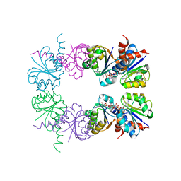
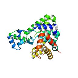
Crystal structure of a frog ependymin related protein
Descriptor: CALCIUM ION, Ependymin-related 1
Authors:Park, S.Y.
Deposit date:2019-03-04
Release date:2019-07-10
Last modified:2019-07-31
Method:X-RAY DIFFRACTION (2 Å)
Cite:Structures of three ependymin-related proteins suggest their function as a hydrophobic molecule binder.
Iucrj, 6, 2019
6JLD
DownloadVisualize
BU of 6jld by Molmil
Crystal structure of a human ependymin related protein
Descriptor: Mammalian ependymin-related protein 1
Authors:Park, S.Y.
Deposit date:2019-03-05
Release date:2019-07-10
Last modified:2019-07-31
Method:X-RAY DIFFRACTION (2 Å)
Cite:Structures of three ependymin-related proteins suggest their function as a hydrophobic molecule binder.
Iucrj, 6, 2019
6JLA
DownloadVisualize
BU of 6jla by Molmil
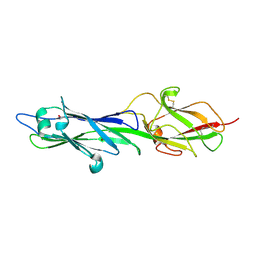
Crystal structure of a mouse ependymin related protein
Descriptor: 2-acetamido-2-deoxy-beta-D-glucopyranose, 2-acetamido-2-deoxy-beta-D-glucopyranose-(1-4)-[alpha-L-fucopyranose-(1-6)]2-acetamido-2-deoxy-beta-D-glucopyranose, Mammalian ependymin-related protein 1
Authors:Park, S.
Deposit date:2019-03-04
Release date:2020-03-04
Last modified:2020-09-16
Method:X-RAY DIFFRACTION (2.4 Å)
Cite:Structures of three ependymin-related proteins suggest their function as a hydrophobic molecule binder.
Iucrj, 6, 2019
3LAM
DownloadVisualize
BU of 3lam by Molmil
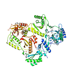
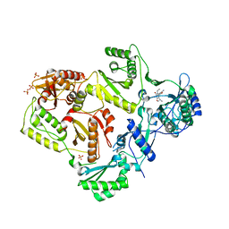
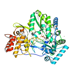
Crystal structure of HIV-1 reverse transcriptase in complex with N1-propyl pyrimidinedione non-nucleoside inhibitor
Descriptor: 3-methyl-5-{[5-(1-methylethyl)-2,6-dioxo-3-propyl-1,2,3,6-tetrahydropyrimidin-4-yl]carbonyl}benzonitrile, HIV Reverse transcriptase, SULFATE ION
Authors:Lansdon, E.B, Mitchell, M.L.
Deposit date:2010-01-06
Release date:2010-02-23
Last modified:2023-09-06
Method:X-RAY DIFFRACTION (2.76 Å)
Cite:N1-Alkyl pyrimidinediones as non-nucleoside inhibitors of HIV-1 reverse transcriptase.
Bioorg.Med.Chem.Lett., 20, 2010
3LAN
DownloadVisualize
BU of 3lan by Molmil
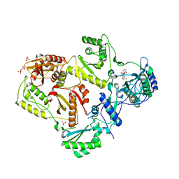
Crystal structure of HIV-1 reverse transcriptase in complex with N1-butyl pyrimidinedione non-nucleoside inhibitor
Descriptor: 3-{[3-butyl-5-(1-methylethyl)-2,6-dioxo-1,2,3,6-tetrahydropyrimidin-4-yl]carbonyl}-5-methylbenzonitrile, HIV Reverse transcriptase, SULFATE ION
Authors:Lansdon, E.B, Mitchell, M.L.
Deposit date:2010-01-06
Release date:2010-02-23
Last modified:2023-09-06
Method:X-RAY DIFFRACTION (2.55 Å)
Cite:N1-Alkyl pyrimidinediones as non-nucleoside inhibitors of HIV-1 reverse transcriptase.
Bioorg.Med.Chem.Lett., 20, 2010
5UJ2
DownloadVisualize
BU of 5uj2 by Molmil
Crystal structure of HCV NS5B genotype 2A JFH-1 isolate with S15G E86Q E87Q C223H V321I mutations and Delta8 neta hairpoin loop deletion in complex with GS-639476 (diphsohate version of GS-9813), Mn2+ and symmetrical primer template 5'-AUAAAUUU
Descriptor: (1S)-1-(4-aminoimidazo[2,1-f][1,2,4]triazin-7-yl)-1,4-anhydro-2-deoxy-2-fluoro-5-O-[(S)-hydroxy(phosphonooxy)phosphoryl]-2-methyl-D-ribitol, CHLORIDE ION, Genome polyprotein, ...
Authors:Edwards, T.E, Fox III, D, Appleby, T.C, Murakami, E, Rey, A, McGrath, M.E.
Deposit date:2017-01-16
Release date:2017-03-22
Last modified:2023-10-04
Method:X-RAY DIFFRACTION (2.9 Å)
Cite:Discovery of a 2'-fluoro-2'-C-methyl C-nucleotide HCV polymerase inhibitor and a phosphoramidate prodrug with favorable properties.
Bioorg. Med. Chem. Lett., 27, 2017
4L00
DownloadVisualize
BU of 4l00 by Molmil
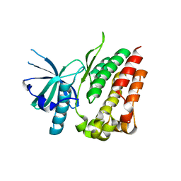
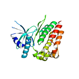
Crystal structure of the apo Jak1 pseudokinase domain
Descriptor: Tyrosine-protein kinase JAK1
Authors:Toms, A.V, Eck, M.J.
Deposit date:2013-05-30
Release date:2013-09-04
Last modified:2024-02-28
Method:X-RAY DIFFRACTION (1.8 Å)
Cite:Structure of a pseudokinase-domain switch that controls oncogenic activation of Jak kinases.
Nat.Struct.Mol.Biol., 20, 2013
4L01
DownloadVisualize
BU of 4l01 by Molmil
Crystal structure of the V658F apo Jak1 pseudokinase domain
Descriptor: Tyrosine-protein kinase JAK1
Authors:Toms, A.V, Rogers, J.M, Eck, M.J.
Deposit date:2013-05-30
Release date:2013-09-04
Last modified:2024-02-28
Method:X-RAY DIFFRACTION (1.9 Å)
Cite:Structure of a pseudokinase-domain switch that controls oncogenic activation of Jak kinases.
Nat.Struct.Mol.Biol., 20, 2013
5T8B
DownloadVisualize
BU of 5t8b by Molmil
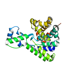
Crystal structure of lpg1496 under 350 MPa
Descriptor: lpg1496
Authors:Huang, Q, Szebenyi, D.
Deposit date:2016-09-07
Release date:2017-08-16
Last modified:2019-12-25
Method:X-RAY DIFFRACTION (1.5 Å)
Cite:Crystal structure of lpg1496 under 350 MPa
To Be Published
5T8C
DownloadVisualize
BU of 5t8c by Molmil
Crystal structure of lpg1496 under 300 MPa
Descriptor: lpg1496
Authors:Huang, Q, Szebenyi, D.
Deposit date:2016-09-07
Release date:2017-08-16
Last modified:2023-10-04
Method:X-RAY DIFFRACTION (1.481 Å)
Cite:Crystal structure of lpg1496 under 300 MPa
To Be Published
2HMU
DownloadVisualize
BU of 2hmu by Molmil
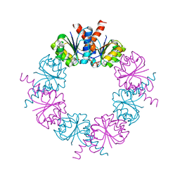
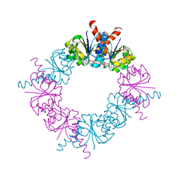
Diamond-shaped octameric ring structure of an RCK domain with ATP bound
Descriptor: ADENOSINE-5'-TRIPHOSPHATE, YuaA protein
Authors:Albright, R.A, Morais-Cabral, J.H.
Deposit date:2006-07-11
Release date:2006-09-26
Last modified:2023-08-30
Method:X-RAY DIFFRACTION (2.25 Å)
Cite:The RCK Domain of the KtrAB K(+) Transporter: Multiple Conformations of an Octameric Ring.
Cell(Cambridge,Mass.), 126, 2006
2HMV
DownloadVisualize
BU of 2hmv by Molmil
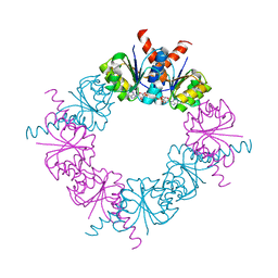
Diamond-shaped octameric ring structure of an RCK domain with ADP bound
Descriptor: ADENOSINE-5'-DIPHOSPHATE, YuaA protein
Authors:Albright, R.A, Morais-Cabral, J.H.
Deposit date:2006-07-11
Release date:2006-09-26
Last modified:2023-08-30
Method:X-RAY DIFFRACTION (2.2 Å)
Cite:The RCK Domain of the KtrAB K(+) Transporter: Multiple Conformations of an Octameric Ring.
Cell(Cambridge,Mass.), 126, 2006
2HMW
DownloadVisualize
BU of 2hmw by Molmil
Square-shaped octameric ring structure of an RCK domain with ATP bound
Descriptor: ADENOSINE-5'-TRIPHOSPHATE, YuaA protein
Authors:Albright, R.A, Morais-Cabral, J.H.
Deposit date:2006-07-11
Release date:2006-09-26
Last modified:2023-08-30
Method:X-RAY DIFFRACTION (3 Å)
Cite:The RCK Domain of the KtrAB K(+) Transporter: Multiple Conformations of an Octameric Ring.
Cell(Cambridge,Mass.), 126, 2006
2HMT
DownloadVisualize
BU of 2hmt by Molmil
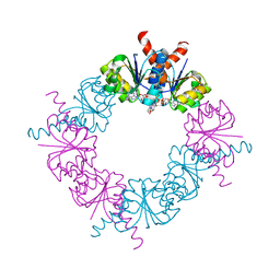
Diamond-shaped octameric ring structure of an RCK domain with NADH bound
Descriptor: 1,4-DIHYDRONICOTINAMIDE ADENINE DINUCLEOTIDE, YuaA protein
Authors:Albright, R.A, Morais-Cabral, J.H.
Deposit date:2006-07-11
Release date:2006-09-26
Last modified:2023-08-30
Method:X-RAY DIFFRACTION (2.2 Å)
Cite:The RCK Domain of the KtrAB K(+) Transporter: Multiple Conformations of an Octameric Ring.
Cell(Cambridge,Mass.), 126, 2006
2HMS
DownloadVisualize
BU of 2hms by Molmil
Rectangular-shaped octameric ring structure of an RCK domain with NADH bound
Descriptor: 1,4-DIHYDRONICOTINAMIDE ADENINE DINUCLEOTIDE, YuaA protein
Authors:Albright, R.A, Morais-Cabral, J.H.
Deposit date:2006-07-11
Release date:2006-09-26
Last modified:2023-08-30
Method:X-RAY DIFFRACTION (2.7 Å)
Cite:The RCK Domain of the KtrAB K(+) Transporter: Multiple Conformations of an Octameric Ring.
Cell(Cambridge,Mass.), 126, 2006
3D93
DownloadVisualize
BU of 3d93 by Molmil
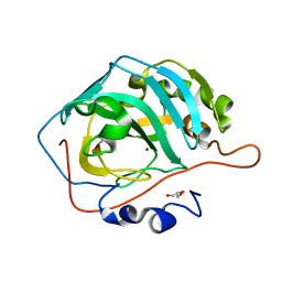
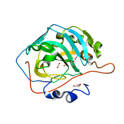
Apo Human carbonic anhydrase II bound with substrate carbon dioxide
Descriptor: CARBON DIOXIDE, GLYCEROL, carbonic anhydrase II
Authors:Domsic, J.F, Avvaru, B.S, McKenna, R.
Deposit date:2008-05-26
Release date:2008-09-02
Last modified:2023-08-30
Method:X-RAY DIFFRACTION (1.1 Å)
Cite:Entrapment of carbon dioxide in the active site of carbonic anhydrase II
J.Biol.Chem., 283, 2008
3D92
DownloadVisualize
BU of 3d92 by Molmil
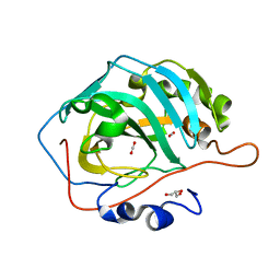
Human carbonic anhydrase II bound with substrate carbon dioxide
Descriptor: CARBON DIOXIDE, GLYCEROL, ZINC ION, ...
Authors:Domsic, J.F, Avvaru, B.S, McKenna, R.
Deposit date:2008-05-26
Release date:2008-09-02
Last modified:2023-08-30
Method:X-RAY DIFFRACTION (1.1 Å)
Cite:Entrapment of carbon dioxide in the active site of carbonic anhydrase II
J.Biol.Chem., 283, 2008

221051

건을2024-06-12부터공개중

PDB statisticsPDBj update infoContact PDBjnumon