2H1T
 
 | |
4QDG
 | |
4Q5K
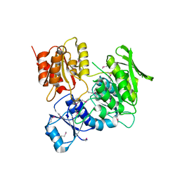
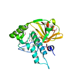 | | Crystal structure of a N-acetylmuramoyl-L-alanine amidase (BACUNI_02947) from Bacteroides uniformis ATCC 8492 at 1.30 A resolution | | Descriptor: | (2R)-2-[[(1R,2S,3R,4R,5R)-4-acetamido-2-[(2S,3R,4R,5S,6R)-3-acetamido-6-(hydroxymethyl)-4,5-bis(oxidanyl)oxan-2-yl]oxy-6,8-dioxabicyclo[3.2.1]octan-3-yl]oxy]propanoic acid, SODIUM ION, Uncharacterized protein | | Authors: | Joint Center for Structural Genomics (JCSG) | | Deposit date: | 2014-04-17 | | Release date: | 2014-05-21 | | Last modified: | 2024-10-16 | | Method: | X-RAY DIFFRACTION (1.3 Å) | | Cite: | Structure-guided functional characterization of DUF1460 reveals a highly specific NlpC/P60 amidase family.
Structure, 22, 2014
|
|
4Q98
 | |
4QB7
 | |
3IRB
 | |
4Q68
 | |
3KHI
 | |
4R03
 | |
4R8O
 | |
4RDB
 | |
3KW0
 | |
3K5J
 | |
3HN7
 | |
3HSA
 | |
3L5O
 | |
3LIU
 | |
3KK7
 | |
3G23
 | |
3DUE
 | |
3FKQ
 | |
3G0T
 | |
3H0N
 | |
3H41
 | |
3GO5
 | |


