Loading
PDBj
MenuPDBj@FacebookPDBj@TwitterPDBj@YouTubewwPDB FoundationwwPDB
RCSB PDBPDBeBMRBAdv. SearchSearch help
Search by PDB author
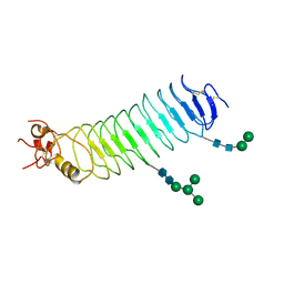
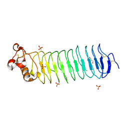
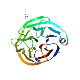
6QHJ
DownloadVisualize
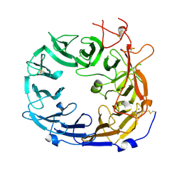
BU of 6qhj by Molmil
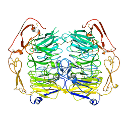
High-resolution crystal structure of calcium- and sodium-bound mouse Olfactomedin-1 beta-propeller domain
Descriptor: 2-acetamido-2-deoxy-beta-D-glucopyranose, CALCIUM ION, GLYCEROL, ...
Authors:Pronker, M.F, van den Hoek, H.G, Janssen, B.J.C.
Deposit date:2019-01-16
Release date:2019-09-11
Last modified:2024-01-24
Method:X-RAY DIFFRACTION (1.25 Å)
Cite:Design and structural characterisation of olfactomedin-1 variants as tools for functional studies.
BMC Mol Cell Biol, 20, 2019
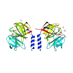
6QM3
DownloadVisualize
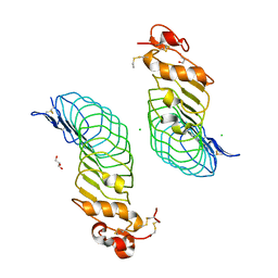
BU of 6qm3 by Molmil
Crystal structure of a calcium- and sodium-bound mouse Olfactomedin-1 disulfide-linked dimer of the Olfactomedin domain and part of coiled coil
Descriptor: 2-acetamido-2-deoxy-beta-D-glucopyranose, ACETATE ION, CACODYLATE ION, ...
Authors:Pronker, M.F, van den Hoek, H.G, Janssen, B.J.C.
Deposit date:2019-02-01
Release date:2019-09-11
Last modified:2024-01-24
Method:X-RAY DIFFRACTION (2 Å)
Cite:Design and structural characterisation of olfactomedin-1 variants as tools for functional studies.
BMC Mol Cell Biol, 20, 2019
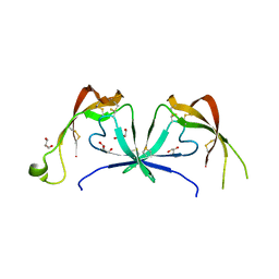
6STE
DownloadVisualize
BU of 6ste by Molmil
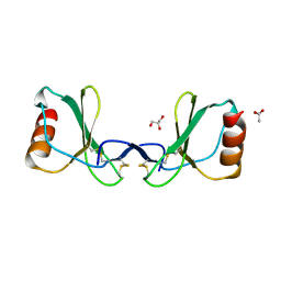
Crystal structure of the tick chemokine-binding protein Evasin-4 (SG 3)
Descriptor: ACETATE ION, DI(HYDROXYETHYL)ETHER, Evasin-4, ...
Authors:Ramirez-Escudero, M, Janssen, B.J.C.
Deposit date:2019-09-10
Release date:2020-09-02
Last modified:2024-01-24
Method:X-RAY DIFFRACTION (1.79 Å)
Cite:Structural characterization of anti-CCL5 activity of the tick salivary protein evasin-4.
J.Biol.Chem., 295, 2020
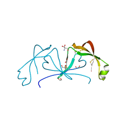
6ST4
DownloadVisualize
BU of 6st4 by Molmil
Crystal structure of the tick chemokine-binding protein Evasin-4 (SG 1)
Descriptor: 1,2-ETHANEDIOL, 2-AMINO-2-HYDROXYMETHYL-PROPANE-1,3-DIOL, Evasin-4
Authors:Ramirez-Escudero, M, Janssen, B.J.C.
Deposit date:2019-09-10
Release date:2020-09-02
Last modified:2020-10-28
Method:X-RAY DIFFRACTION (1.29 Å)
Cite:Structural characterization of anti-CCL5 activity of the tick salivary protein evasin-4.
J.Biol.Chem., 295, 2020
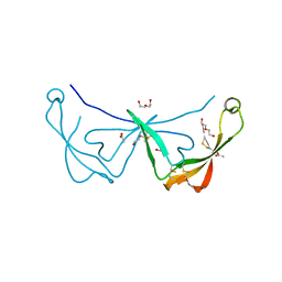
6STC
DownloadVisualize
BU of 6stc by Molmil
Crystal structure of the tick chemokine-binding protein Evasin-4 (SG 2)
Descriptor: ACETATE ION, DI(HYDROXYETHYL)ETHER, Evasin-4
Authors:Ramirez-Escudero, M, Janssen, B.J.C.
Deposit date:2019-09-10
Release date:2020-09-02
Last modified:2024-01-24
Method:X-RAY DIFFRACTION (1.69 Å)
Cite:Structural characterization of anti-CCL5 activity of the tick salivary protein evasin-4.
J.Biol.Chem., 295, 2020
6STK
DownloadVisualize
BU of 6stk by Molmil
Crystal structure of the CC-chemokine 5 (CCL5) E66S mutation
Descriptor: ACETATE ION, C-C motif chemokine 5, GLYCEROL
Authors:Ramirez-Escudero, M, Janssen, B.J.C.
Deposit date:2019-09-10
Release date:2020-09-02
Last modified:2024-01-24
Method:X-RAY DIFFRACTION (1.52 Å)
Cite:Structural characterization of anti-CCL5 activity of the tick salivary protein evasin-4.
J.Biol.Chem., 295, 2020
5NMT
DownloadVisualize
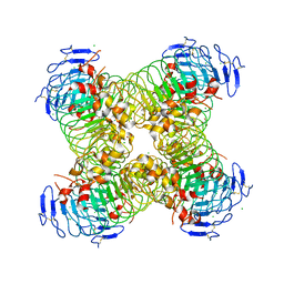
BU of 5nmt by Molmil
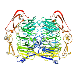
Dimer structure of Sortilin ectodomain crystal form 1, 2.3A
Descriptor: 2-acetamido-2-deoxy-beta-D-glucopyranose, 2-acetamido-2-deoxy-beta-D-glucopyranose-(1-4)-2-acetamido-2-deoxy-beta-D-glucopyranose, CHLORIDE ION, ...
Authors:Leloup, N.O.L, Janssen, B.J.C.
Deposit date:2017-04-07
Release date:2017-11-29
Last modified:2024-01-17
Method:X-RAY DIFFRACTION (2.3 Å)
Cite:Low pH-induced conformational change and dimerization of sortilin triggers endocytosed ligand release.
Nat Commun, 8, 2017
5O0M
DownloadVisualize
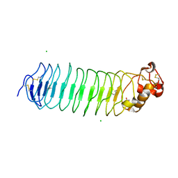
BU of 5o0m by Molmil
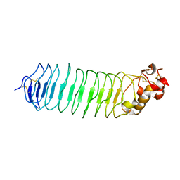
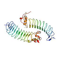
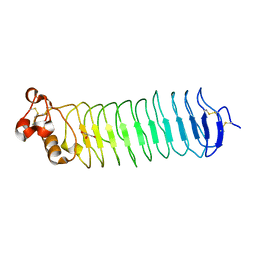
Deglycosylated Nogo Receptor with native disulfide structure 3
Descriptor: 2-acetamido-2-deoxy-beta-D-glucopyranose, Reticulon-4 receptor
Authors:Pronker, M.F, Janssen, B.J.C.
Deposit date:2017-05-16
Release date:2017-10-18
Last modified:2024-01-17
Method:X-RAY DIFFRACTION (1.9 Å)
Cite:Nogo Receptor crystal structures with a native disulfide pattern suggest a novel mode of self-interaction.
Acta Crystallogr D Struct Biol, 73, 2017
5NNJ
DownloadVisualize
BU of 5nnj by Molmil
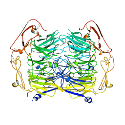
Dimer structure of Sortilin ectodomain crystal form 3, 4.0 Angstrom
Descriptor: 2-acetamido-2-deoxy-beta-D-glucopyranose, Sortilin, beta-D-mannopyranose-(1-4)-2-acetamido-2-deoxy-beta-D-glucopyranose-(1-4)-2-acetamido-2-deoxy-beta-D-glucopyranose
Authors:Leloup, N.O.L, Janssen, B.J.C.
Deposit date:2017-04-09
Release date:2017-11-29
Last modified:2024-01-17
Method:X-RAY DIFFRACTION (4 Å)
Cite:Low pH-induced conformational change and dimerization of sortilin triggers endocytosed ligand release.
Nat Commun, 8, 2017
5NMR
DownloadVisualize
BU of 5nmr by Molmil
Monomeric mouse Sortilin extracellular domain
Descriptor: 2-acetamido-2-deoxy-beta-D-glucopyranose, 2-acetamido-2-deoxy-beta-D-glucopyranose-(1-4)-2-acetamido-2-deoxy-beta-D-glucopyranose, CALCIUM ION, ...
Authors:Leloup, N.O.L, Loessl, P, Meijer, D.H.M, Heck, A.J.R, Thies-Weesie, D.M.E, Janssen, B.J.C.
Deposit date:2017-04-07
Release date:2017-12-06
Last modified:2024-01-17
Method:X-RAY DIFFRACTION (2.1 Å)
Cite:Low pH-induced conformational change and dimerization of sortilin triggers endocytosed ligand release.
Nat Commun, 8, 2017
5O0K
DownloadVisualize
BU of 5o0k by Molmil
Deglycosylated Nogo Receptor with native disulfide structure
Descriptor: 2-acetamido-2-deoxy-beta-D-glucopyranose, CHLORIDE ION, GLYCEROL, ...
Authors:Pronker, M.F, Janssen, B.J.C.
Deposit date:2017-05-16
Release date:2017-10-18
Last modified:2024-01-17
Method:X-RAY DIFFRACTION (2.3 Å)
Cite:Nogo Receptor crystal structures with a native disulfide pattern suggest a novel mode of self-interaction.
Acta Crystallogr D Struct Biol, 73, 2017
5O0R
DownloadVisualize
BU of 5o0r by Molmil
Deglycosylated Nogo Receptor with native disulfide structure
Descriptor: 2-acetamido-2-deoxy-beta-D-glucopyranose, CHLORIDE ION, GLYCEROL, ...
Authors:Pronker, M.F, Janssen, B.J.C.
Deposit date:2017-05-16
Release date:2017-11-08
Last modified:2024-01-17
Method:X-RAY DIFFRACTION (2.5 Å)
Cite:Nogo Receptor crystal structures with a native disulfide pattern suggest a novel mode of self-interaction.
Acta Crystallogr D Struct Biol, 73, 2017
5O0O
DownloadVisualize
BU of 5o0o by Molmil
Deglycosylated Nogo Receptor with native disulfide structure 5
Descriptor: 2-acetamido-2-deoxy-beta-D-glucopyranose, CHLORIDE ION, GLYCEROL, ...
Authors:Pronker, M.F, Janssen, B.J.C.
Deposit date:2017-05-16
Release date:2017-10-18
Last modified:2024-01-17
Method:X-RAY DIFFRACTION (2.2 Å)
Cite:Nogo Receptor crystal structures with a native disulfide pattern suggest a novel mode of self-interaction.
Acta Crystallogr D Struct Biol, 73, 2017
5NNI
DownloadVisualize
BU of 5nni by Molmil
Dimer structure of Sortilin ectodomain crystal form 2, 3.2 Angstrom
Descriptor: 2-acetamido-2-deoxy-beta-D-glucopyranose, 2-acetamido-2-deoxy-beta-D-glucopyranose-(1-4)-2-acetamido-2-deoxy-beta-D-glucopyranose, Sortilin, ...
Authors:Leloup, N.O.L, Janssen, B.J.C.
Deposit date:2017-04-09
Release date:2017-11-29
Last modified:2024-01-17
Method:X-RAY DIFFRACTION (3.21 Å)
Cite:Low pH-induced conformational change and dimerization of sortilin triggers endocytosed ligand release.
Nat Commun, 8, 2017
5O0L
DownloadVisualize
BU of 5o0l by Molmil
Deglycosylated Nogo Receptor with native disulfide structure 2
Descriptor: 2-acetamido-2-deoxy-beta-D-glucopyranose, CHLORIDE ION, Reticulon-4 receptor
Authors:Pronker, M.F, Janssen, B.J.C.
Deposit date:2017-05-16
Release date:2017-10-18
Last modified:2024-01-17
Method:X-RAY DIFFRACTION (2.511 Å)
Cite:Nogo Receptor crystal structures with a native disulfide pattern suggest a novel mode of self-interaction.
Acta Crystallogr D Struct Biol, 73, 2017
5O0P
DownloadVisualize
BU of 5o0p by Molmil
Deglycosylated Nogo Receptor with native disulfide structure 6
Descriptor: 2-acetamido-2-deoxy-beta-D-glucopyranose, GLYCEROL, Reticulon-4 receptor, ...
Authors:Pronker, M.F, Janssen, B.J.C.
Deposit date:2017-05-16
Release date:2017-10-18
Last modified:2024-01-17
Method:X-RAY DIFFRACTION (2 Å)
Cite:Nogo Receptor crystal structures with a native disulfide pattern suggest a novel mode of self-interaction.
Acta Crystallogr D Struct Biol, 73, 2017
5O0Z
DownloadVisualize
BU of 5o0z by Molmil
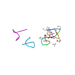
Structure of laspartomycin C in complex with geranyl-phosphate
Descriptor: ACETIC ACID, CALCIUM ION, CHLORIDE ION, ...
Authors:Vlieg, H.C, Kleijn, L.H.J, Martin, N.I, Janssen, B.J.C.
Deposit date:2017-05-17
Release date:2017-11-15
Last modified:2019-10-16
Method:X-RAY DIFFRACTION (1.28 Å)
Cite:A High-Resolution Crystal Structure that Reveals Molecular Details of Target Recognition by the Calcium-Dependent Lipopeptide Antibiotic Laspartomycin C.
Angew. Chem. Int. Ed. Engl., 56, 2017
5O0N
DownloadVisualize
BU of 5o0n by Molmil
Deglycosylated Nogo Receptor with native disulfide structure 4
Descriptor: 2-acetamido-2-deoxy-beta-D-glucopyranose-(1-4)-2-acetamido-2-deoxy-beta-D-glucopyranose, CHLORIDE ION, Reticulon-4 receptor, ...
Authors:Pronker, M.F, Tas, R.P, Vlieg, H.C, Janssen, B.J.C.
Deposit date:2017-05-16
Release date:2017-10-18
Last modified:2024-01-17
Method:X-RAY DIFFRACTION (2.5 Å)
Cite:Nogo Receptor crystal structures with a native disulfide pattern suggest a novel mode of self-interaction.
Acta Crystallogr D Struct Biol, 73, 2017
5O0Q
DownloadVisualize
BU of 5o0q by Molmil
Deglycosylated Nogo Receptor with native disulfide structure
Descriptor: 2-acetamido-2-deoxy-beta-D-glucopyranose, CHLORIDE ION, GLYCEROL, ...
Authors:Pronker, M.F, Janssen, B.J.C.
Deposit date:2017-05-16
Release date:2017-10-18
Last modified:2024-01-17
Method:X-RAY DIFFRACTION (2.5 Å)
Cite:Nogo Receptor crystal structures with a native disulfide pattern suggest a novel mode of self-interaction.
Acta Crystallogr D Struct Biol, 73, 2017
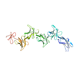
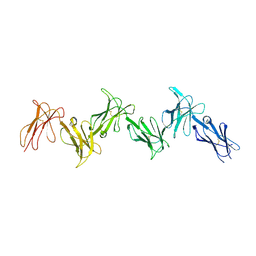
6GRQ
DownloadVisualize
BU of 6grq by Molmil
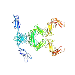
Paired immunoglobulin-like receptor B (PirB) or Leukocyte immunoglobulin-like receptor subfamily B member 3 (LILRB3) full extracellular domain
Descriptor: 2-acetamido-2-deoxy-beta-D-glucopyranose, 2-acetamido-2-deoxy-beta-D-glucopyranose-(1-4)-2-acetamido-2-deoxy-beta-D-glucopyranose, Paired immunoglobulin-like receptor B
Authors:Vlieg, H.C, Huizinga, E.G, Janssen, B.J.C.
Deposit date:2018-06-12
Release date:2019-02-06
Last modified:2024-01-17
Method:X-RAY DIFFRACTION (3.3 Å)
Cite:Structure and flexibility of the extracellular region of the PirB receptor.
J.Biol.Chem., 294, 2019
6GRT
DownloadVisualize
BU of 6grt by Molmil
Paired immunoglobulin-like receptor B (PirB) or Leukocyte immunoglobulin-like receptor subfamily B member 3 (LILRB3) full extracellular domain
Descriptor: 2-acetamido-2-deoxy-beta-D-glucopyranose, Paired immunoglobulin-like receptor B, alpha-D-mannopyranose-(1-3)-[alpha-D-mannopyranose-(1-6)]beta-D-mannopyranose-(1-4)-2-acetamido-2-deoxy-beta-D-glucopyranose-(1-4)-2-acetamido-2-deoxy-beta-D-glucopyranose, ...
Authors:Vlieg, H.C, Huizinga, E.G, Janssen, B.J.C.
Deposit date:2018-06-12
Release date:2019-02-06
Last modified:2024-01-17
Method:X-RAY DIFFRACTION (4.504 Å)
Cite:Structure and flexibility of the extracellular region of the PirB receptor.
J.Biol.Chem., 294, 2019
6GRS
DownloadVisualize
BU of 6grs by Molmil
Paired immunoglobulin-like receptor B (PirB) or Leukocyte immunoglobulin-like receptor subfamily B member 3 (LILRB3) full extracellular domain
Descriptor: 2-acetamido-2-deoxy-beta-D-glucopyranose, 2-acetamido-2-deoxy-beta-D-glucopyranose-(1-4)-2-acetamido-2-deoxy-beta-D-glucopyranose, Paired immunoglobulin-like receptor B
Authors:Vlieg, H.C, Huizinga, E.G, Janssen, B.J.C.
Deposit date:2018-06-12
Release date:2019-02-06
Last modified:2024-01-17
Method:X-RAY DIFFRACTION (3.4 Å)
Cite:Structure and flexibility of the extracellular region of the PirB receptor.
J.Biol.Chem., 294, 2019
6AP8
DownloadVisualize
BU of 6ap8 by Molmil
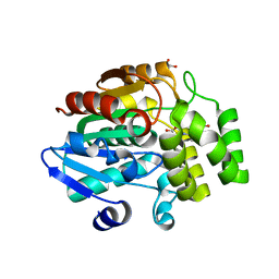
Crystal Structure of rice D14 bound to 2-(2-methyl-3-nitroanilino)benzoic acid
Descriptor: 2-[(2-methyl-3-nitrophenyl)amino]benzoic acid, GLYCEROL, Strigolactone esterase D14
Authors:Hamiaux, C.
Deposit date:2017-08-17
Release date:2018-03-21
Last modified:2023-10-04
Method:X-RAY DIFFRACTION (1.27 Å)
Cite:Inhibition of strigolactone receptors byN-phenylanthranilic acid derivatives: Structural and functional insights.
J. Biol. Chem., 293, 2018
6AP6
DownloadVisualize
BU of 6ap6 by Molmil
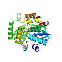
Crystal Structure of DAD2 in complex with tolfenamic acid
Descriptor: 2-[(3-chloro-2-methylphenyl)amino]benzoic acid, Probable strigolactone esterase DAD2
Authors:Hamiaux, C.
Deposit date:2017-08-17
Release date:2018-03-21
Last modified:2023-10-04
Method:X-RAY DIFFRACTION (1.65 Å)
Cite:Inhibition of strigolactone receptors byN-phenylanthranilic acid derivatives: Structural and functional insights.
J. Biol. Chem., 293, 2018
6AP7
DownloadVisualize
BU of 6ap7 by Molmil
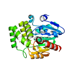
Crystal Structure of DAD2 in complex with 2-(2-methyl-3-nitroanilino)benzoic acid
Descriptor: 2-[(2-methyl-3-nitrophenyl)amino]benzoic acid, GLYCEROL, Probable strigolactone esterase DAD2
Authors:Hamiaux, C.
Deposit date:2017-08-17
Release date:2018-03-21
Last modified:2023-10-04
Method:X-RAY DIFFRACTION (1.51 Å)
Cite:Inhibition of strigolactone receptors byN-phenylanthranilic acid derivatives: Structural and functional insights.
J. Biol. Chem., 293, 2018

222926

건을2024-07-24부터공개중

PDB statisticsPDBj update infoContact PDBjnumon