Loading
PDBj
MenuPDBj@FacebookPDBj@TwitterPDBj@YouTubewwPDB FoundationwwPDB
RCSB PDBPDBeBMRBAdv. SearchSearch help
Search by PDB author
4J9H
DownloadVisualize
BU of 4j9h by Molmil
Crystal structure of the ABL-SH3 domain complexed with the designed high-affinity peptide ligand P7 at pH 8
Descriptor: P7, SULFATE ION, Tyrosine-protein kinase ABL1
Authors:Camara-Artigas, A.
Deposit date:2013-02-16
Release date:2014-01-29
Last modified:2023-09-20
Method:X-RAY DIFFRACTION (1.7 Å)
Cite:Crystal structure of the ABL-SH3 domain complexed with the designed high-affinity peptide ligand P7
To be Published
4J9C
DownloadVisualize
BU of 4j9c by Molmil
Crystal structure of the Abl-SH3 domain H59Q-N96T mutant complexed with the designed high-affinity peptide ligand P17
Descriptor: DI(HYDROXYETHYL)ETHER, P17, TRIETHYLENE GLYCOL, ...
Authors:Camara-Artigas, A.
Deposit date:2013-02-16
Release date:2014-01-29
Last modified:2023-09-20
Method:X-RAY DIFFRACTION (1.051 Å)
Cite:Crystal structure of the Abl-SH3 domain H59Q-N96T mutant complexed with the designed high-affinity peptide ligand P17
To be Published
4JJC
DownloadVisualize
BU of 4jjc by Molmil
Crystal structure of the Abl-SH3 domain at pH5
Descriptor: DI(HYDROXYETHYL)ETHER, Tyrosine-protein kinase ABL1
Authors:Camara-Artigas, A, Martin-Garcia, J.M.
Deposit date:2013-03-07
Release date:2014-03-12
Last modified:2023-11-08
Method:X-RAY DIFFRACTION (1.6 Å)
Cite:Crystal structure of the Abl-SH3 domain at pH5
To be Published
4J9E
DownloadVisualize
BU of 4j9e by Molmil
Crystal structure of the N114A mutant of the Abl-SH3 domain complexed with the high affinity peptide P17
Descriptor: P17, SULFATE ION, Tyrosine-protein kinase ABL1
Authors:Camara-Artigas, A.
Deposit date:2013-02-16
Release date:2014-01-29
Last modified:2023-09-20
Method:X-RAY DIFFRACTION (1.4 Å)
Cite:Crystal structure of the N114A mutant of the Abl-SH3 domain complexed with the high affinity peptide P17
To be Published
4J9B
DownloadVisualize
BU of 4j9b by Molmil
Crystal structure of the Abl-SH3 domain H59Q-N96T mutant
Descriptor: DI(HYDROXYETHYL)ETHER, Tyrosine-protein kinase ABL1
Authors:Camara-Artigas, A.
Deposit date:2013-02-16
Release date:2014-01-29
Last modified:2023-09-20
Method:X-RAY DIFFRACTION (1.702 Å)
Cite:Crystal structure of the Abl-SH3 domain H59Q-N96T mutant
To be Published
4J9D
DownloadVisualize
BU of 4j9d by Molmil
Crystal structure of the N114A mutant of the Abl-SH3 domain complexed with the high affinity peptide P0
Descriptor: P0, SULFATE ION, Tyrosine-protein kinase ABL1
Authors:Camara-Artigas, A.
Deposit date:2013-02-16
Release date:2014-01-29
Last modified:2023-09-20
Method:X-RAY DIFFRACTION (1.5 Å)
Cite:Crystal structure of the N114A mutant of the Abl-SH3 domain complexed with the high affinity peptide P0
To be Published
4JJB
DownloadVisualize
BU of 4jjb by Molmil
Crystal structure of the Abl-SH3 domain at pH3
Descriptor: DI(HYDROXYETHYL)ETHER, SULFATE ION, Tyrosine-protein kinase ABL1
Authors:Camara-Artigas, A, Martin-Garcia, J.M.
Deposit date:2013-03-07
Release date:2014-03-12
Last modified:2023-11-08
Method:X-RAY DIFFRACTION (1.65 Å)
Cite:Crystal structure of the Abl-SH3 domain at pH3
To be Published
2KQ0
DownloadVisualize
BU of 2kq0 by Molmil
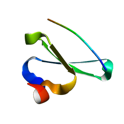
Human NEDD4 3rd WW Domain Complex with Ebola Zaire Virus Matrix Protein VP40 Derived Peptide ILPTAPPEYMEA
Descriptor: 12-mer from Matrix protein VP40, E3 ubiquitin-protein ligase NEDD4
Authors:Iglesias-Bexiga, M, Macias, M, Bonet, R, Blanco, F.J, Cobos, E.S, Luque, I.
Deposit date:2009-10-23
Release date:2010-11-03
Last modified:2024-05-01
Method:SOLUTION NMR
Cite:Human NEDD4 3rd WW Domain Complex with Ebola Zaire Virus Matrix Protein VP40 Derived Peptide
To be Published
6GIS
DownloadVisualize
BU of 6gis by Molmil
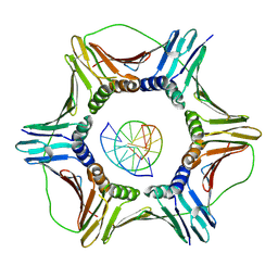
Structural basis of human clamp sliding on DNA
Descriptor: DNA (5'-D(P*AP*TP*AP*CP*GP*AP*TP*GP*GP*G)-3'), DNA (5'-D(P*CP*CP*CP*AP*TP*CP*GP*TP*AP*T)-3'), Proliferating cell nuclear antigen
Authors:De March, M, Merino, N, Barrera-Vilarmau, S, Crehuet, R, Onesti, S, Blanco, F.J, De Biasio, A.
Deposit date:2018-05-15
Release date:2018-05-30
Last modified:2024-01-17
Method:X-RAY DIFFRACTION (2.82 Å)
Cite:Structural basis of human PCNA sliding on DNA.
Nat Commun, 8, 2017
4ZTD
DownloadVisualize
BU of 4ztd by Molmil
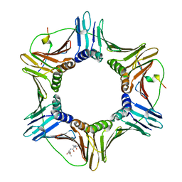
Crystal Structure of Human PCNA in complex with a TRAIP peptide
Descriptor: ALA-GLY-ALA-GLY-ALA, ALA-PHE-GLN-ALA-LYS-LEU-ASP-THR-PHE-LEU-TRP-SER, Proliferating cell nuclear antigen
Authors:Montoya, G, Mortuza, G.B, Blanco, F.J, Ibanez de Opakua, A.
Deposit date:2015-05-14
Release date:2015-12-16
Last modified:2024-01-10
Method:X-RAY DIFFRACTION (2.199 Å)
Cite:TRAIP is a PCNA-binding ubiquitin ligase that protects genome stability after replication stress.
J.Cell Biol., 212, 2016
<12


224201

건을2024-08-28부터공개중

PDB statisticsPDBj update infoContact PDBjnumon