Loading
PDBj
MenuPDBj@FacebookPDBj@TwitterPDBj@YouTubewwPDB FoundationwwPDB
RCSB PDBPDBeBMRBAdv. SearchSearch help
Search by PDB author
3KUS
DownloadVisualize
BU of 3kus by Molmil
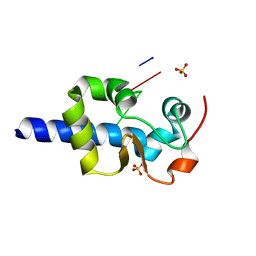
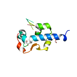
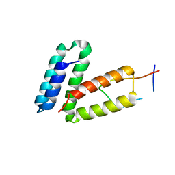
Crystal structure of the MLLE domain of poly(A)-binding protein in complex with the binding region of Paip2
Descriptor: 4-(2-HYDROXYETHYL)-1-PIPERAZINE ETHANESULFONIC ACID, GLYCEROL, PAIP2 protein, ...
Authors:Kozlov, G, Gehring, K.
Deposit date:2009-11-27
Release date:2010-02-09
Last modified:2023-09-06
Method:X-RAY DIFFRACTION (1.4 Å)
Cite:Molecular Determinants of PAM2 Recognition by the MLLE Domain of Poly(A)-Binding Protein.
J.Mol.Biol., 397, 2010
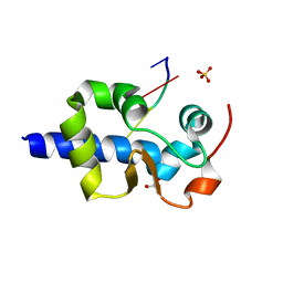
3KUI
DownloadVisualize
BU of 3kui by Molmil
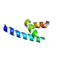
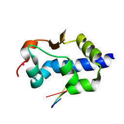
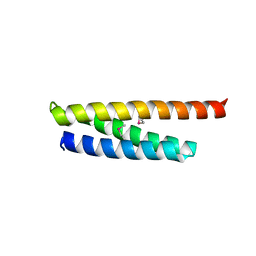
Crystal structure of C-terminal domain of PABPC1 in complex with binding region of eRF3a
Descriptor: GSPT1 protein, Polyadenylate-binding protein 1, SULFATE ION, ...
Authors:Kozlov, G, Gehring, K.
Deposit date:2009-11-27
Release date:2010-05-12
Last modified:2023-09-06
Method:X-RAY DIFFRACTION (2.3 Å)
Cite:Molecular basis of eRF3 recognition by the MLLE domain of poly(A)-binding protein.
Plos One, 5, 2010
3KUR
DownloadVisualize
BU of 3kur by Molmil
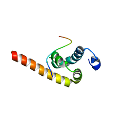
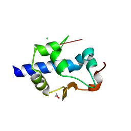
Crystal structure of the MLLE domain of poly(A)-binding protein
Descriptor: CHLORIDE ION, Polyadenylate-binding protein 1
Authors:Kozlov, G, Gehring, K.
Deposit date:2009-11-27
Release date:2010-02-09
Last modified:2023-09-06
Method:X-RAY DIFFRACTION (2.5 Å)
Cite:Molecular Determinants of PAM2 Recognition by the MLLE Domain of Poly(A)-Binding Protein.
J.Mol.Biol., 397, 2010
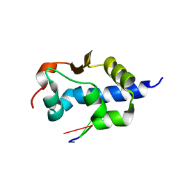
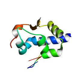
4G3O
DownloadVisualize
BU of 4g3o by Molmil
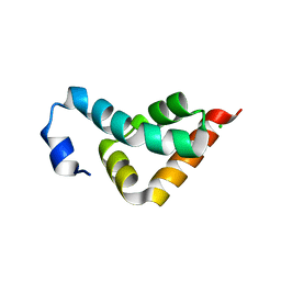
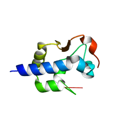
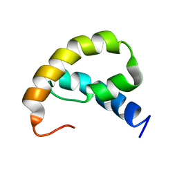
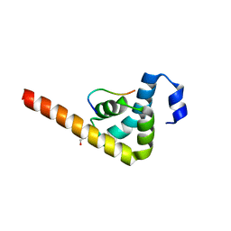
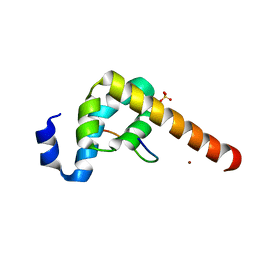
Crystal structure of the CUE domain of the E3 ubiquitin ligase AMFR (gp78)
Descriptor: E3 ubiquitin-protein ligase AMFR
Authors:Kozlov, G, LePage, K, Gehring, K.
Deposit date:2012-07-15
Release date:2012-07-25
Last modified:2023-09-13
Method:X-RAY DIFFRACTION (1.6 Å)
Cite:Crystal structure of the CUE domain of the E3 ubiquitin ligase AMFR (gp78)
To be Published
3KUT
DownloadVisualize
BU of 3kut by Molmil
Crystal structure of the MLLE domain of poly(A)-binding protein in complex with the binding region of Paip2
Descriptor: CHLORIDE ION, PAIP2 protein, Polyadenylate-binding protein 1
Authors:Kozlov, G, Gehring, K.
Deposit date:2009-11-27
Release date:2010-02-09
Last modified:2023-09-06
Method:X-RAY DIFFRACTION (1.5 Å)
Cite:Molecular Determinants of PAM2 Recognition by the MLLE Domain of Poly(A)-Binding Protein.
J.Mol.Biol., 397, 2010
7SOT
DownloadVisualize
BU of 7sot by Molmil
LaM domain of human LARP1 in complex with AAAAAA oligonucleotide
Descriptor: Isoform 2 of La-related protein 1, RNA (5'-R(P*AP*AP*AP*AP*AP*A)-3'), SULFATE ION
Authors:Kozlov, G, Gehring, K.
Deposit date:2021-11-01
Release date:2022-08-03
Last modified:2023-10-18
Method:X-RAY DIFFRACTION (1.52 Å)
Cite:Structural basis of 3'-end poly(A) RNA recognition by LARP1.
Nucleic Acids Res., 50, 2022
7SOV
DownloadVisualize
BU of 7sov by Molmil
LaM domain of human LARP1 in complex with AAAAAAAAAAA RNA polynucleotide
Descriptor: GLYCEROL, Isoform 2 of La-related protein 1, RNA (5'-R(P*AP*AP*AP*AP*AP*AP*AP*AP*AP*AP*A)-3'), ...
Authors:Kozlov, G, Gehring, K.
Deposit date:2021-11-01
Release date:2022-08-03
Last modified:2023-10-18
Method:X-RAY DIFFRACTION (1.45 Å)
Cite:Structural basis of 3'-end poly(A) RNA recognition by LARP1.
Nucleic Acids Res., 50, 2022
7SOQ
DownloadVisualize
BU of 7soq by Molmil
LaM domain of human LARP1 in complex with AAA RNA
Descriptor: Isoform 2 of La-related protein 1, RNA (5'-R(*AP*AP*A)-3')
Authors:Kozlov, G, Gehring, K.
Deposit date:2021-11-01
Release date:2022-08-03
Last modified:2023-10-18
Method:X-RAY DIFFRACTION (1.15 Å)
Cite:Structural basis of 3'-end poly(A) RNA recognition by LARP1.
Nucleic Acids Res., 50, 2022
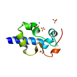
7SOO
DownloadVisualize
BU of 7soo by Molmil
LaM domain of human LARP1
Descriptor: Isoform 2 of La-related protein 1, SODIUM ION, SULFATE ION
Authors:Kozlov, G, Gehring, K.
Deposit date:2021-11-01
Release date:2022-08-03
Last modified:2023-10-18
Method:X-RAY DIFFRACTION (1.65 Å)
Cite:Structural basis of 3'-end poly(A) RNA recognition by LARP1.
Nucleic Acids Res., 50, 2022
7SOP
DownloadVisualize
BU of 7sop by Molmil
LaM domain of human LARP1 in complex with AAAUAA RNA
Descriptor: Isoform 2 of La-related protein 1, RNA (5'-R(P*AP*AP*AP*UP*AP*A)-3')
Authors:Kozlov, G, Gehring, K.
Deposit date:2021-11-01
Release date:2022-08-03
Last modified:2023-10-18
Method:X-RAY DIFFRACTION (1.55 Å)
Cite:Structural basis of 3'-end poly(A) RNA recognition by LARP1.
Nucleic Acids Res., 50, 2022
7SOR
DownloadVisualize
BU of 7sor by Molmil
LaM domain of human LARP1 in complex with AAA RNA
Descriptor: 1,2-ETHANEDIOL, CHLORIDE ION, Isoform 2 of La-related protein 1, ...
Authors:Kozlov, G, Gehring, K.
Deposit date:2021-11-01
Release date:2022-08-03
Last modified:2023-10-18
Method:X-RAY DIFFRACTION (1.35 Å)
Cite:Structural basis of 3'-end poly(A) RNA recognition by LARP1.
Nucleic Acids Res., 50, 2022
7SOS
DownloadVisualize
BU of 7sos by Molmil
LaM domain of human LARP1 in complex with AAAA RNA
Descriptor: Isoform 2 of La-related protein 1, POTASSIUM ION, RNA (5'-R(*AP*AP*AP*A)-3')
Authors:Kozlov, G, Gehring, K.
Deposit date:2021-11-01
Release date:2022-08-03
Last modified:2023-10-18
Method:X-RAY DIFFRACTION (1.25 Å)
Cite:Structural basis of 3'-end poly(A) RNA recognition by LARP1.
Nucleic Acids Res., 50, 2022
7SOU
DownloadVisualize
BU of 7sou by Molmil
LaM domain of human LARP1 in complex with AAAAAA polynucleotide
Descriptor: Isoform 2 of La-related protein 1, RNA (5'-R(P*AP*AP*AP*AP*AP*A)-3')
Authors:Kozlov, G, Gehring, K.
Deposit date:2021-11-01
Release date:2022-08-03
Last modified:2023-10-18
Method:X-RAY DIFFRACTION (1.45 Å)
Cite:Structural basis of 3'-end poly(A) RNA recognition by LARP1.
Nucleic Acids Res., 50, 2022
7SOW
DownloadVisualize
BU of 7sow by Molmil
LaM domain of human LARP1 in complex with UUUUUU
Descriptor: 1,2-ETHANEDIOL, Isoform 2 of La-related protein 1, RNA (5'-R(P*UP*UP*UP*UP*UP*U)-3')
Authors:Kozlov, G, Gehring, K.
Deposit date:2021-11-01
Release date:2022-11-09
Last modified:2024-10-30
Method:X-RAY DIFFRACTION (1.3 Å)
Cite:Enhanced binding of guanylated poly(A) RNA by the LaM domain of LARP1.
Rna Biol., 21, 2024
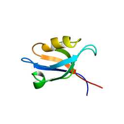
3PDZ
DownloadVisualize
BU of 3pdz by Molmil
SOLUTION STRUCTURE OF THE PDZ2 DOMAIN FROM HUMAN PHOSPHATASE HPTP1E
Descriptor: PROTEIN (TYROSINE PHOSPHATASE (PTP-BAS, TYPE 1))
Authors:Kozlov, G, Gehring, K, Ekiel, I.
Deposit date:1999-05-10
Release date:2000-03-17
Last modified:2023-12-27
Method:SOLUTION NMR
Cite:Solution structure of the PDZ2 domain from human phosphatase hPTP1E and its interactions with C-terminal peptides from the Fas receptor.
Biochemistry, 39, 2000
1R6H
DownloadVisualize
BU of 1r6h by Molmil
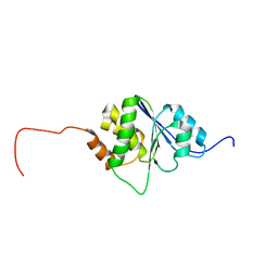
Solution Structure of human PRL-3
Descriptor: protein tyrosine phosphatase type IVA, member 3 isoform 1
Authors:Kozlov, G, Gehring, K, Ekiel, I.
Deposit date:2003-10-15
Release date:2004-01-13
Last modified:2024-05-22
Method:SOLUTION NMR
Cite:Structural Insights into Molecular Function of the Metastasis-associated Phosphatase PRL-3.
J.Biol.Chem., 279, 2004
1RRZ
DownloadVisualize
BU of 1rrz by Molmil
Solution structure of GlgS protein from E. coli
Descriptor: Glycogen synthesis protein glgS
Authors:Kozlov, G, Gehring, K, Montreal-Kingston Bacterial Structural Genomics Initiative (BSGI)
Deposit date:2003-12-09
Release date:2004-06-01
Last modified:2024-05-22
Method:SOLUTION NMR
Cite:Structure of GlgS from Escherichia coli suggests a role in protein-protein interactions.
BMC Biol., 2, 2004
3O0W
DownloadVisualize
BU of 3o0w by Molmil
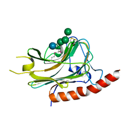
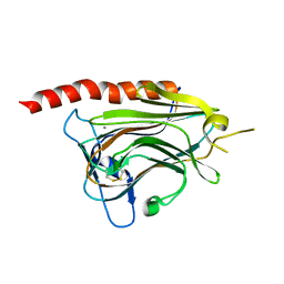
Structural basis of carbohydrate recognition by calreticulin
Descriptor: CALCIUM ION, Calreticulin, alpha-D-glucopyranose-(1-3)-alpha-D-mannopyranose-(1-2)-alpha-D-mannopyranose-(1-2)-alpha-D-mannopyranose
Authors:Kozlov, G, Gehring, K.
Deposit date:2010-07-20
Release date:2010-09-29
Last modified:2020-07-29
Method:X-RAY DIFFRACTION (1.95 Å)
Cite:Structural basis of carbohydrate recognition by calreticulin.
J.Biol.Chem., 285, 2010
3O0V
DownloadVisualize
BU of 3o0v by Molmil
Crystal structure of the calreticulin lectin domain
Descriptor: CALCIUM ION, Calreticulin
Authors:Kozlov, G, Gehring, K.
Deposit date:2010-07-20
Release date:2010-09-29
Last modified:2024-11-06
Method:X-RAY DIFFRACTION (2.3 Å)
Cite:Structural basis of carbohydrate recognition by calreticulin.
J.Biol.Chem., 285, 2010
3O10
DownloadVisualize
BU of 3o10 by Molmil
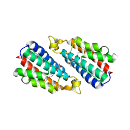
Crystal structure of the HEPN domain from human sacsin
Descriptor: MALONATE ION, Sacsin
Authors:Kozlov, G, Gehring, K.
Deposit date:2010-07-20
Release date:2011-03-30
Last modified:2014-07-30
Method:X-RAY DIFFRACTION (1.9 Å)
Cite:Structural basis of defects in the sacsin HEPN domain responsible for autosomal recessive spastic ataxia of Charlevoix-Saguenay (ARSACS).
J.Biol.Chem., 286, 2011
1SSL
DownloadVisualize
BU of 1ssl by Molmil
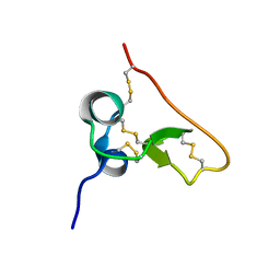
Solution structure of the PSI domain from the Met receptor
Descriptor: Hepatocyte growth factor receptor
Authors:Kozlov, G, Perreault, A, Schrag, J.D, Cygler, M, Gehring, K, Ekiel, I.
Deposit date:2004-03-24
Release date:2004-10-12
Last modified:2024-10-30
Method:SOLUTION NMR
Cite:Insights into function of PSI domains from structure of the Met receptor PSI domain.
Biochem.Biophys.Res.Commun., 321, 2004
1RWU
DownloadVisualize
BU of 1rwu by Molmil
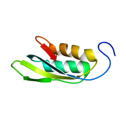
Solution structure of conserved protein YbeD from E. coli
Descriptor: Hypothetical UPF0250 protein ybeD
Authors:Kozlov, G, Arrowsmith, C.H, Gehring, K, Northeast Structural Genomics Consortium (NESG)
Deposit date:2003-12-17
Release date:2004-12-21
Last modified:2024-05-22
Method:SOLUTION NMR
Cite:Structural similarity of YbeD protein from Escherichia coli to allosteric regulatory domains
J.Bacteriol., 186, 2004
5V90
DownloadVisualize
BU of 5v90 by Molmil
Crystal structure of ERp29 D-domain in complex with the P-domain of calreticulin
Descriptor: Calreticulin, Endoplasmic reticulum resident protein 29, GLYCEROL
Authors:Kozlov, G, Munoz-Escobar, J, Gehring, K.
Deposit date:2017-03-22
Release date:2017-06-21
Last modified:2023-10-04
Method:X-RAY DIFFRACTION (3.255 Å)
Cite:Mapping the ER Interactome: The P Domains of Calnexin and Calreticulin as Plurivalent Adapters for Foldases and Chaperones.
Structure, 25, 2017
5WD8
DownloadVisualize
BU of 5wd8 by Molmil
Crystal structure of Legionella pneumophila effector lpg2328
Descriptor: Lem22
Authors:Kozlov, G, Wong, K, Gehring, K, Montreal-Kingston Bacterial Structural Genomics Initiative (BSGI)
Deposit date:2017-07-04
Release date:2017-11-29
Last modified:2024-10-30
Method:X-RAY DIFFRACTION (1.944 Å)
Cite:Crystal structure of the Legionella effector Lem22.
Proteins, 86, 2018
5VRQ
DownloadVisualize
BU of 5vrq by Molmil
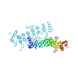
Crystal structure of Legionella pneumophila effector AnkC
Descriptor: Ankyrin repeat-containing protein
Authors:Kozlov, G, Wong, K, Wang, W, Skubak, P, Munoz-Escobar, J, Liu, Y, Pannu, N.S, Gehring, K, Montreal-Kingston Bacterial Structural Genomics Initiative (BSGI)
Deposit date:2017-05-11
Release date:2017-11-29
Last modified:2024-03-13
Method:X-RAY DIFFRACTION (3.205 Å)
Cite:Ankyrin repeats as a dimerization module.
Biochem. Biophys. Res. Commun., 495, 2018

227111

건을2024-11-06부터공개중

PDB statisticsPDBj update infoContact PDBjnumon