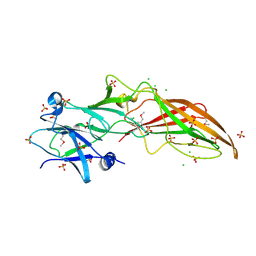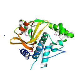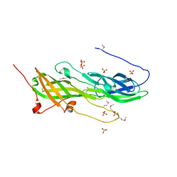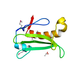
Loading
PDBj
MenuPDBj@FacebookPDBj@TwitterPDBj@YouTubewwPDB FoundationwwPDB
RCSB PDBPDBeBMRBAdv. SearchSearch help
Search by PDB author
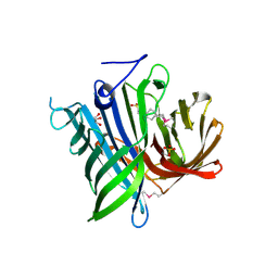
2IAY
DownloadVisualize
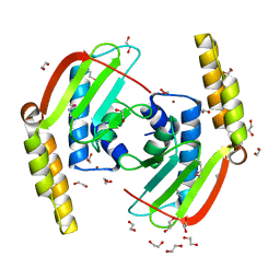
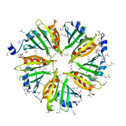
BU of 2iay by Molmil
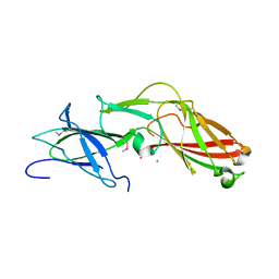
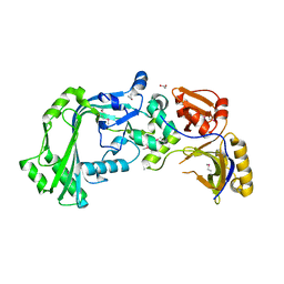
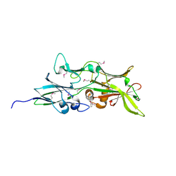
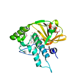
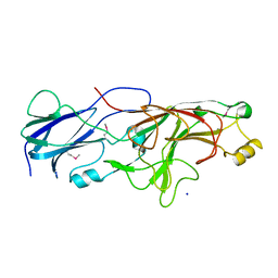
Crystal structure of a duf1831 family protein (lp2179) from lactobacillus plantarum at 1.20 A resolution
Descriptor: CHLORIDE ION, GLYCEROL, Hypothetical protein
Authors:Joint Center for Structural Genomics (JCSG)
Deposit date:2006-09-08
Release date:2006-10-10
Last modified:2023-01-25
Method:X-RAY DIFFRACTION (1.2 Å)
Cite:Structure of LP2179, the first representative of Pfam family PF08866, suggests a new fold with a role in amino-acid metabolism.
Acta Crystallogr.,Sect.F, 66, 2010
2ICH
DownloadVisualize
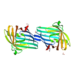
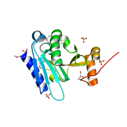
BU of 2ich by Molmil
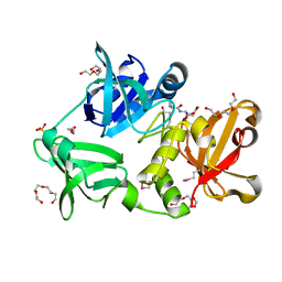
CRYSTAL STRUCTURE OF A PUTATIVE ATTH (NE1406) FROM NITROSOMONAS EUROPAEA AT 2.00 A RESOLUTION
Descriptor: 2-[N-CYCLOHEXYLAMINO]ETHANE SULFONIC ACID, GLYCEROL, Putative AttH, ...
Authors:Joint Center for Structural Genomics (JCSG)
Deposit date:2006-09-12
Release date:2006-10-03
Last modified:2023-01-25
Method:X-RAY DIFFRACTION (2 Å)
Cite:Structure of the first representative of Pfam family PF09410 (DUF2006) reveals a structural signature of the calycin superfamily that suggests a role in lipid metabolism.
Acta Crystallogr.,Sect.F, 66, 2010
2GLZ
DownloadVisualize
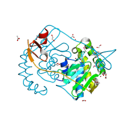
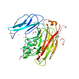
BU of 2glz by Molmil
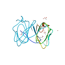
Crystal structure of a formylmethanofuran dehydrogenase subunit e-like protein (dhaf_2992) from desulfitobacterium hafniense dcb-2 at 1.45 A resolution
Descriptor: 1,2-ETHANEDIOL, NICKEL (II) ION, ZINC ION, ...
Authors:Joint Center for Structural Genomics (JCSG)
Deposit date:2006-04-05
Release date:2006-04-18
Last modified:2023-01-25
Method:X-RAY DIFFRACTION (1.45 Å)
Cite:Structures of three members of Pfam PF02663 (FmdE) implicated in microbial methanogenesis reveal a conserved alpha+beta core domain and an auxiliary C-terminal treble-clef zinc finger.
Acta Crystallogr.,Sect.F, 66, 2010
2H1T
DownloadVisualize
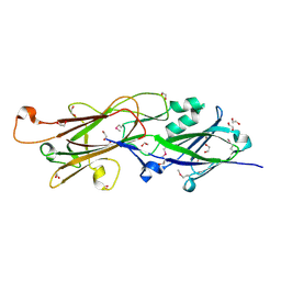
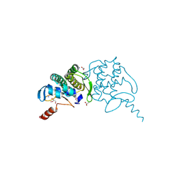
BU of 2h1t by Molmil
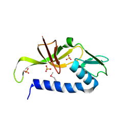
Crystal structure of a duf1089 family protein (pa1994) from pseudomonas aeruginosa at 1.80 A resolution
Descriptor: (4S)-2-METHYL-2,4-PENTANEDIOL, 1,2-ETHANEDIOL, Hypothetical protein
Authors:Joint Center for Structural Genomics (JCSG)
Deposit date:2006-05-16
Release date:2006-05-30
Last modified:2023-01-25
Method:X-RAY DIFFRACTION (1.8 Å)
Cite:The structure of the first representative of Pfam family PF06475 reveals a new fold with possible involvement in glycolipid metabolism.
Acta Crystallogr.,Sect.F, 66, 2010
2GVI
DownloadVisualize
BU of 2gvi by Molmil
Crystal structure of a putative formylmethanofuran dehydrogenase subunit e (ta1109) from thermoplasma acidophilum at 1.87 A resolution
Descriptor: 1,2-ETHANEDIOL, ACETIC ACID, UNKNOWN LIGAND, ...
Authors:Joint Center for Structural Genomics (JCSG)
Deposit date:2006-05-02
Release date:2006-06-13
Last modified:2023-01-25
Method:X-RAY DIFFRACTION (1.87 Å)
Cite:Structures of three members of Pfam PF02663 (FmdE) implicated in microbial methanogenesis reveal a conserved alpha+beta core domain and an auxiliary C-terminal treble-clef zinc finger.
Acta Crystallogr.,Sect.F, 66, 2010
4RDB
DownloadVisualize
BU of 4rdb by Molmil
Crystal structure of an immunoreactive 32 kDa antigen PG49 (PG_0181) from Porphyromonas gingivalis W83 at 1.45 A resolution (PSI Community Target, Nakayama)
Descriptor: 1,2-ETHANEDIOL, ACETONE, Immunoreactive 32 kDa antigen PG49
Authors:Joint Center for Structural Genomics (JCSG)
Deposit date:2014-09-18
Release date:2014-12-03
Last modified:2023-02-01
Method:X-RAY DIFFRACTION (1.45 Å)
Cite:A Distinct Type of Pilus from the Human Microbiome.
Cell, 165, 2016
4R8O
DownloadVisualize
BU of 4r8o by Molmil
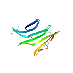
Crystal structure of a DUF3836 family protein (BVU_1206) from Bacteroides vulgatus ATCC 8482 at 2.50 A resolution
Descriptor: Uncharacterized protein
Authors:Joint Center for Structural Genomics (JCSG)
Deposit date:2014-09-02
Release date:2014-09-17
Last modified:2024-10-09
Method:X-RAY DIFFRACTION (2.5 Å)
Cite:Structures of single-layer beta-sheet proteins evolved from beta-hairpin repeats.
Protein Sci., 28, 2019
4R03
DownloadVisualize
BU of 4r03 by Molmil
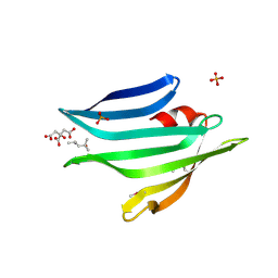
Crystal structure of a DUF3836 family protein (BDI_3222) from Parabacteroides distasonis ATCC 8503 at 1.50 A resolution
Descriptor: (4S)-2-METHYL-2,4-PENTANEDIOL, CITRIC ACID, PHOSPHATE ION, ...
Authors:Joint Center for Structural Genomics (JCSG)
Deposit date:2014-07-29
Release date:2014-08-27
Last modified:2023-02-01
Method:X-RAY DIFFRACTION (1.5 Å)
Cite:Structures of single-layer beta-sheet proteins evolved from beta-hairpin repeats.
Protein Sci., 28, 2019
4JG5
DownloadVisualize
BU of 4jg5 by Molmil
Crystal structure of a putative cell adhesion protein (BDI_3519) from Parabacteroides distasonis ATCC 8503 at 2.34 A resolution (PSI Community Target, Nakayama)
Descriptor: Putative cell adhesion protein
Authors:Joint Center for Structural Genomics (JCSG)
Deposit date:2013-02-28
Release date:2013-03-20
Last modified:2023-02-01
Method:X-RAY DIFFRACTION (2.34 Å)
Cite:A Distinct Type of Pilus from the Human Microbiome.
Cell, 165, 2016
4JRF
DownloadVisualize
BU of 4jrf by Molmil
Crystal structure of a putative cell adhesion protein (BACOVA_01548) from Bacteroides ovatus ATCC 8483 at 1.98 A resolution (PSI Community Target, Nakayama)
Descriptor: CALCIUM ION, CHLORIDE ION, SULFATE ION, ...
Authors:Joint Center for Structural Genomics (JCSG)
Deposit date:2013-03-21
Release date:2013-04-03
Last modified:2023-02-01
Method:X-RAY DIFFRACTION (1.98 Å)
Cite:A Distinct Type of Pilus from the Human Microbiome.
Cell, 165, 2016
4K4K
DownloadVisualize
BU of 4k4k by Molmil
Crystal structure of a putative cell adhesion protein (BACUNI_00621) from Bacteroides uniformis ATCC 8492 at 1.67 A resolution
Descriptor: CHLORIDE ION, Putative cell adhesion protein
Authors:Joint Center for Structural Genomics (JCSG)
Deposit date:2013-04-12
Release date:2013-05-01
Last modified:2023-02-01
Method:X-RAY DIFFRACTION (1.67 Å)
Cite:A Distinct Type of Pilus from the Human Microbiome.
Cell, 165, 2016
3H41
DownloadVisualize
BU of 3h41 by Molmil
CRYSTAL STRUCTURE OF A NLPC/P60 FAMILY PROTEIN (BCE_2878) FROM BACILLUS CEREUS ATCC 10987 AT 1.79 A RESOLUTION
Descriptor: ALANINE, D-GLUTAMIC ACID, NLP/P60 family protein, ...
Authors:Joint Center for Structural Genomics (JCSG)
Deposit date:2009-04-17
Release date:2009-05-05
Last modified:2023-02-01
Method:X-RAY DIFFRACTION (1.79 Å)
Cite:Structure of the gamma-D-glutamyl-L-diamino acid endopeptidase YkfC from Bacillus cereus in complex with L-Ala-[gamma]-D-Glu: insights into substrate recognition by NlpC/P60 cysteine peptidases
Acta Crystallogr.,Sect.F, 66, 2010
3H50
DownloadVisualize
BU of 3h50 by Molmil
CRYSTAL STRUCTURE OF A TETRACENOMYCIN POLYKETIDE SYNTHESIS PROTEIN (TCMJ) FROM XANTHOMONAS CAMPESTRIS PV. CAMPESTRIS AT 1.60 A RESOLUTION
Descriptor: ACETATE ION, Tetracenomycin polyketide synthesis protein, ZINC ION
Authors:Joint Center for Structural Genomics (JCSG)
Deposit date:2009-04-21
Release date:2009-05-05
Last modified:2023-02-01
Method:X-RAY DIFFRACTION (1.6 Å)
Cite:Conformational changes associated with the binding of zinc acetate at the putative active site of XcTcmJ, a cupin from Xanthomonas campestris pv. campestris.
Acta Crystallogr.,Sect.F, 66, 2010
3IRB
DownloadVisualize
BU of 3irb by Molmil
Crystal structure of protein with unknown function from DUF35 family (13815350) from SULFOLOBUS SOLFATARICUS at 1.80 A resolution
Descriptor: ACETIC ACID, SULFATE ION, ZINC ION, ...
Authors:Joint Center for Structural Genomics (JCSG)
Deposit date:2009-08-21
Release date:2009-09-01
Last modified:2023-02-01
Method:X-RAY DIFFRACTION (1.8 Å)
Cite:The structure of SSO2064, the first representative of Pfam family PF01796, reveals a novel two-domain zinc-ribbon OB-fold architecture with a potential acyl-CoA-binding role.
Acta Crystallogr.,Sect.F, 66, 2010
3HSA
DownloadVisualize
BU of 3hsa by Molmil
Crystal structure of pleckstrin homology domain (YP_926556.1) from SHEWANELLA AMAZONENSIS SB2B at 1.99 A resolution
Descriptor: DI(HYDROXYETHYL)ETHER, GLYCEROL, pleckstrin homology domain
Authors:Joint Center for Structural Genomics (JCSG)
Deposit date:2009-06-10
Release date:2009-06-23
Last modified:2023-02-01
Method:X-RAY DIFFRACTION (1.99 Å)
Cite:Bacterial pleckstrin homology domains: a prokaryotic origin for the PH domain.
J.Mol.Biol., 396, 2010
3K5J
DownloadVisualize
BU of 3k5j by Molmil
Crystal structure of Putative SUFU (suppressor of fused protein) homolog (YP_208451.1) from Neisseria gonorrhoeae FA 1090 at 1.40 A resolution
Descriptor: GLYCEROL, SULFATE ION, Suppressor of fused family protein
Authors:Joint Center for Structural Genomics (JCSG)
Deposit date:2009-10-07
Release date:2010-01-26
Last modified:2019-07-17
Method:X-RAY DIFFRACTION (1.4 Å)
Cite:The crystal structure of a bacterial Sufu-like protein defines a novel group of bacterial proteins that are similar to the N-terminal domain of human Sufu.
Protein Sci., 19, 2010
3HBZ
DownloadVisualize
BU of 3hbz by Molmil
Crystal structure of a putative glycoside hydrolase (bt_2081) from bacteroides thetaiotaomicron vpi-5482 at 2.05 A resolution
Descriptor: ACETATE ION, CACODYLATE ION, CALCIUM ION, ...
Authors:Joint Center for Structural Genomics (JCSG)
Deposit date:2009-05-05
Release date:2009-05-26
Last modified:2023-02-01
Method:X-RAY DIFFRACTION (2.05 Å)
Cite:Structure of Bacteroides thetaiotaomicron BT2081 at 2.05 A resolution: the first structural representative of a new protein family that may play a role in carbohydrate metabolism.
Acta Crystallogr.,Sect.F, 66, 2010
3H0N
DownloadVisualize
BU of 3h0n by Molmil
Crystal structure of a duf1470 family protein (jann_2411) from jannaschia sp. ccs1 at 1.45 A resolution
Descriptor: ACETATE ION, GLYCEROL, NICKEL (II) ION, ...
Authors:Joint Center for Structural Genomics (JCSG)
Deposit date:2009-04-09
Release date:2009-04-21
Last modified:2023-02-01
Method:X-RAY DIFFRACTION (1.45 Å)
Cite:The structure of Jann_2411 (DUF1470) from Jannaschia sp. at 1.45  resolution reveals a new fold (the ABATE domain) and suggests its possible role as a transcription regulator.
Acta Crystallogr.,Sect.F, 66, 2010
3KK7
DownloadVisualize
BU of 3kk7 by Molmil
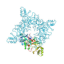
Crystal structure of Putative cell invasion protein with MAC/Perforin domain (NP_812351.1) from BACTERIODES THETAIOTAOMICRON VPI-5482 at 2.46 A resolution
Descriptor: (4S)-2-METHYL-2,4-PENTANEDIOL, 1,2-ETHANEDIOL, CHLORIDE ION, ...
Authors:Joint Center for Structural Genomics (JCSG)
Deposit date:2009-11-04
Release date:2009-11-24
Last modified:2023-02-01
Method:X-RAY DIFFRACTION (2.46 Å)
Cite:Structure of a membrane-attack complex/perforin (MACPF) family protein from the human gut symbiont Bacteroides thetaiotaomicron.
Acta Crystallogr.,Sect.F, 66, 2010
4QDG
DownloadVisualize
BU of 4qdg by Molmil
Crystal structure of a putative adhesin (BT2657) from Bacteroides thetaiotaomicron VPI-5482 at 2.20 A resolution (PSI Community Target, Nakayama)
Descriptor: 1,2-ETHANEDIOL, MAGNESIUM ION, Putative adhesin
Authors:Joint Center for Structural Genomics (JCSG)
Deposit date:2014-05-13
Release date:2014-06-11
Last modified:2023-02-01
Method:X-RAY DIFFRACTION (2.2 Å)
Cite:A Distinct Type of Pilus from the Human Microbiome.
Cell, 165, 2016
4Q5K
DownloadVisualize
BU of 4q5k by Molmil
Crystal structure of a N-acetylmuramoyl-L-alanine amidase (BACUNI_02947) from Bacteroides uniformis ATCC 8492 at 1.30 A resolution
Descriptor: (2R)-2-[[(1R,2S,3R,4R,5R)-4-acetamido-2-[(2S,3R,4R,5S,6R)-3-acetamido-6-(hydroxymethyl)-4,5-bis(oxidanyl)oxan-2-yl]oxy-6,8-dioxabicyclo[3.2.1]octan-3-yl]oxy]propanoic acid, SODIUM ION, Uncharacterized protein
Authors:Joint Center for Structural Genomics (JCSG)
Deposit date:2014-04-17
Release date:2014-05-21
Last modified:2023-12-06
Method:X-RAY DIFFRACTION (1.3 Å)
Cite:Structure-guided functional characterization of DUF1460 reveals a highly specific NlpC/P60 amidase family.
Structure, 22, 2014
4Q98
DownloadVisualize
BU of 4q98 by Molmil
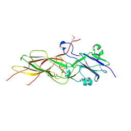
Crystal structure of a fimbrilin (fimA) from Porphyromonas gingivalis W83 at 1.30 A resolution (PSI Community Target, Nakayama)
Descriptor: Major fimbrial subunit protein, SODIUM ION
Authors:Joint Center for Structural Genomics (JCSG)
Deposit date:2014-04-29
Release date:2014-06-04
Last modified:2023-02-01
Method:X-RAY DIFFRACTION (1.3 Å)
Cite:A Distinct Type of Pilus from the Human Microbiome.
Cell, 165, 2016
4QB7
DownloadVisualize
BU of 4qb7 by Molmil
Crystal structure of a fimbrial protein (BVU_2522) from Bacteroides vulgatus ATCC 8482 at 2.55 A resolution
Descriptor: CHLORIDE ION, SULFATE ION, Uncharacterized protein
Authors:Joint Center for Structural Genomics (JCSG)
Deposit date:2014-05-06
Release date:2014-07-02
Last modified:2023-02-01
Method:X-RAY DIFFRACTION (2.55 Å)
Cite:A Distinct Type of Pilus from the Human Microbiome.
Cell, 165, 2016
4Q68
DownloadVisualize
BU of 4q68 by Molmil
Crystal structure of a N-acetylmuramoyl-L-alanine amidase (BACUNI_02947) from Bacteroides uniformis ATCC 8492 at 1.07 A resolution
Descriptor: 2-acetamido-2-deoxy-beta-D-glucopyranose, SODIUM ION, Uncharacterized protein
Authors:Joint Center for Structural Genomics (JCSG)
Deposit date:2014-04-21
Release date:2014-05-07
Last modified:2023-12-06
Method:X-RAY DIFFRACTION (1.07 Å)
Cite:Structure-guided functional characterization of DUF1460 reveals a highly specific NlpC/P60 amidase family.
Structure, 22, 2014
5CAG
DownloadVisualize
BU of 5cag by Molmil
Crystal structure of a putative adhesin (BACOVA_02677) from Bacteroides ovatus ATCC 8483 at 3.00 A resolution (PSI Community Target, Nakayama)
Descriptor: SULFATE ION, Uncharacterized protein
Authors:Joint Center for Structural Genomics (JCSG), Nakayama, K.
Deposit date:2015-06-29
Release date:2015-10-14
Last modified:2020-04-22
Method:X-RAY DIFFRACTION (3 Å)
Cite:A Distinct Type of Pilus from the Human Microbiome.
Cell, 165, 2016

225946

건을2024-10-09부터공개중

PDB statisticsPDBj update infoContact PDBjnumon