Loading
PDBj
MenuPDBj@FacebookPDBj@TwitterPDBj@YouTubewwPDB FoundationwwPDB
RCSB PDBPDBeBMRBAdv. SearchSearch help
Search by PDB author
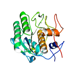
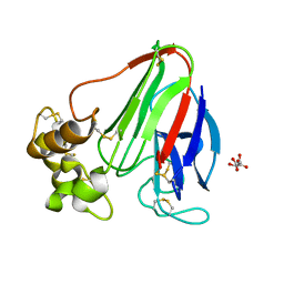
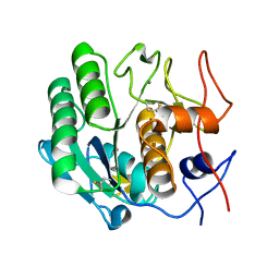
3DE1
DownloadVisualize
BU of 3de1 by Molmil
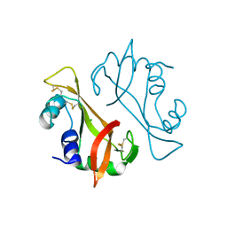
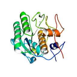
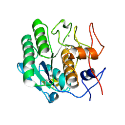
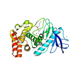
Proteinase K by LB nanotemplate method after the third step of high X-Ray dose on ESRF ID23-1 beamline
Descriptor: CALCIUM ION, Proteinase K
Authors:Pechkova, E, Tripathi, S.K, Nicolini, C.
Deposit date:2008-06-07
Release date:2009-06-09
Last modified:2024-10-09
Method:X-RAY DIFFRACTION (2 Å)
Cite:Radiation stability of proteinase K crystals grown by LB nanotemplate method
J.Struct.Biol., 168, 2009
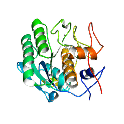
3DDZ
DownloadVisualize
BU of 3ddz by Molmil
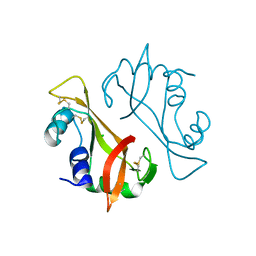
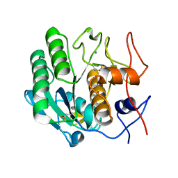
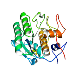
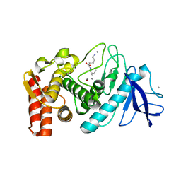
Proteinase K by LB nanotemplate method after the first step of high X-Ray dose on ESRF ID23-1 beamline
Descriptor: CALCIUM ION, Proteinase K
Authors:Pechkova, E, Tripathi, S.K, Nicolini, C.
Deposit date:2008-06-07
Release date:2009-06-09
Last modified:2024-11-06
Method:X-RAY DIFFRACTION (1.7 Å)
Cite:Radiation stability of proteinase K crystals grown by LB nanotemplate method
J.Struct.Biol., 168, 2009
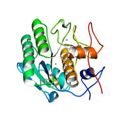
3D9Q
DownloadVisualize
BU of 3d9q by Molmil
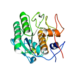
Proteinase K by LB nanotemplate method before high X-Ray dose on ESRF ID23-1 beamline
Descriptor: CALCIUM ION, Proteinase K
Authors:Pechkova, E, Tripathi, S.K, Nicolini, C.
Deposit date:2008-05-27
Release date:2009-06-02
Last modified:2024-11-06
Method:X-RAY DIFFRACTION (1.43 Å)
Cite:Radiation stability of proteinase K crystals grown by LB nanotemplate method
J.Struct.Biol., 168, 2009
3DE0
DownloadVisualize
BU of 3de0 by Molmil
Proteinase K by LB nanotemplate method after the second step of high X-Ray dose on ESRF ID23-1 beamline
Descriptor: CALCIUM ION, Proteinase K
Authors:Pechkova, E, Tripathi, S.K, Nicolini, C.
Deposit date:2008-06-07
Release date:2009-06-09
Last modified:2023-11-01
Method:X-RAY DIFFRACTION (1.9 Å)
Cite:Radiation stability of proteinase K crystals grown by LB nanotemplate method
J.Struct.Biol., 168, 2009
3I6H
DownloadVisualize
BU of 3i6h by Molmil
Ribonuclease A by LB nanotemplate method before high X-Ray dose on ESRF ID14-2 beamline
Descriptor: CHLORIDE ION, Ribonuclease pancreatic
Authors:Pechkova, E, Tripathi, S.K, Ravelli, R, McSweeney, S, Nicolini, C.
Deposit date:2009-07-07
Release date:2010-07-07
Last modified:2024-10-16
Method:X-RAY DIFFRACTION (1.3 Å)
Cite:Atomic structure and radiation resistance of langmuir-blodgett protein crystals
To be Published
3I34
DownloadVisualize
BU of 3i34 by Molmil
Proteinase K by LB Nanotemplate Method after high X-Ray dose on ID14-2 Beamline at ESRF
Descriptor: CALCIUM ION, MERCURY (II) ION, Proteinase K
Authors:Pechkova, E, Tripathi, S.K, Ravelli, R, McSweeney, S, Nicolini, C.
Deposit date:2009-06-30
Release date:2010-06-30
Last modified:2024-10-30
Method:X-RAY DIFFRACTION (1 Å)
Cite:Radiation damage study of Proteinase K at ID14-2 beamline at ESRF
To be Published
3I6F
DownloadVisualize
BU of 3i6f by Molmil
Ribonuclease A by Classical hanging drop method before high X-Ray dose on ESRF ID14-2 beamline
Descriptor: CHLORIDE ION, Ribonuclease pancreatic
Authors:Pechkova, E, Tripathi, S.K, Ravelli, R, McSweeney, S, Nicolini, C.
Deposit date:2009-07-07
Release date:2010-07-07
Last modified:2023-11-01
Method:X-RAY DIFFRACTION (1.3 Å)
Cite:Atomic structure and radiation resistance of langmuir-blodgett protein crystals
To be Published
4OXK
DownloadVisualize
BU of 4oxk by Molmil
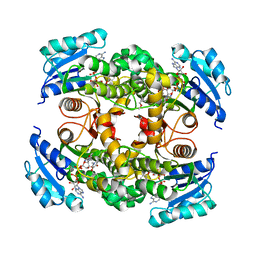
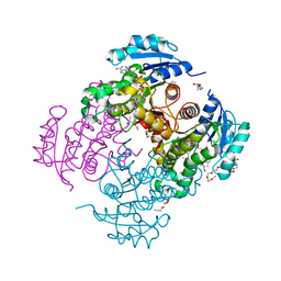
Multiple binding modes of inhibitor PT155 to the Mycobacterium tuberculosis enoyl-ACP reductase InhA within a tetramer
Descriptor: 3,6,9,12,15-pentaoxaoctadecan-17-amine, 5-(4-amino-2-methylphenoxy)-2-hexyl-4-hydroxy-1-methylpyridinium, Enoyl-[acyl-carrier-protein] reductase [NADH], ...
Authors:Li, H.J, Pan, P, Lai, C.T, Liu, N, Garcia-Diaz, M, Simmerling, C, Tonge, P.J.
Deposit date:2014-02-05
Release date:2014-04-30
Last modified:2023-09-27
Method:X-RAY DIFFRACTION (1.8429 Å)
Cite:A Structural and Energetic Model for the Slow-Onset Inhibition of the Mycobacterium tuberculosis Enoyl-ACP Reductase InhA.
Acs Chem.Biol., 9, 2014
3I67
DownloadVisualize
BU of 3i67 by Molmil
Ribonuclease A by LB nanotemplate method after high X-Ray dose on ESRF ID14-2 beamline
Descriptor: CHLORIDE ION, Ribonuclease pancreatic
Authors:Pechkova, E, Tripathi, S.K, Ravelli, R, McSweeney, S, Nicolini, C.
Deposit date:2009-07-06
Release date:2010-07-07
Last modified:2023-11-01
Method:X-RAY DIFFRACTION (1.3 Å)
Cite:Atomic structure and radiation resistance of langmuir-blodgett protein crystals
To be Published
4DJ1
DownloadVisualize
BU of 4dj1 by Molmil
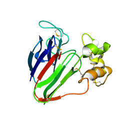
Thaumatin I by Langmuir-Blodgett Hanging Drop Method at 1.98A resolution for Unique Water Distribution
Descriptor: Thaumatin I
Authors:Pechkova, E, Sivozhelezov, V, Belmonte, L, Nicolini, C.
Deposit date:2012-02-01
Release date:2012-07-18
Last modified:2024-10-30
Method:X-RAY DIFFRACTION (1.98 Å)
Cite:Unique water distribution of Langmuir-Blodgett versus classical crystals.
J.Struct.Biol., 180, 2012
3DVQ
DownloadVisualize
BU of 3dvq by Molmil
Proteinase K by LB nanotemplate method before high X-Ray dose on ESRF ID14-2 beamline
Descriptor: CALCIUM ION, Proteinase K
Authors:Pechkova, E, Tripathi, S.K, Nicolini, C.
Deposit date:2008-07-20
Release date:2009-07-07
Last modified:2023-11-01
Method:X-RAY DIFFRACTION (1.02 Å)
Cite:Radiation damage in protein structural characterization by Synchrotron Radiation: State of the art and Nanotechnology-based perspective
To be Published
3DW1
DownloadVisualize
BU of 3dw1 by Molmil
Proteinase K by LB nanotemplate method after the third step high X-Ray dose on ESRF ID14-2 beamline
Descriptor: CALCIUM ION, Proteinase K
Authors:Pechkova, E, Tripathi, S.K, Nicolini, C.
Deposit date:2008-07-21
Release date:2009-07-07
Last modified:2024-10-09
Method:X-RAY DIFFRACTION (1.03 Å)
Cite:Radiation damage in protein structural characterization by Synchrotron Radiation: State of the art and Nanotechnology-based perspective
To be Published
3IJU
DownloadVisualize
BU of 3iju by Molmil
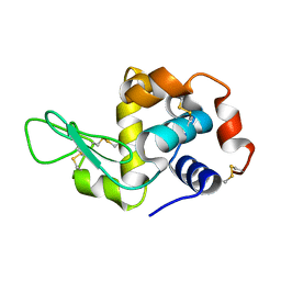
Chicken egg white lysozyme by highly ordered APA (Anodic Porous Alumina) nanotemplate crystallization method
Descriptor: Lysozyme C
Authors:Pechkova, E, Tripathi, S.K, Nicolini, C.
Deposit date:2009-08-05
Release date:2010-08-11
Last modified:2023-11-01
Method:X-RAY DIFFRACTION (1.6 Å)
Cite:Comparison of lysozyme crystals grown by APA and classical hanging drop method
To be Published
3DZN
DownloadVisualize
BU of 3dzn by Molmil
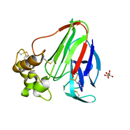
Thaumatin by LB nanotemplate method before high X-Ray dose on ESRF ID29 beamline
Descriptor: L(+)-TARTARIC ACID, Thaumatin-1
Authors:Tripathi, S, Pechkova, E, Nicolini, C.
Deposit date:2008-07-30
Release date:2009-07-07
Last modified:2024-10-30
Method:X-RAY DIFFRACTION (1.51 Å)
Cite:Radiation damage in protein structural characterization by Synchrotron Radiation: State of the art and Nanotechnology-based perspective
To be Published
3E0A
DownloadVisualize
BU of 3e0a by Molmil
Thaumatin by Classical hanging drop method after high X-Ray dose on ESRF ID29 beamline
Descriptor: L(+)-TARTARIC ACID, Thaumatin-1
Authors:Tripathi, S, Pechkova, E, Nicolini, C.
Deposit date:2008-07-31
Release date:2009-07-07
Last modified:2023-11-01
Method:X-RAY DIFFRACTION (1.51 Å)
Cite:Radiation damage in protein structural characterization by Synchrotron Radiation: State of the art and Nanotechnology-based perspective
To be Published
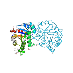
4MKN
DownloadVisualize
BU of 4mkn by Molmil
Crystal structure of chloroplastic triosephosphate isomerase from Chlamydomonas reinhardtii at 1.1 A of resolution
Descriptor: (4R)-2-METHYLPENTANE-2,4-DIOL, (4S)-2-METHYL-2,4-PENTANEDIOL, Triosephosphate isomerase
Authors:Fermani, S, Sciabolini, C, Zaffagnini, M, Lemaire, S.D.
Deposit date:2013-09-05
Release date:2014-01-01
Last modified:2023-09-20
Method:X-RAY DIFFRACTION (1.1 Å)
Cite:High-Resolution Crystal Structure and Redox Properties of Chloroplastic Triosephosphate Isomerase from Chlamydomonas reinhardtii.
Mol Plant, 7, 2014
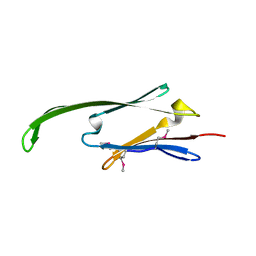
2Y22
DownloadVisualize
BU of 2y22 by Molmil
Human AlphaB-crystallin Domain (residues 67-157)
Descriptor: ALPHA-CRYSTALLIN B
Authors:Naylor, C.E, Bagneris, C, Clark, A.R, Keep, N.H, Slingsby, C.
Deposit date:2010-12-13
Release date:2011-03-02
Last modified:2024-10-09
Method:X-RAY DIFFRACTION (3.7 Å)
Cite:Crystal Structure of R120G Disease Mutant of Human Alphab-Crystallin Domain Dimer Shows Closure of a Groove
J.Mol.Biol., 408, 2011
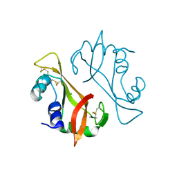
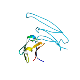
2Y1Y
DownloadVisualize
BU of 2y1y by Molmil
Human alphaB crystallin ACD(residues 71-157)
Descriptor: (4R)-2-METHYLPENTANE-2,4-DIOL, ALPHA-CRYSTALLIN B CHAIN,
Authors:Naylor, C.E, Bagneris, C, Clark, A.R, Keep, N.H, Slingsby, C.
Deposit date:2010-12-10
Release date:2011-03-02
Last modified:2023-12-20
Method:X-RAY DIFFRACTION (2 Å)
Cite:Crystal Structure of R120G Disease Mutant of Human Alphab-Crystallin Domain Dimer Shows Closure of a Groove
J.Mol.Biol., 408, 2011
3I1H
DownloadVisualize
BU of 3i1h by Molmil
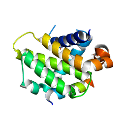
Crystal structure of human BFL-1 in complex with BAK BH3 peptide
Descriptor: Apoptosis regulator BAK, Protein BFL-1
Authors:Guan, R, Xiao, R, Zhao, L, Acton, T, White, E, Gelinas, C, Montelione, G.T, Northeast Structural Genomics Consortium (NESG)
Deposit date:2009-06-26
Release date:2009-07-14
Last modified:2023-09-06
Method:X-RAY DIFFRACTION (2.2 Å)
Cite:crystal structure of human BFL-1 in complex with BAK BH3 peptide
To be Published
3O5N
DownloadVisualize
BU of 3o5n by Molmil
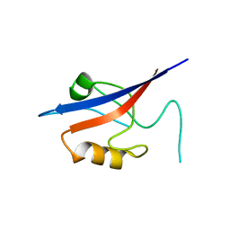
Tetrahydroquinoline carboxylates are potent inhibitors of the Shank PDZ domain, a putative target in autism disorders
Descriptor: (3aS,4R,9bR)-9-nitro-3a,4,5,9b-tetrahydro-3H-cyclopenta[c]quinoline-4,6-dicarboxylic acid, SH3 and multiple ankyrin repeat domains protein 3
Authors:Saupe, J, Roske, Y, Schillinger, C, Kamdem, N, Radetzki, S, Diehl, A, Oschkinat, H, Krause, G, Heinemann, U, Rademann, J.
Deposit date:2010-07-28
Release date:2011-06-15
Last modified:2024-02-21
Method:X-RAY DIFFRACTION (1.83 Å)
Cite:Discovery, structure-activity relationship studies, and crystal structure of nonpeptide inhibitors bound to the shank3 PDZ domain.
Chemmedchem, 6, 2011
3DE3
DownloadVisualize
BU of 3de3 by Molmil
Proteinase K by Classical hanging drop method before high X-Ray dose on ESRF ID23-1 beamline
Descriptor: CALCIUM ION, Proteinase K
Authors:Pechkova, E, Tripathi, S.K, Nicolini, C.
Deposit date:2008-06-08
Release date:2009-06-09
Last modified:2011-07-13
Method:X-RAY DIFFRACTION (1.43 Å)
Cite:Radiation stability of proteinase K crystals grown by LB nanotemplate method
J.Struct.Biol., 168, 2009
3DE5
DownloadVisualize
BU of 3de5 by Molmil
roteinase K by Classical hanging drop method after the second step of high X-Ray dose on ESRF ID23-1 beamline
Descriptor: CALCIUM ION, Proteinase K
Authors:Pechkova, E, Tripathi, S.K, Nicolini, C.
Deposit date:2008-06-08
Release date:2009-06-09
Last modified:2024-10-30
Method:X-RAY DIFFRACTION (2.1 Å)
Cite:Radiation stability of proteinase K crystals grown by LB nanotemplate method
J.Struct.Biol., 168, 2009
3DO1
DownloadVisualize
BU of 3do1 by Molmil
Thermolysin by Classical hanging drop method before high X-Ray dose on ESRF ID14-2 beamline
Descriptor: CALCIUM ION, LYSINE, Thermolysin, ...
Authors:Pechkova, E, Tripathi, S.K, Nicolini, C.
Deposit date:2008-07-03
Release date:2009-07-07
Last modified:2023-11-01
Method:X-RAY DIFFRACTION (1.33 Å)
Cite:Radiation damage in protein structural characterization by Synchrotron Radiation: State of the art and Nanotechnology-based perspective
To be Published
3DO0
DownloadVisualize
BU of 3do0 by Molmil
Thermolysin by classical hanging drop method after high X-Ray dose on esrf ID14-2 beamline
Descriptor: CALCIUM ION, LYSINE, Thermolysin, ...
Authors:Pechkova, E, Tripathi, S.K, Nicolini, C.
Deposit date:2008-07-03
Release date:2009-07-07
Last modified:2024-03-20
Method:X-RAY DIFFRACTION (1.36 Å)
Cite:Radiation damage in protein structural characterization by Synchrotron Radiation: State of the art and Nanotechnology-based perspective
To be Published
4OXN
DownloadVisualize
BU of 4oxn by Molmil
Substrate-like binding mode of inhibitor PT155 to the Mycobacterium tuberculosis enoyl-ACP reductase InhA
Descriptor: 3,6,9,12,15-pentaoxaoctadecan-17-amine, 4-(2-HYDROXYETHYL)-1-PIPERAZINE ETHANESULFONIC ACID, 5-(4-amino-2-methylphenoxy)-2-hexyl-4-hydroxy-1-methylpyridinium, ...
Authors:Li, H.J, Pan, P, Lai, C.T, Liu, N, Garcia-Diaz, M, Simmerling, C, Tonge, P.J.
Deposit date:2014-02-05
Release date:2014-04-30
Last modified:2023-09-27
Method:X-RAY DIFFRACTION (2.2926 Å)
Cite:A Structural and Energetic Model for the Slow-Onset Inhibition of the Mycobacterium tuberculosis Enoyl-ACP Reductase InhA.
Acs Chem.Biol., 9, 2014

227111

건을2024-11-06부터공개중

PDB statisticsPDBj update infoContact PDBjnumon