Loading
PDBj
MenuPDBj@FacebookPDBj@TwitterPDBj@YouTubewwPDB FoundationwwPDB
RCSB PDBPDBeBMRBAdv. SearchSearch help
Search by PDB author
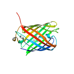
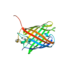
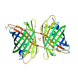
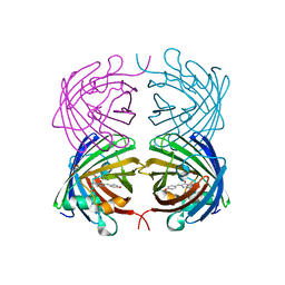
4RYW
DownloadVisualize
BU of 4ryw by Molmil
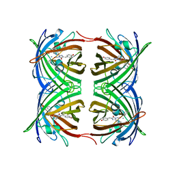
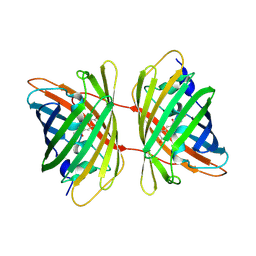
Crystal structure of the photoconverted green fluorescent protein NowGFP_conv (the variant of cyan Cerulean) at pH 7.0
Descriptor: GLYCEROL, NowGFP_conv
Authors:Pletnev, V.Z, Pletneva, N.V, Pletnev, S.V.
Deposit date:2014-12-17
Release date:2015-09-02
Last modified:2023-12-06
Method:X-RAY DIFFRACTION (2.5 Å)
Cite:Structure of the green fluorescent protein NowGFP with an anionic tryptophan-based chromophore.
Acta Crystallogr.,Sect.D, 71, 2015
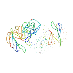
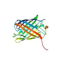
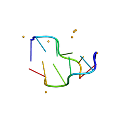
5EBI
DownloadVisualize
BU of 5ebi by Molmil
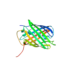
Crystal structure of a DNA-RNA chimera in complex with Ba2+ ions: a case of unusual multi-domain twinning
Descriptor: BARIUM ION, DNA/RNA (5'-D(*C)-R(P*G)-D(P*C)-R(P*G)-D(P*C)-R(P*G)-3')
Authors:Gilski, M, Drozdzal, P, Kierzek, R, Jaskolski, M.
Deposit date:2015-10-19
Release date:2016-02-10
Last modified:2024-01-10
Method:X-RAY DIFFRACTION (1.09 Å)
Cite:Atomic resolution structure of a chimeric DNA-RNA Z-type duplex in complex with Ba(2+) ions: a case of complicated multi-domain twinning.
Acta Crystallogr D Struct Biol, 72, 2016
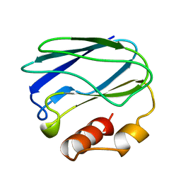
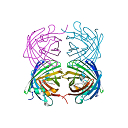
1PZC
DownloadVisualize
BU of 1pzc by Molmil
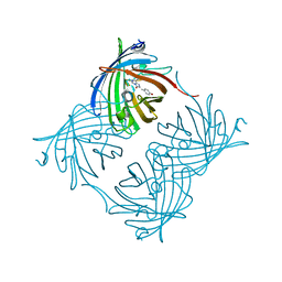
APO-PSEUDOAZURIN (METAL FREE PROTEIN)
Descriptor: PSEUDOAZURIN
Authors:Petratos, K.
Deposit date:1995-02-22
Release date:1995-09-15
Last modified:2024-02-14
Method:X-RAY DIFFRACTION (1.85 Å)
Cite:The crystal structure of apo-pseudoazurin from Alcaligenes faecalis S-6.
Febs Lett., 368, 1995
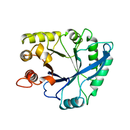
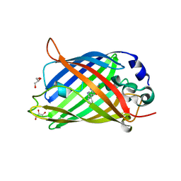
1NAR
DownloadVisualize
BU of 1nar by Molmil
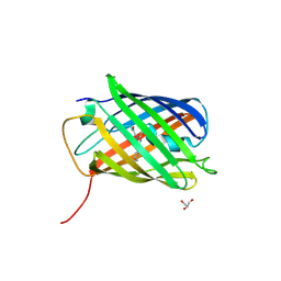
CRYSTAL STRUCTURE OF NARBONIN REFINED AT 1.8 ANGSTROMS RESOLUTION
Descriptor: NARBONIN
Authors:Hennig, M, Schlesier, B, Wilson, K.S.
Deposit date:1993-09-10
Release date:1994-01-31
Last modified:2024-02-14
Method:X-RAY DIFFRACTION (1.8 Å)
Cite:Crystal structure of narbonin at 1.8 A resolution.
Acta Crystallogr.,Sect.D, 51, 1995
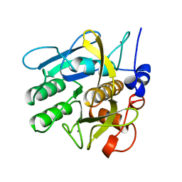
1SVN
DownloadVisualize
BU of 1svn by Molmil
SAVINASE
Descriptor: CALCIUM ION, SAVINASE (TM)
Authors:Betzel, C, Klupsch, S, Papendorf, G, Hastrup, S, Branner, S, Wilson, K.S.
Deposit date:1995-09-01
Release date:1996-10-14
Last modified:2024-03-06
Method:X-RAY DIFFRACTION (1.4 Å)
Cite:Crystal structure of the alkaline proteinase Savinase from Bacillus lentus at 1.4 A resolution.
J.Mol.Biol., 223, 1992
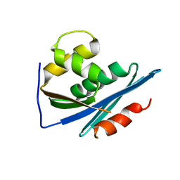
1RDH
DownloadVisualize
BU of 1rdh by Molmil
CRYSTALLOGRAPHIC ANALYSES OF AN ACTIVE HIV-1 RIBONUCLEASE H DOMAIN SHOW STRUCTURAL FEATURES THAT DISTINGUISH IT FROM THE INACTIVE FORM
Descriptor: HIV-1 REVERSE TRANSCRIPTASE (RIBONUCLEASE H DOMAIN)
Authors:Finzel, B.C, Chattopadhyay, D, Einspahr, H.M.
Deposit date:1993-03-05
Release date:1994-05-31
Last modified:2024-02-14
Method:X-RAY DIFFRACTION (2.8 Å)
Cite:Crystallographic analyses of an active HIV-1 ribonuclease H domain show structural features that distinguish it from the inactive form.
Acta Crystallogr.,Sect.D, 49, 1993
2OGR
DownloadVisualize
BU of 2ogr by Molmil
Crystal Structure of Yellow Fluorescent Protein from Zoanthus sp. at 1.8 A Resolution
Descriptor: FLUORESCENT PROTEIN FP538
Authors:Pletneva, N.V, Pletnev, S.V, Tikhonova, T.V, Pletnev, V.Z.
Deposit date:2007-01-08
Release date:2007-09-25
Last modified:2023-12-27
Method:X-RAY DIFFRACTION (1.8 Å)
Cite:Three-dimensional structure of yellow fluorescent protein zYFP538 from Zoanthus sp. at the resolution 1.8 angstrom
Bioorg.Khim., 33
3U8A
DownloadVisualize
BU of 3u8a by Molmil
Crystal structure of monomeric reversibly photoswitchable red fluorescent protein rsTagRFP in the OFF state
Descriptor: Fluorescent protein rsTagRFP
Authors:Pletnev, S.
Deposit date:2011-10-16
Release date:2012-02-22
Last modified:2012-05-09
Method:X-RAY DIFFRACTION (1.783 Å)
Cite:A structural basis for reversible photoswitching of absorbance spectra in red fluorescent protein rsTagRFP.
J.Mol.Biol., 417, 2012
2OJK
DownloadVisualize
BU of 2ojk by Molmil
Crystal Structure of Green Fluorescent Protein from Zoanthus sp at 2.2 A Resolution
Descriptor: GFP-like fluorescent chromoprotein FP506
Authors:Pletneva, N.V, Pletnev, S.V, Tikhonova, T.V, Pletnev, V.Z.
Deposit date:2007-01-12
Release date:2007-09-25
Last modified:2023-11-15
Method:X-RAY DIFFRACTION (2.2 Å)
Cite:Refined crystal structures of red and green fluorescent proteins from the button polyp Zoanthus.
Acta Crystallogr.,Sect.D, 63, 2007
3U8C
DownloadVisualize
BU of 3u8c by Molmil
Crystal structure of monomeric reversibly photoswitchable red fluorescent protein rsTagRFP in the ON state
Descriptor: Fluorescent protein rsTagRFP, GLYCEROL
Authors:Pletnev, S.
Deposit date:2011-10-16
Release date:2012-02-22
Last modified:2012-05-09
Method:X-RAY DIFFRACTION (2.188 Å)
Cite:A structural basis for reversible photoswitching of absorbance spectra in red fluorescent protein rsTagRFP.
J.Mol.Biol., 417, 2012
2OXI
DownloadVisualize
BU of 2oxi by Molmil
REFINED CRYSTAL STRUCTURE OF CU-SUBSTITUTED ALCOHOL DEHYDROGENASE AT 2.1 ANGSTROMS RESOLUTION
Descriptor: ALCOHOL DEHYDROGENASE, COPPER (II) ION, DIMETHYL SULFOXIDE, ...
Authors:Al-Karadaghi, S, Cedergren-Zeppezauer, E.S.
Deposit date:1993-11-08
Release date:1994-01-31
Last modified:2024-02-21
Method:X-RAY DIFFRACTION (2.1 Å)
Cite:Refined structure of Cu-substituted alcohol dehydrogenase at 2.1 A resolution.
Acta Crystallogr.,Sect.D, 51, 1995
3BXC
DownloadVisualize
BU of 3bxc by Molmil
Monomeric Far-red Fluorescent Protein mKate Crystallized at pH 9.0
Descriptor: Far-red fluorescent protein mKate
Authors:Pletnev, S, Pletneva, N, Pletnev, V.
Deposit date:2008-01-12
Release date:2008-07-22
Last modified:2019-07-24
Method:X-RAY DIFFRACTION (2.6 Å)
Cite:A Crystallographic Study of Bright Far-Red Fluorescent Protein mKate Reveals pH-induced cis-trans Isomerization of the Chromophore.
J.Biol.Chem., 283, 2008
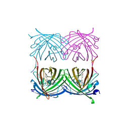
3BXB
DownloadVisualize
BU of 3bxb by Molmil
Monomeric Far-red Fluorescent Protein mKate Crystallized at pH 7.0
Descriptor: Far-red fluorescent protein mKate
Authors:Pletnev, S, Pletneva, N, Pletnev, V.
Deposit date:2008-01-12
Release date:2008-07-22
Last modified:2019-07-24
Method:X-RAY DIFFRACTION (2.6 Å)
Cite:A Crystallographic Study of Bright Far-Red Fluorescent Protein mKate Reveals pH-induced cis-trans Isomerization of the Chromophore.
J.Biol.Chem., 283, 2008
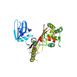
3EE1
DownloadVisualize
BU of 3ee1 by Molmil
Novel fold of VirA, a type III secretion system effector protein from Shigella flexneri
Descriptor: Effector protein virA
Authors:Davis, J.S.
Deposit date:2008-09-03
Release date:2008-12-16
Last modified:2017-10-25
Method:X-RAY DIFFRACTION (3.01 Å)
Cite:Novel fold of VirA, a type III secretion system effector protein from Shigella flexneri
Protein Sci., 17, 2008
3GB3
DownloadVisualize
BU of 3gb3 by Molmil
X-ray structure of genetically encoded photosensitizer KillerRed in native form
Descriptor: KillerRed, SULFATE ION
Authors:Pletnev, S, Pletneva, N.V, Pletnev, V.Z.
Deposit date:2009-02-18
Release date:2009-09-08
Last modified:2024-10-09
Method:X-RAY DIFFRACTION (1.75 Å)
Cite:Structural basis for phototoxicity of the genetically encoded photosensitizer KillerRed.
J.Biol.Chem., 284, 2009
2CST
DownloadVisualize
BU of 2cst by Molmil
CRYSTAL STRUCTURE OF THE CLOSED FORM OF CHICKEN CYTOSOLIC ASPARTATE AMINOTRANSFERASE AT 1.9 ANGSTROMS RESOLUTION
Descriptor: ASPARTATE AMINOTRANSFERASE, MALEIC ACID, PYRIDOXAL-5'-PHOSPHATE
Authors:Malashkevich, V.N, Strokopytov, B.V, Borisov, V.V.
Deposit date:1994-09-06
Release date:1994-11-30
Last modified:2017-11-29
Method:X-RAY DIFFRACTION (1.9 Å)
Cite:Crystal structure of the closed form of chicken cytosolic aspartate aminotransferase at 1.9 A resolution.
J.Mol.Biol., 247, 1995
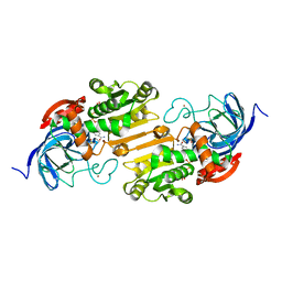
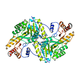
1B5F
DownloadVisualize
BU of 1b5f by Molmil
NATIVE CARDOSIN A FROM CYNARA CARDUNCULUS L.
Descriptor: PROTEIN (CARDOSIN A), alpha-D-mannopyranose-(1-3)-[alpha-D-mannopyranose-(1-6)]beta-D-mannopyranose-(1-4)-2-acetamido-2-deoxy-beta-D-glucopyranose-(1-4)-[alpha-L-fucopyranose-(1-3)]2-acetamido-2-deoxy-beta-D-glucopyranose, alpha-D-mannopyranose-(1-3)-beta-D-mannopyranose-(1-4)-2-acetamido-2-deoxy-beta-D-glucopyranose-(1-4)-[alpha-L-fucopyranose-(1-3)]2-acetamido-2-deoxy-beta-D-glucopyranose, ...
Authors:Frazao, C, Bento, I, Carrondo, M.A.
Deposit date:1999-01-06
Release date:1999-01-13
Last modified:2024-10-30
Method:X-RAY DIFFRACTION (1.72 Å)
Cite:Crystal structure of cardosin A, a glycosylated and Arg-Gly-Asp-containing aspartic proteinase from the flowers of Cynara cardunculus L.
J.Biol.Chem., 274, 1999
2ICR
DownloadVisualize
BU of 2icr by Molmil
Red fluorescent protein zRFP574 from Zoanthus sp.
Descriptor: Red fluorescent protein zoanRFP, SULFATE ION
Authors:Pletnev, S, Pletneva, N, Tikhonova, T, Pletnev, V.
Deposit date:2006-09-13
Release date:2007-10-02
Last modified:2023-11-15
Method:X-RAY DIFFRACTION (1.51 Å)
Cite:Refined crystal structures of red and green fluorescent proteins from the button polyp Zoanthus.
Acta Crystallogr.,Sect.D, 63, 2007
2BD1
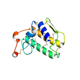
DownloadVisualize
BU of 2bd1 by Molmil
A possible role of the second calcium ion in interfacial binding: Atomic and medium resolution crystal structures of the quadruple mutant of phospholipase A2
Descriptor: (4S)-2-METHYL-2,4-PENTANEDIOL, CALCIUM ION, Phospholipase A2
Authors:Sekar, K, Velmurugan, D, Tsai, M.D.
Deposit date:2005-10-19
Release date:2006-07-04
Last modified:2024-10-30
Method:X-RAY DIFFRACTION (1.9 Å)
Cite:Suggestive evidence for the involvement of the second calcium and surface loop in interfacial binding: monoclinic and trigonal crystal structures of a quadruple mutant of phospholipase A(2).
Acta Crystallogr.,Sect.D, 62, 2006
1C51
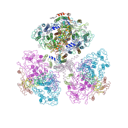
DownloadVisualize
BU of 1c51 by Molmil
PHOTOSYNTHETIC REACTION CENTER AND CORE ANTENNA SYSTEM (TRIMERIC), ALPHA CARBON ONLY
Descriptor: CHLOROPHYLL A, IRON/SULFUR CLUSTER, PHYLLOQUINONE, ...
Authors:Klukas, O, Schubert, W.D, Jordan, P, Krauss, N, Fromme, P, Witt, H.T, Saenger, W.
Deposit date:1999-10-21
Release date:2000-03-31
Last modified:2023-12-27
Method:X-RAY DIFFRACTION (4 Å)
Cite:Photosystem I, an improved model of the stromal subunits PsaC, PsaD, and PsaE.
J.Biol.Chem., 274, 1999
3GL4
DownloadVisualize
BU of 3gl4 by Molmil
X-ray structure of photobleached killerred
Descriptor: KillerRed
Authors:Pletnev, S, Pletneva, N.V, Pletnev, V.Z.
Deposit date:2009-03-11
Release date:2009-09-08
Last modified:2023-11-15
Method:X-RAY DIFFRACTION (2.15 Å)
Cite:Structural basis for phototoxicity of the genetically encoded photosensitizer KillerRed.
J.Biol.Chem., 284, 2009
2ALD
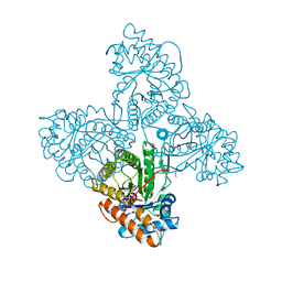
DownloadVisualize
BU of 2ald by Molmil
HUMAN MUSCLE ALDOLASE
Descriptor: FRUCTOSE-BISPHOSPHATE ALDOLASE
Authors:Dalby, A.R, Littlechild, J.A.
Deposit date:1998-10-21
Release date:1999-04-20
Last modified:2024-02-14
Method:X-RAY DIFFRACTION (2.1 Å)
Cite:Crystal structure of human muscle aldolase complexed with fructose 1,6-bisphosphate: mechanistic implications.
Protein Sci., 8, 1999
4JEO
DownloadVisualize
BU of 4jeo by Molmil
Crystal structure of red fluorescent protein lanRFPdam exposed to prolonged X-ray irradiation
Descriptor: GLYCEROL, Red fluorescent protein blFP-R5
Authors:Pletnev, V.Z, Pletneva, N.V, Pletnev, S.V.
Deposit date:2013-02-27
Release date:2013-09-18
Last modified:2023-12-06
Method:X-RAY DIFFRACTION (2.35 Å)
Cite:Structure of the red fluorescent protein from a lancelet (Branchiostoma lanceolatum): a novel GYG chromophore covalently bound to a nearby tyrosine.
Acta Crystallogr.,Sect.D, 69, 2013
4KPI
DownloadVisualize
BU of 4kpi by Molmil
Rotational order-disorder structure of reversibly photoswitchable red fluorescent protein rsTagRFP
Descriptor: Reversibly photoswitchable red fluorescent protein rsTagRFP
Authors:Pletnev, S.
Deposit date:2013-05-13
Release date:2014-02-26
Last modified:2023-12-06
Method:X-RAY DIFFRACTION (1.58 Å)
Cite:The rotational order-disorder structure of the reversibly photoswitchable red fluorescent protein rsTagRFP.
Acta Crystallogr.,Sect.D, 70, 2014
4JGE
DownloadVisualize
BU of 4jge by Molmil
Crystal structure of red fluorescent gene-engineered variant with improved folding - lanRFP_delS83 (Branchiostoma lanceolatum)
Descriptor: GLYCEROL, Red fluorescent protein blFP-R5
Authors:Pletnev, V.Z, Pletneva, N.V, Pletnev, S.V.
Deposit date:2013-03-01
Release date:2013-09-18
Last modified:2023-12-06
Method:X-RAY DIFFRACTION (1.94 Å)
Cite:Structure of the red fluorescent protein from a lancelet (Branchiostoma lanceolatum): a novel GYG chromophore covalently bound to a nearby tyrosine.
Acta Crystallogr.,Sect.D, 69, 2013

226707

건을2024-10-30부터공개중

PDB statisticsPDBj update infoContact PDBjnumon