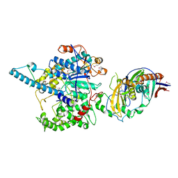
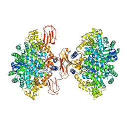
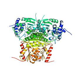
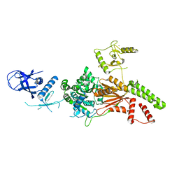
1G8L
 
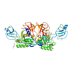 | | CRYSTAL STRUCTURE OF ESCHERICHIA COLI MOEA | | 分子名称: | GLYCEROL, MOLYBDOPTERIN BIOSYNTHESIS MOEA PROTEIN | | 著者 | Xiang, S, Nichols, J, Rajagopalan, K.V, Schindelin, H. | | 登録日 | 2000-11-17 | | 公開日 | 2001-05-02 | | 最終更新日 | 2024-02-07 | | 実験手法 | X-RAY DIFFRACTION (1.95 Å) | | 主引用文献 | The crystal structure of Escherichia coli MoeA and its relationship to the multifunctional protein gephyrin.
Structure, 9, 2001
|
|
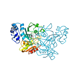
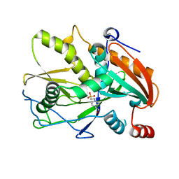
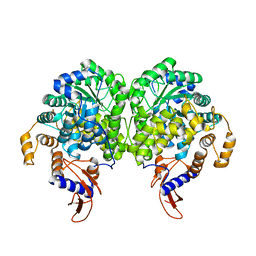
1G8R
 | | MOEA | | 分子名称: | GLYCEROL, MOLYBDOPTERIN BIOSYNTHESIS MOEA PROTEIN | | 著者 | Xiang, S, Nichols, J, Rajagopalan, K.V, Schindelin, H. | | 登録日 | 2000-11-20 | | 公開日 | 2001-05-02 | | 最終更新日 | 2023-08-09 | | 実験手法 | X-RAY DIFFRACTION (2.65 Å) | | 主引用文献 | The crystal structure of Escherichia coli MoeA and its relationship to the multifunctional protein gephyrin.
Structure, 9, 2001
|
|
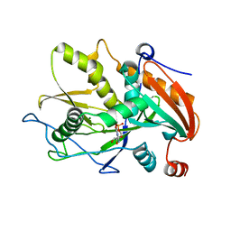
1ALN
 | |
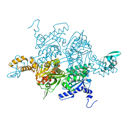
2DFK
 | | Crystal structure of the CDC42-Collybistin II complex | | 分子名称: | GLYCEROL, SULFATE ION, cell division cycle 42 isoform 1, ... | | 著者 | Xiang, S, Kim, E.Y, Connelly, J.J, Nassar, N, Kirsch, J, Winking, J, Schwarz, G, Schindelin, H. | | 登録日 | 2006-03-02 | | 公開日 | 2006-05-02 | | 最終更新日 | 2023-10-25 | | 実験手法 | X-RAY DIFFRACTION (2.15 Å) | | 主引用文献 | The Crystal Structure of Cdc42 in Complex with Collybistin II, a Gephyrin-interacting Guanine Nucleotide Exchange Factor.
J.Mol.Biol., 359, 2006
|
|
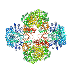
1AF2
 | |
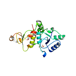
1CTT
 | | TRANSITION-STATE SELECTIVITY FOR A SINGLE OH GROUP DURING CATALYSIS BY CYTIDINE DEAMINASE | | 分子名称: | 3,4-DIHYDRO-1H-PYRIMIDIN-2-ONE NUCLEOSIDE, CYTIDINE DEAMINASE, ZINC ION | | 著者 | Xiang, S, Short, S.A, Wolfenden, R, Carter, C.W. | | 登録日 | 1995-02-11 | | 公開日 | 1995-05-08 | | 最終更新日 | 2024-02-07 | | 実験手法 | X-RAY DIFFRACTION (2.2 Å) | | 主引用文献 | Transition-state selectivity for a single hydroxyl group during catalysis by cytidine deaminase.
Biochemistry, 34, 1995
|
|
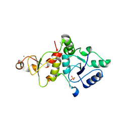
1CTU
 | | TRANSITION-STATE SELECTIVITY FOR A SINGLE OH GROUP DURING CATALYSIS BY CYTIDINE DEAMINASE | | 分子名称: | 4-HYDROXY-3,4-DIHYDRO-ZEBULARINE, CYTIDINE DEAMINASE, ZINC ION | | 著者 | Xiang, S, Short, S.A, Wolfenden, R, Carter, C.W. | | 登録日 | 1995-02-11 | | 公開日 | 1995-05-08 | | 最終更新日 | 2024-02-07 | | 実験手法 | X-RAY DIFFRACTION (2.3 Å) | | 主引用文献 | Transition-state selectivity for a single hydroxyl group during catalysis by cytidine deaminase.
Biochemistry, 34, 1995
|
|
3FQG
 | | Crystal Structure of the S. pombe Rai1 | | 分子名称: | MAGNESIUM ION, Protein din1 | | 著者 | Xiang, S, Tong, L. | | 登録日 | 2009-01-07 | | 公開日 | 2009-02-03 | | 最終更新日 | 2024-02-21 | | 実験手法 | X-RAY DIFFRACTION (2 Å) | | 主引用文献 | Structure and function of the 5'-->3' exoribonuclease Rat1 and its activating partner Rai1.
Nature, 458, 2009
|
|
3FQD
 | |
3FQI
 | | Crystal Structure of the Mouse Dom3Z | | 分子名称: | 4-(2-HYDROXYETHYL)-1-PIPERAZINE ETHANESULFONIC ACID, MAGNESIUM ION, Protein Dom3Z | | 著者 | Xiang, S, Tong, L. | | 登録日 | 2009-01-07 | | 公開日 | 2009-02-03 | | 最終更新日 | 2024-02-21 | | 実験手法 | X-RAY DIFFRACTION (2.013 Å) | | 主引用文献 | Structure and function of the 5'-->3' exoribonuclease Rat1 and its activating partner Rai1.
Nature, 458, 2009
|
|
3FQJ
 | |
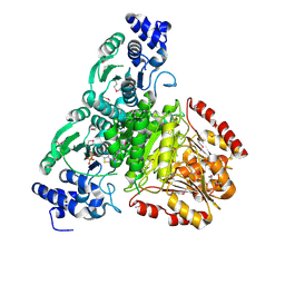
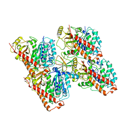
3BG5
 | | Crystal Structure of Staphylococcus Aureus Pyruvate Carboxylase | | 分子名称: | 5-(HEXAHYDRO-2-OXO-1H-THIENO[3,4-D]IMIDAZOL-6-YL)PENTANAL, ADENOSINE-5'-TRIPHOSPHATE, MANGANESE (II) ION, ... | | 著者 | Xiang, S, Tong, L. | | 登録日 | 2007-11-26 | | 公開日 | 2008-02-26 | | 最終更新日 | 2023-11-15 | | 実験手法 | X-RAY DIFFRACTION (2.8 Å) | | 主引用文献 | Crystal structures of human and Staphylococcus aureus pyruvate carboxylase and molecular insights into the carboxyltransfer reaction.
Nat.Struct.Mol.Biol., 15, 2008
|
|
3BG3
 | |
3BG9
 | |
3K8X
 | | Crystal structure of the carboxyltransferase domain of acetyl-coenzyme A carboxylase in complex with tepraloxydim | | 分子名称: | (5S)-2-[(1E)-N-{[(2E)-3-chloroprop-2-en-1-yl]oxy}propanimidoyl]-3-hydroxy-5-(tetrahydro-2H-pyran-4-yl)cyclohex-2-en-1-one, Acetyl-CoA carboxylase | | 著者 | Xiang, S, Callaghan, M.M, Watson, K.G, Tong, L. | | 登録日 | 2009-10-15 | | 公開日 | 2009-12-01 | | 最終更新日 | 2024-02-21 | | 実験手法 | X-RAY DIFFRACTION (2.3 Å) | | 主引用文献 | A different mechanism for the inhibition of the carboxyltransferase domain of acetyl-coenzyme A carboxylase by tepraloxydim.
Proc.Natl.Acad.Sci.USA, 106, 2009
|
|
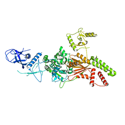
2O1S
 | | 1-deoxy-D-xylulose 5-phosphate synthase (DXS) from Escherichia coli | | 分子名称: | 1-deoxy-D-xylulose-5-phosphate synthase, DIPHOSPHATE, MAGNESIUM ION, ... | | 著者 | Xiang, S, Usunow, G, Lange, G, Busch, M, Tong, L. | | 登録日 | 2006-11-29 | | 公開日 | 2006-12-26 | | 最終更新日 | 2023-12-27 | | 実験手法 | X-RAY DIFFRACTION (2.4 Å) | | 主引用文献 | Crystal Structure of 1-Deoxy-D-xylulose 5-Phosphate Synthase, a Crucial Enzyme for Isoprenoids Biosynthesis.
J.Biol.Chem., 282, 2007
|
|
2O1X
 | | 1-deoxy-D-xylulose 5-phosphate synthase (DXS) from Deinococcus radiodurans | | 分子名称: | 1-deoxy-D-xylulose-5-phosphate synthase, MAGNESIUM ION, THIAMINE DIPHOSPHATE | | 著者 | Xiang, S, Usunow, G, Lange, G, Busch, M, Tong, L. | | 登録日 | 2006-11-29 | | 公開日 | 2006-12-26 | | 最終更新日 | 2023-12-27 | | 実験手法 | X-RAY DIFFRACTION (2.9 Å) | | 主引用文献 | Crystal Structure of 1-Deoxy-D-xylulose 5-Phosphate Synthase, a Crucial Enzyme for Isoprenoids Biosynthesis.
J.Biol.Chem., 282, 2007
|
|
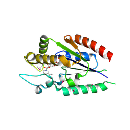
8IR2
 | | Crystal structure of the SLF1 BRCT domain in complex with a Rad18 peptide containing pS442 and pS444 | | 分子名称: | CHLORIDE ION, ISOPROPYL ALCOHOL, MAGNESIUM ION, ... | | 著者 | Xiang, S, Huang, W, Qiu, F. | | 登録日 | 2023-03-17 | | 公開日 | 2023-11-15 | | 実験手法 | X-RAY DIFFRACTION (1.75 Å) | | 主引用文献 | Structural insights into Rad18 targeting by the SLF1 BRCT domains.
J.Biol.Chem., 299, 2023
|
|
8IR4
 | | Crystal structure of the SLF1 BRCT domain in complex with a Rad18 peptide containing pS442 | | 分子名称: | CHLORIDE ION, ISOPROPYL ALCOHOL, MAGNESIUM ION, ... | | 著者 | Xiang, S, Huang, W, Qiu, F. | | 登録日 | 2023-03-17 | | 公開日 | 2023-11-15 | | 実験手法 | X-RAY DIFFRACTION (1.62 Å) | | 主引用文献 | Structural insights into Rad18 targeting by the SLF1 BRCT domains.
J.Biol.Chem., 299, 2023
|
|
5ZK1
 | | Crystal Structure of the CRTC2(SeMet)-CREB-CRE complex | | 分子名称: | CREB-regulated transcription coactivator 2, Cyclic AMP-responsive element-binding protein 1, DNA (5'-D(*CP*TP*TP*GP*GP*CP*TP*GP*AP*CP*GP*TP*CP*AP*GP*CP*CP*AP*AP*G)-3'), ... | | 著者 | Xiang, S, Zhai, L, Valencia-Swain, J. | | 登録日 | 2018-03-22 | | 公開日 | 2018-06-20 | | 最終更新日 | 2023-11-22 | | 実験手法 | X-RAY DIFFRACTION (3.05 Å) | | 主引用文献 | Structural Insights into the CRTC2-CREB Complex Assembly on CRE.
J. Mol. Biol., 430, 2018
|
|
5ZKO
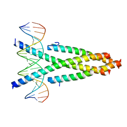 | | Crystal structure of the CRTC2-CREB-CRE complex | | 分子名称: | CREB-regulated transcription coactivator 2, Cyclic AMP-responsive element-binding protein 1, DNA (5'-D(*CP*TP*TP*GP*GP*CP*TP*GP*AP*CP*GP*TP*CP*AP*GP*CP*CP*AP*AP*G)-3') | | 著者 | Xiang, S, Zhai, L, Valecia-Swain, J. | | 登録日 | 2018-03-24 | | 公開日 | 2018-06-20 | | 最終更新日 | 2023-11-22 | | 実験手法 | X-RAY DIFFRACTION (3.05 Å) | | 主引用文献 | Structural Insights into the CRTC2-CREB Complex Assembly on CRE.
J. Mol. Biol., 430, 2018
|
|
2QG6
 | |
6QUS
 | | HsCKK (human CAMSAP1) decorated 13pf taxol-GDP microtubule | | 分子名称: | Calmodulin-regulated spectrin-associated protein 1, GUANOSINE-5'-DIPHOSPHATE, GUANOSINE-5'-TRIPHOSPHATE, ... | | 著者 | Atherton, J.M, Luo, Y, Xiang, S, Yang, C, Jiang, K, Stangier, M, Vemu, A, Cook, A, Wang, S, Roll-Mecak, A, Steinmetz, M.O, Akhmanova, A, Baldus, M, Moores, C.A. | | 登録日 | 2019-02-28 | | 公開日 | 2019-11-27 | | 最終更新日 | 2024-05-15 | | 実験手法 | ELECTRON MICROSCOPY (3.7 Å) | | 主引用文献 | Structural determinants of microtubule minus end preference in CAMSAP CKK domains.
Nat Commun, 10, 2019
|
|
6L8O
 | |
6L8N
 | | Crystal structure of the K. lactis Rad5 | | 分子名称: | DNA repair protein RAD5, ZINC ION | | 著者 | Shen, M, Xiang, S. | | 登録日 | 2019-11-06 | | 公開日 | 2020-11-11 | | 最終更新日 | 2024-04-03 | | 実験手法 | X-RAY DIFFRACTION (3.6 Å) | | 主引用文献 | Structural basis for the multi-activity factor Rad5 in replication stress tolerance.
Nat Commun, 12, 2021
|
|


