9IUZ
 
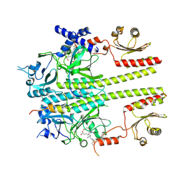 | | Constitutively active mutant(Y276H) of Arabidopsis phytochrome B(phyB) in complex with phytochrome-interacting factor 6(PIF6) | | 分子名称: | 3-[5-[[(3~{R},4~{R})-3-ethyl-4-methyl-5-oxidanylidene-3,4-dihydropyrrol-2-yl]methyl]-2-[[5-[(4-ethyl-3-methyl-5-oxidanylidene-pyrrol-2-yl)methyl]-3-(3-hydroxy-3-oxopropyl)-4-methyl-1~{H}-pyrrol-2-yl]methyl]-4-methyl-1~{H}-pyrrol-3-yl]propanoic acid, Phytochrome B, Phytochrome-interacting factor 6 | | 著者 | Wang, Z, Wang, W, Zhao, D, Song, Y, Xu, B, Zhao, J, Wang, J. | | 登録日 | 2024-07-22 | | 公開日 | 2024-10-02 | | 最終更新日 | 2024-10-09 | | 実験手法 | ELECTRON MICROSCOPY (3.19 Å) | | 主引用文献 | Light-induced remodeling of phytochrome B enables signal transduction by phytochrome-interacting factor.
Cell, 2024
|
|
8YB4
 
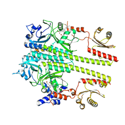 | | Pfr conformer of Arabidopsis thaliana phytochrome B in complex with phytochrome-interacting factor 6 | | 分子名称: | 3-[5-[[(3~{R},4~{R})-3-ethyl-4-methyl-5-oxidanylidene-3,4-dihydropyrrol-2-yl]methyl]-2-[[5-[(4-ethyl-3-methyl-5-oxidanylidene-pyrrol-2-yl)methyl]-3-(3-hydroxy-3-oxopropyl)-4-methyl-1~{H}-pyrrol-2-yl]methyl]-4-methyl-1~{H}-pyrrol-3-yl]propanoic acid, phytochrome B, phytochrome-interacting factor 6 | | 著者 | Wang, Z, Wang, W, Zhao, D, Song, Y, Xu, B, Zhao, J, Wang, J. | | 登録日 | 2024-02-11 | | 公開日 | 2024-10-02 | | 最終更新日 | 2024-10-09 | | 実験手法 | ELECTRON MICROSCOPY (3.1 Å) | | 主引用文献 | Light-induced remodeling of phytochrome B enables signal transduction by phytochrome-interacting factor.
Cell, 2024
|
|
4NA4
 
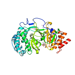 | | Crystal structure of mouse poly(ADP-ribose) glycohydrolase (PARG) catalytic domain with ADP-HPD | | 分子名称: | 5'-O-[(S)-{[(S)-{[(2R,3R,4S)-3,4-DIHYDROXYPYRROLIDIN-2-YL]METHOXY}(HYDROXY)PHOSPHORYL]OXY}(HYDROXY)PHOSPHORYL]ADENOSINE, IODIDE ION, Poly(ADP-ribose) glycohydrolase | | 著者 | Wang, Z, Cheng, Z, Xu, W. | | 登録日 | 2013-10-21 | | 公開日 | 2014-01-29 | | 最終更新日 | 2024-10-30 | | 実験手法 | X-RAY DIFFRACTION (2.5 Å) | | 主引用文献 | Crystallographic and biochemical analysis of the mouse poly(ADP-ribose) glycohydrolase.
Plos One, 9, 2014
|
|
7QA2
 
 | |
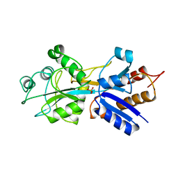
1IXI
 
 | |
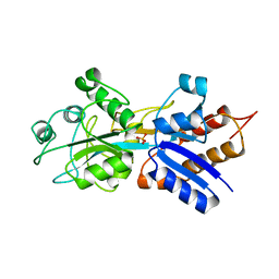
1IXG
 
 | |
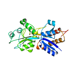
1IXH
 
 | |
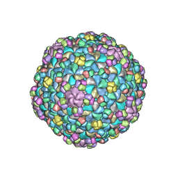
5WK1
 
 | |
8JRN
 
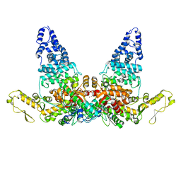 | | Structure of E6AP-E6 complex in Att1 state | | 分子名称: | Protein E6, Ubiquitin-protein ligase E3A, ZINC ION | | 著者 | Wang, Z, Yu, X. | | 登録日 | 2023-06-17 | | 公開日 | 2024-06-05 | | 実験手法 | ELECTRON MICROSCOPY (2.6 Å) | | 主引用文献 | Structural insights into the functional mechanism of the ubiquitin ligase E6AP.
Nat Commun, 15, 2024
|
|
8JRQ
 
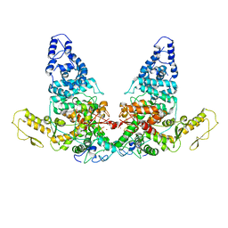 | | Structure of E6AP-E6 complex in Det1 state | | 分子名称: | Protein E6, Ubiquitin-protein ligase E3A, ZINC ION | | 著者 | Wang, Z, Yu, X. | | 登録日 | 2023-06-17 | | 公開日 | 2024-06-05 | | 実験手法 | ELECTRON MICROSCOPY (4.15 Å) | | 主引用文献 | Structural insights into the functional mechanism of the ubiquitin ligase E6AP.
Nat Commun, 15, 2024
|
|
8JRP
 
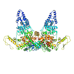 | | Structure of E6AP-E6 complex in Att3 state | | 分子名称: | Protein E6, Ubiquitin-protein ligase E3A, ZINC ION | | 著者 | Wang, Z, Yu, X. | | 登録日 | 2023-06-17 | | 公開日 | 2024-06-05 | | 実験手法 | ELECTRON MICROSCOPY (3.58 Å) | | 主引用文献 | Structural insights into the functional mechanism of the ubiquitin ligase E6AP.
Nat Commun, 15, 2024
|
|
8JRR
 
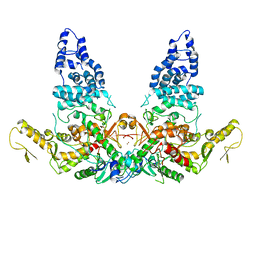 | | Structure of E6AP-E6 complex in Det2 state | | 分子名称: | Protein E6, Ubiquitin-protein ligase E3A, ZINC ION | | 著者 | Wang, Z, Yu, X. | | 登録日 | 2023-06-17 | | 公開日 | 2024-06-05 | | 実験手法 | ELECTRON MICROSCOPY (4.35 Å) | | 主引用文献 | Structural insights into the functional mechanism of the ubiquitin ligase E6AP.
Nat Commun, 15, 2024
|
|
8JRO
 
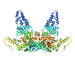 | | Structure of E6AP-E6 complex in Att2 state | | 分子名称: | Protein E6, Ubiquitin-protein ligase E3A, ZINC ION | | 著者 | Wang, Z, Yu, X. | | 登録日 | 2023-06-17 | | 公開日 | 2024-06-05 | | 実験手法 | ELECTRON MICROSCOPY (3.01 Å) | | 主引用文献 | Structural insights into the functional mechanism of the ubiquitin ligase E6AP.
Nat Commun, 15, 2024
|
|
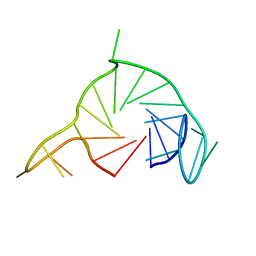
7Q48
 
 | |
7QIO
 
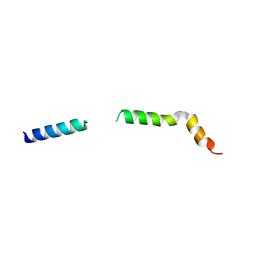 | | Homology model of myosin neck domain in skeletal sarcomere | | 分子名称: | Myosin light chain 1/3, skeletal muscle isoform, Myosin regulatory light chain 2, ... | | 著者 | Wang, Z, Grange, M, Pospich, S, Wagner, T, Kho, A.L, Gautel, M, Raunser, S. | | 登録日 | 2021-12-15 | | 公開日 | 2022-02-16 | | 最終更新日 | 2024-07-17 | | 実験手法 | ELECTRON MICROSCOPY (9 Å) | | 主引用文献 | Structures from intact myofibrils reveal mechanism of thin filament regulation through nebulin.
Science, 375, 2022
|
|
7QIN
 
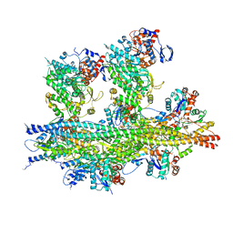 | | In situ structure of actomyosin complex in skeletal sarcomere | | 分子名称: | ADENOSINE-5'-DIPHOSPHATE, Actin, alpha skeletal muscle, ... | | 著者 | Wang, Z, Grange, M, Pospich, S, Wagner, T, Kho, A.L, Gautel, M, Raunser, S. | | 登録日 | 2021-12-15 | | 公開日 | 2022-02-16 | | 最終更新日 | 2022-03-02 | | 実験手法 | ELECTRON MICROSCOPY (6.6 Å) | | 主引用文献 | Structures from intact myofibrils reveal mechanism of thin filament regulation through nebulin.
Science, 375, 2022
|
|
7QIM
 
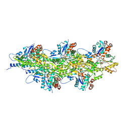 | | In situ structure of nebulin bound to actin filament in skeletal sarcomere | | 分子名称: | ACTS protein, ADENOSINE-5'-DIPHOSPHATE, MAGNESIUM ION, ... | | 著者 | Wang, Z, Grange, M, Pospich, S, Wagner, T, Kho, A.L, Gautel, M, Raunser, S. | | 登録日 | 2021-12-15 | | 公開日 | 2022-03-16 | | 実験手法 | ELECTRON MICROSCOPY (4.5 Å) | | 主引用文献 | Structures from intact myofibrils reveal mechanism of thin filament regulation through nebulin.
Science, 375, 2022
|
|
7Q6L
 
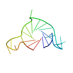 | |
4RF6
 
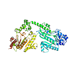 | | Crystal structure of double-domain arginine kinase from Anthopleura japonicas | | 分子名称: | Arginine kinase | | 著者 | Wang, Z, Qiao, Z, Ye, S, Zhang, R. | | 登録日 | 2014-09-25 | | 公開日 | 2015-04-08 | | 最終更新日 | 2023-09-20 | | 実験手法 | X-RAY DIFFRACTION (1.95 Å) | | 主引用文献 | Structure of a double-domain phosphagen kinase reveals an asymmetric arrangement of the tandem domains.
Acta Crystallogr.,Sect.D, 71, 2015
|
|
4RF8
 
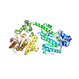 | | Crystal structure of double-domain arginine kinase from Anthopleura japonicas in complex with ADP | | 分子名称: | 4-(2-HYDROXYETHYL)-1-PIPERAZINE ETHANESULFONIC ACID, ADENOSINE-5'-DIPHOSPHATE, Arginine kinase, ... | | 著者 | Wang, Z, Qiao, Z, Ye, S, Zhang, R. | | 登録日 | 2014-09-25 | | 公開日 | 2015-04-08 | | 最終更新日 | 2023-09-20 | | 実験手法 | X-RAY DIFFRACTION (2.17 Å) | | 主引用文献 | Structure of a double-domain phosphagen kinase reveals an asymmetric arrangement of the tandem domains.
Acta Crystallogr.,Sect.D, 71, 2015
|
|
4RF7
 
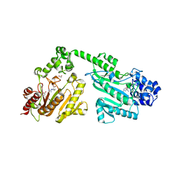 | | Crystal structure of double-domain arginine kinase from Anthopleura japonicas in complex with substrate L-arginine | | 分子名称: | ACETATE ION, ARGININE, Arginine kinase | | 著者 | Wang, Z, Qiao, Z, Ye, S, Zhang, R. | | 登録日 | 2014-09-25 | | 公開日 | 2015-04-08 | | 最終更新日 | 2023-09-20 | | 実験手法 | X-RAY DIFFRACTION (2.1 Å) | | 主引用文献 | Structure of a double-domain phosphagen kinase reveals an asymmetric arrangement of the tandem domains.
Acta Crystallogr.,Sect.D, 71, 2015
|
|
5NG5
 
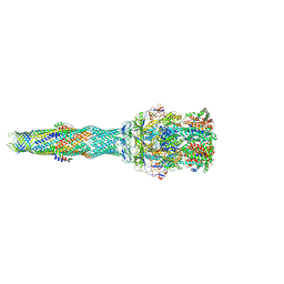 | | multi-drug efflux; membrane transport; RND superfamily; Drug resistance | | 分子名称: | 6-[2-(3,4-dimethoxyphenyl)ethylsulfanyl]-8-[4-(2-methoxyethyl)piperazin-1-yl]-3,3-dimethyl-1,4-dihydropyrano[3,4-c]pyridine-5-carbonitrile, Multidrug efflux pump accessory protein AcrZ, Multidrug efflux pump subunit AcrA, ... | | 著者 | Wang, Z, Fan, G, Hryc, C.F, Blaza, J.N, Serysheva, I.I, Schmid, M.F, Chiu, W, Luisi, B.F, Du, D. | | 登録日 | 2017-03-16 | | 公開日 | 2017-04-19 | | 最終更新日 | 2017-08-02 | | 実験手法 | ELECTRON MICROSCOPY (6.5 Å) | | 主引用文献 | An allosteric transport mechanism for the AcrAB-TolC Multidrug Efflux Pump.
Elife, 6, 2017
|
|
3LPY
 
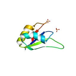 | | Crystal structure of the RRM domain of CyP33 | | 分子名称: | 4-(2-HYDROXYETHYL)-1-PIPERAZINE ETHANESULFONIC ACID, Peptidyl-prolyl cis-trans isomerase E, SULFATE ION | | 著者 | Wang, Z, Patel, D.J. | | 登録日 | 2010-02-07 | | 公開日 | 2010-07-07 | | 最終更新日 | 2024-02-21 | | 実験手法 | X-RAY DIFFRACTION (2 Å) | | 主引用文献 | Pro isomerization in MLL1 PHD3-bromo cassette connects H3K4me readout to CyP33 and HDAC-mediated repression.
Cell(Cambridge,Mass.), 141, 2010
|
|
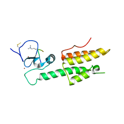
3LQH
 
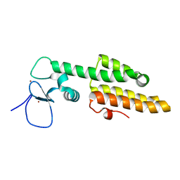 | |
3LQJ
 
 | |
