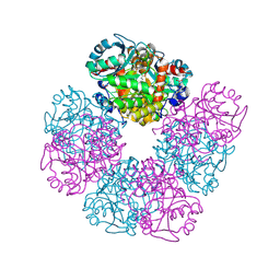
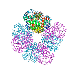
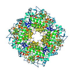
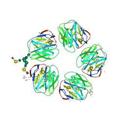
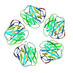
1GYK
 
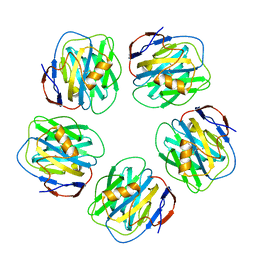 | | Serum Amyloid P Component co-crystallised with MOBDG at neutral pH | | 分子名称: | CALCIUM ION, METHYL 4,6-O-[(1R)-1-CARBOXYETHYLIDENE]-BETA-D-GALACTOPYRANOSIDE, SERUM AMYLOID P-COMPONENT | | 著者 | Thompson, D, Pepys, M.B, Tickle, I, Wood, S.P. | | 登録日 | 2002-04-25 | | 公開日 | 2003-05-22 | | 最終更新日 | 2023-12-13 | | 実験手法 | X-RAY DIFFRACTION (2.2 Å) | | 主引用文献 | The Structures of Crystalline Complexes of Human Serum Amyloid P Component with its Carbohydrate Ligand, the Cyclic Pyruvate Acetal of Galactose
J.Mol.Biol., 320, 2002
|
|
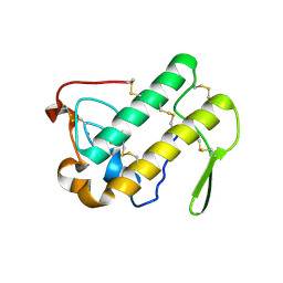
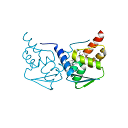
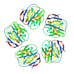
1B09
 | |
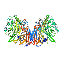
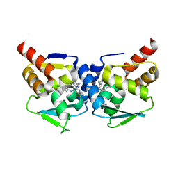
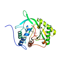
6GL0
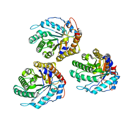 | | Structure of ZgEngAGH5_4 in complex with a cellotriose | | 分子名称: | Endoglucanase, family GH5, MAGNESIUM ION, ... | | 著者 | Dorival, J, Ruppert, S, Gunnoo, M, Orlowski, A, Chapelais, M, Dabin, J, Labourel, A, Thompson, D, Michel, G, Czjzek, M, Genicot, S. | | 登録日 | 2018-05-22 | | 公開日 | 2018-10-31 | | 最終更新日 | 2024-01-17 | | 実験手法 | X-RAY DIFFRACTION (2.2 Å) | | 主引用文献 | The laterally acquired GH5ZgEngAGH5_4from the marine bacteriumZobellia galactanivoransis dedicated to hemicellulose hydrolysis.
Biochem. J., 475, 2018
|
|
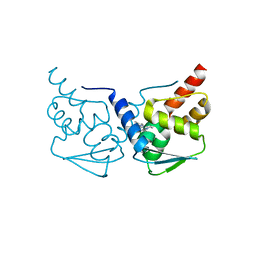
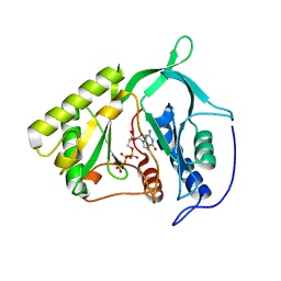
6GL2
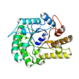 | | Structure of ZgEngAGH5_4 wild type at 1.2 Angstrom resolution | | 分子名称: | Endoglucanase, family GH5, IMIDAZOLE | | 著者 | Dorival, J, Ruppert, S, Gunnoo, M, Orlowski, A, Chapelais, M, Dabin, J, Labourel, A, Thompson, D, Michel, G, Czjzek, M, Genicot, S. | | 登録日 | 2018-05-22 | | 公開日 | 2018-10-31 | | 最終更新日 | 2024-01-17 | | 実験手法 | X-RAY DIFFRACTION (1.96 Å) | | 主引用文献 | The laterally acquired GH5ZgEngAGH5_4from the marine bacteriumZobellia galactanivoransis dedicated to hemicellulose hydrolysis.
Biochem. J., 475, 2018
|
|
2BJI
 | | High Resolution Structure of myo-Inositol Monophosphatase, The Target of Lithium Therapy | | 分子名称: | INOSITOL-1(OR 4)-MONOPHOSPHATASE, MAGNESIUM ION | | 著者 | Gill, R, Mohammed, F, Badyal, R, Coates, L, Erskine, P, Thompson, D, Cooper, J, Gore, M, Wood, S. | | 登録日 | 2005-02-03 | | 公開日 | 2005-02-11 | | 最終更新日 | 2023-12-13 | | 実験手法 | X-RAY DIFFRACTION (1.24 Å) | | 主引用文献 | High-resolution structure of myo-inositol monophosphatase, the putative target of lithium therapy.
Acta Crystallogr. D Biol. Crystallogr., 61, 2005
|
|
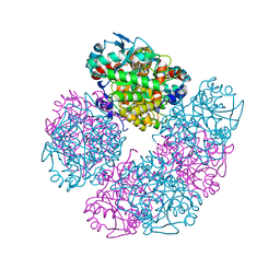
5HNR
 | | The X-ray structure of octameric human native 5-aminolaevulinic acid dehydratase. | | 分子名称: | DELTA-AMINO VALERIC ACID, Delta-aminolevulinic acid dehydratase, SULFATE ION, ... | | 著者 | Mills-Davies, N.L, Thompson, D, Shoolingin-Jordan, P.M, Erskine, P.T, Cooper, J.B. | | 登録日 | 2016-01-18 | | 公開日 | 2016-01-27 | | 最終更新日 | 2024-01-10 | | 実験手法 | X-RAY DIFFRACTION (2.83 Å) | | 主引用文献 | Structural studies of substrate and product complexes of 5-aminolaevulinic acid dehydratase from humans, Escherichia coli and the hyperthermophile Pyrobaculum calidifontis.
Acta Crystallogr D Struct Biol, 73, 2017
|
|
1N28
 | | Crystal structure of the H48Q mutant of human group IIA phospholipase A2 | | 分子名称: | CALCIUM ION, Phospholipase A2, membrane associated | | 著者 | Edwards, S.H, Thompson, D, Baker, S.F, Wood, S.P, Wilton, D.C. | | 登録日 | 2002-10-22 | | 公開日 | 2003-10-28 | | 最終更新日 | 2021-10-27 | | 実験手法 | X-RAY DIFFRACTION (1.5 Å) | | 主引用文献 | The crystal structure of the H48Q active site mutant of human group IIA secreted phospholipase A2 at 1.5 A resolution provides an insight into the catalytic mechanism
Biochemistry, 41, 2002
|
|
1N29
 | | Crystal structure of the N1A mutant of human group IIA phospholipase A2 | | 分子名称: | CALCIUM ION, Phospholipase A2, membrane associated | | 著者 | Edwards, S.H, Thompson, D, Baker, S.F, Wood, S.P, Wilton, D.C. | | 登録日 | 2002-10-22 | | 公開日 | 2003-10-28 | | 最終更新日 | 2021-10-27 | | 実験手法 | X-RAY DIFFRACTION (2.6 Å) | | 主引用文献 | The crystal structure of the H48Q active site mutant of human group IIA secreted phospholipase A2 at 1.5 A resolution provides an insight into the catalytic mechanism
Biochemistry, 41, 2002
|
|
1H4J
 | | Methylobacterium extorquens methanol dehydrogenase D303E mutant | | 分子名称: | CALCIUM ION, Methanol dehydrogenase [cytochrome c] subunit 1, Methanol dehydrogenase [cytochrome c] subunit 2, ... | | 著者 | Mohammed, F, Gill, R, Thompson, D, Cooper, J.B, Wood, S.P, Afolabi, P.R, Anthony, C. | | 登録日 | 2001-05-11 | | 公開日 | 2001-08-23 | | 最終更新日 | 2024-05-01 | | 実験手法 | X-RAY DIFFRACTION (3 Å) | | 主引用文献 | Site-Directed Mutagenesis and X-Ray Crystallography of the Pqq-Containing Quinoprotein Methanol Dehydrogenase and its Electron Acceptor, Cytochrome C(L)(,)
Biochemistry, 40, 2001
|
|
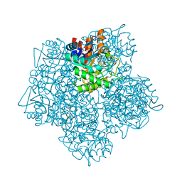
1E51
 | | Crystal structure of native human erythrocyte 5-aminolaevulinic acid dehydratase | | 分子名称: | 3-[5-(AMINOMETHYL)-4-(CARBOXYMETHYL)-1H-PYRROL-3-YL]PROPANOIC ACID, DELTA-AMINOLEVULINIC ACID DEHYDRATASE, SULFATE ION, ... | | 著者 | Mills-Davies, N.L, Thompson, D, Cooper, J.B, Shoolingin-Jordan, P.M. | | 登録日 | 2000-07-13 | | 公開日 | 2001-07-12 | | 最終更新日 | 2023-12-13 | | 実験手法 | X-RAY DIFFRACTION (2.83 Å) | | 主引用文献 | The Crystal Structure of Human Ala-Dehydratase
To be Published
|
|
5LZL
 | | Pyrobaculum calidifontis 5-aminolaevulinic acid dehydratase | | 分子名称: | Delta-aminolevulinic acid dehydratase, ZINC ION | | 著者 | Azim, N, Erskine, P.T, Guo, J, Cooper, J.B. | | 登録日 | 2016-09-30 | | 公開日 | 2016-10-12 | | 最終更新日 | 2024-01-17 | | 実験手法 | X-RAY DIFFRACTION (3.47 Å) | | 主引用文献 | Structural studies of substrate and product complexes of 5-aminolaevulinic acid dehydratase from humans, Escherichia coli and the hyperthermophile Pyrobaculum calidifontis.
Acta Crystallogr D Struct Biol, 73, 2017
|
|
5MW2
 | | CRYSTAL STRUCTURE OF BCL-6 BTB-domain with BI-3802 | | 分子名称: | 2-[6-[[5-chloranyl-2-[(3~{S},5~{R})-3,5-dimethylpiperidin-1-yl]pyrimidin-4-yl]amino]-1-methyl-2-oxidanylidene-quinolin-3-yl]oxy-~{N}-methyl-ethanamide, B-cell lymphoma 6 protein | | 著者 | Bader, G, Flotzinger, G, Weiss-Puxbaum, A, Zoephel, A. | | 登録日 | 2017-01-18 | | 公開日 | 2017-10-04 | | 最終更新日 | 2024-05-08 | | 実験手法 | X-RAY DIFFRACTION (2.35 Å) | | 主引用文献 | Chemically Induced Degradation of the Oncogenic Transcription Factor BCL6.
Cell Rep, 20, 2017
|
|
5MW6
 | |
5MWD
 | | Crystal structure of the BCL6 BTB-domain with compound 2 | | 分子名称: | 5-[[5-chloranyl-2-(3,5-dimethylpyrazol-1-yl)pyrimidin-4-yl]amino]-1,3-dihydroindol-2-one, B-cell lymphoma 6 protein | | 著者 | Bader, G, Flotzinger, G, Weiss-Puxbaum, A, Zoephel, A. | | 登録日 | 2017-01-18 | | 公開日 | 2017-10-04 | | 最終更新日 | 2024-05-08 | | 実験手法 | X-RAY DIFFRACTION (1.85 Å) | | 主引用文献 | Chemically Induced Degradation of the Oncogenic Transcription Factor BCL6.
Cell Rep, 20, 2017
|
|
5HMS
 | | X-ray structure of human recombinant 5-aminolaevulinic acid dehydratase (hrALAD). | | 分子名称: | Delta-aminolevulinic acid dehydratase, ZINC ION | | 著者 | Butler, D, Erskine, P.T, Cooper, J.B, Shoolingin-Jordan, P.M. | | 登録日 | 2016-01-17 | | 公開日 | 2016-01-27 | | 最終更新日 | 2024-01-10 | | 実験手法 | X-RAY DIFFRACTION (2.8 Å) | | 主引用文献 | Structural studies of substrate and product complexes of 5-aminolaevulinic acid dehydratase from humans, Escherichia coli and the hyperthermophile Pyrobaculum calidifontis.
Acta Crystallogr D Struct Biol, 73, 2017
|
|
5MHB
 | | Product-Complex of E.coli 5-Amino Laevulinic Acid Dehydratase | | 分子名称: | 3-[5-(AMINOMETHYL)-4-(CARBOXYMETHYL)-1H-PYRROL-3-YL]PROPANOIC ACID, Delta-aminolevulinic acid dehydratase, GLYCEROL, ... | | 著者 | Norton, E, Erskine, P.T, Shoolingin-Jordan, P.M, Cooper, J.B. | | 登録日 | 2016-11-23 | | 公開日 | 2016-12-07 | | 最終更新日 | 2024-01-17 | | 実験手法 | X-RAY DIFFRACTION (2.1 Å) | | 主引用文献 | Structural studies of substrate and product complexes of 5-aminolaevulinic acid dehydratase from humans, Escherichia coli and the hyperthermophile Pyrobaculum calidifontis.
Acta Crystallogr D Struct Biol, 73, 2017
|
|
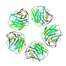
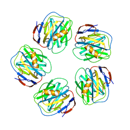
4AVV
 | | Structure of CPHPC bound to Serum Amyloid P Component | | 分子名称: | (2R)-1-[6-[(2R)-2-carboxypyrrolidin-1-yl]-6-oxidanylidene-hexanoyl]pyrrolidine-2-carboxylic acid, 2-acetamido-2-deoxy-beta-D-glucopyranose, 2-acetamido-2-deoxy-beta-D-glucopyranose-(1-4)-2-acetamido-2-deoxy-beta-D-glucopyranose, ... | | 著者 | Kolstoe, S.E, Jenvey, M.C, Wood, S.P. | | 登録日 | 2012-05-29 | | 公開日 | 2013-06-19 | | 最終更新日 | 2023-12-20 | | 実験手法 | X-RAY DIFFRACTION (1.6 Å) | | 主引用文献 | Interaction of Serum Amyloid P Component with Hexanoyl Bis(D-Proline) (Cphpc)
Acta Crystallogr.,Sect.D, 70, 2014
|
|
4AVT
 | | Structure of CPHPC bound to Serum Amyloid P Component | | 分子名称: | (2R)-1-[6-[(2R)-2-carboxypyrrolidin-1-yl]-6-oxidanylidene-hexanoyl]pyrrolidine-2-carboxylic acid, 2-acetamido-2-deoxy-beta-D-glucopyranose, CALCIUM ION, ... | | 著者 | Kolstoe, S.E, Purvis, A, Wood, S.P. | | 登録日 | 2012-05-29 | | 公開日 | 2013-06-19 | | 最終更新日 | 2023-12-20 | | 実験手法 | X-RAY DIFFRACTION (3.2 Å) | | 主引用文献 | Interaction of Serum Amyloid P Component with Hexanoyl Bis(D-Proline) (Cphpc)
Acta Crystallogr.,Sect.D, 70, 2014
|
|
4AYU
 | | Structure of N-Acetyl-D-Proline bound to serum amyloid P component | | 分子名称: | 2-acetamido-2-deoxy-beta-D-glucopyranose, CALCIUM ION, GLYCEROL, ... | | 著者 | Hughes, P, Kolstoe, S.E, Wood, S.P. | | 登録日 | 2012-06-22 | | 公開日 | 2013-07-10 | | 最終更新日 | 2023-12-20 | | 実験手法 | X-RAY DIFFRACTION (1.5 Å) | | 主引用文献 | Interaction of Serum Amyloid P Component with Hexanoyl Bis(D-Proline) (Cphpc)
Acta Crystallogr.,Sect.D, 70, 2014
|
|
4AVS
 | |
2IRY
 | |
2IRX
 | |
2IRU
 | |


