8RDX
 
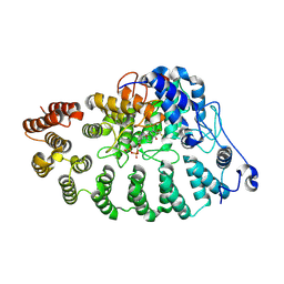 | | PGGtase I in complex with probe BAY-6092 | | 分子名称: | (5~{R})-5-(2-methoxyphenyl)-9-[(2~{R})-3,3,3-tris(fluoranyl)-2-methoxy-2-phenyl-propanoyl]-3,9-diazaspiro[5.5]undecan-2-one, CHLORIDE ION, DIPHOSPHATE, ... | | 著者 | Steuber, H. | | 登録日 | 2023-12-08 | | 公開日 | 2024-02-14 | | 最終更新日 | 2024-04-10 | | 実験手法 | X-RAY DIFFRACTION (3.67 Å) | | 主引用文献 | Discovery of YAP1/TAZ pathway inhibitors through phenotypic screening with potent anti-tumor activity via blockade of Rho-GTPase signaling.
Cell Chem Biol, 2024
|
|
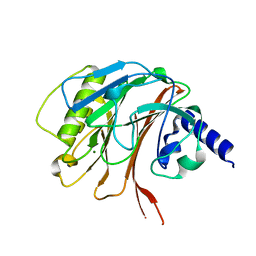
3OLM
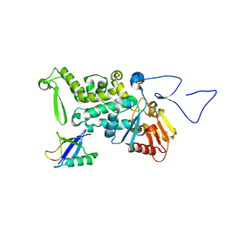 | | Structure and Function of a Ubiquitin Binding Site within the Catalytic Domain of a HECT Ubiquitin Ligase | | 分子名称: | E3 ubiquitin-protein ligase RSP5, Ubiquitin | | 著者 | Kim, H.C, Steffen, A, Chen, J, Huibregtse, J.M. | | 登録日 | 2010-08-26 | | 公開日 | 2011-03-23 | | 最終更新日 | 2023-09-06 | | 実験手法 | X-RAY DIFFRACTION (2.495 Å) | | 主引用文献 | Structure and function of a HECT domain ubiquitin-binding site.
Embo Rep., 12, 2011
|
|
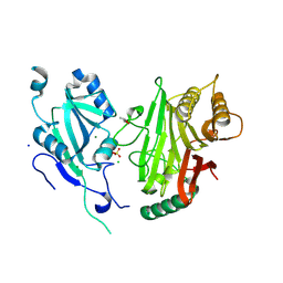
4YDH
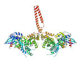 | | The structure of human FMNL1 N-terminal domains bound to Cdc42 | | 分子名称: | Cell division control protein 42 homolog, Formin-like protein 1, MAGNESIUM ION, ... | | 著者 | Kuhn, S, Anand, K, Geyer, M. | | 登録日 | 2015-02-22 | | 公開日 | 2015-05-13 | | 最終更新日 | 2024-01-10 | | 実験手法 | X-RAY DIFFRACTION (3.8 Å) | | 主引用文献 | The structure of FMNL2-Cdc42 yields insights into the mechanism of lamellipodia and filopodia formation.
Nat Commun, 6, 2015
|
|
4YC7
 | |
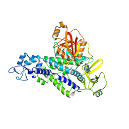
6YHM
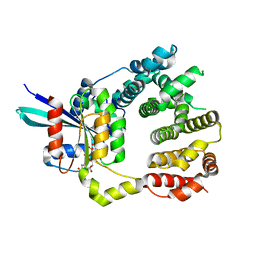
 | | Crystal structure of the C-terminal domain of CNFy from Yersinia pseudotuberculosis | | 分子名称: | Cytotoxic necrotizing factor, MAGNESIUM ION | | 著者 | Lukat, P, Gazdag, E.M, Heidler, T.V, Blankenfeldt, W. | | 登録日 | 2020-03-30 | | 公開日 | 2020-12-30 | | 最終更新日 | 2024-01-24 | | 実験手法 | X-RAY DIFFRACTION (1.13 Å) | | 主引用文献 | Crystal structure of bacterial cytotoxic necrotizing factor CNF Y reveals molecular building blocks for intoxication.
Embo J., 40, 2021
|
|
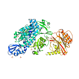
6YHN
 | | Crystal structure of domains 4-5 of CNFy from Yersinia pseudotuberculosis | | 分子名称: | (R,R)-2,3-BUTANEDIOL, CHLORIDE ION, Cytotoxic necrotizing factor, ... | | 著者 | Lukat, P, Gazdag, E.M, Heidler, T.V, Blankenfeldt, W. | | 登録日 | 2020-03-30 | | 公開日 | 2020-12-30 | | 最終更新日 | 2024-05-01 | | 実験手法 | X-RAY DIFFRACTION (1.8 Å) | | 主引用文献 | Crystal structure of bacterial cytotoxic necrotizing factor CNF Y reveals molecular building blocks for intoxication.
Embo J., 40, 2021
|
|
6YHL
 | |
6YHK
 | | Crystal structure of full-length CNFy (C866S) from Yersinia pseudotuberculosis | | 分子名称: | CHLORIDE ION, Cytotoxic necrotizing factor, SULFATE ION | | 著者 | Lukat, P, Gazdag, E.M, Heidler, T.V, Blankenfeldt, W. | | 登録日 | 2020-03-30 | | 公開日 | 2020-12-30 | | 最終更新日 | 2024-05-01 | | 実験手法 | X-RAY DIFFRACTION (2.7 Å) | | 主引用文献 | Crystal structure of bacterial cytotoxic necrotizing factor CNF Y reveals molecular building blocks for intoxication.
Embo J., 40, 2021
|
|


