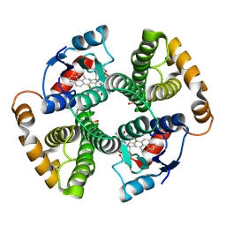
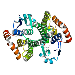
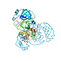
6CQK
 
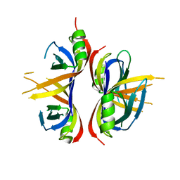 | | Crystal Structure of mitochondrial single-stranded DNA binding proteins from S. cerevisiae, Rim1 (Form1) | | 分子名称: | SsDNA-binding protein essential for mitochondrial genome maintenance | | 著者 | Singh, S.P, Kukshal, V, Bona, P.D, Lytle, A.K, Edwin, A, Galletto, R. | | 登録日 | 2018-03-15 | | 公開日 | 2018-05-30 | | 最終更新日 | 2023-10-04 | | 実験手法 | X-RAY DIFFRACTION (2.8 Å) | | 主引用文献 | The mitochondrial single-stranded DNA binding protein from S. cerevisiae, Rim1, does not form stable homo-tetramers and binds DNA as a dimer of dimers.
Nucleic Acids Res., 46, 2018
|
|
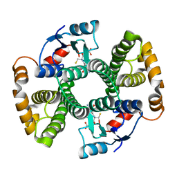
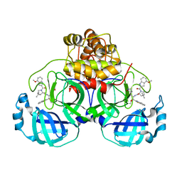
6CQM
 | | Crystal Structure of mitochondrial single-stranded DNA binding proteins from S. cerevisiae, Rim1 (Form2) | | 分子名称: | Single-stranded DNA-binding protein RIM1, mitochondrial | | 著者 | Singh, S.P, Kukshal, V, Bona, P.D, Lytle, A.K, Edwin, A, Galletto, R. | | 登録日 | 2018-03-15 | | 公開日 | 2018-05-30 | | 最終更新日 | 2024-03-13 | | 実験手法 | X-RAY DIFFRACTION (3 Å) | | 主引用文献 | The mitochondrial single-stranded DNA binding protein from S. cerevisiae, Rim1, does not form stable homo-tetramers and binds DNA as a dimer of dimers.
Nucleic Acids Res., 46, 2018
|
|
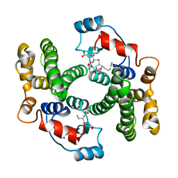
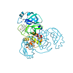
6CQO
 | | Crystal Structure of mitochondrial single-stranded DNA binding proteins from S. cerevisiae (SeMet Labeled), Rim1 (Form2) | | 分子名称: | Single-stranded DNA-binding protein RIM1, mitochondrial | | 著者 | Singh, S.P, Kukshal, V, Bona, P.D, Lytle, A.K, Edwin, A, Galletto, R. | | 登録日 | 2018-03-15 | | 公開日 | 2018-05-30 | | 最終更新日 | 2020-02-26 | | 実験手法 | X-RAY DIFFRACTION (2.8 Å) | | 主引用文献 | The mitochondrial single-stranded DNA binding protein from S. cerevisiae, Rim1, does not form stable homo-tetramers and binds DNA as a dimer of dimers.
Nucleic Acids Res., 46, 2018
|
|
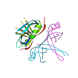
5GO1
 | | Structural, Functional characterization and discovery of novel inhibitors of Leishmania amazonensis Nucleoside Diphosphatase Kinase (NDK) | | 分子名称: | Nucleoside diphosphate kinase | | 著者 | Mishra, A.K, Agnihotri, P, Singh, S.P, Pratap, J.V. | | 登録日 | 2016-07-26 | | 公開日 | 2017-07-26 | | 最終更新日 | 2023-11-08 | | 実験手法 | X-RAY DIFFRACTION (2.5 Å) | | 主引用文献 | Discovery of novel inhibitors for Leishmania nucleoside diphosphatase kinase (NDK) based on its structural and functional characterization.
J. Comput. Aided Mol. Des., 31, 2017
|
|
1F3B
 | | CRYSTAL STRUCTURE OF MGSTA1-1 IN COMPLEX WITH GLUTATHIONE CONJUGATE OF BENZO[A]PYRENE EPOXIDE | | 分子名称: | 2-AMINO-4-[1-(CARBOXYMETHYL-CARBAMOYL)-2-(9-HYDROXY-7,8-DIOXO-7,8,9,10-TETRAHYDRO-BENZO[DEF]CHRYSEN-10-YLSULFANYL)-ETHYLCARBAMOYL]-BUTYRIC ACID, GLUTATHIONE S-TRANSFERASE YA CHAIN | | 著者 | Gu, Y, Singh, S.V, Ji, X. | | 登録日 | 2000-06-01 | | 公開日 | 2000-10-18 | | 最終更新日 | 2023-11-15 | | 実験手法 | X-RAY DIFFRACTION (2 Å) | | 主引用文献 | Residue R216 and catalytic efficiency of a murine class alpha glutathione S-transferase toward benzo[a]pyrene 7(R),8(S)-diol 9(S), 10(R)-epoxide.
Biochemistry, 39, 2000
|
|
1F3A
 | | CRYSTAL STRUCTURE OF MGSTA1-1 IN COMPLEX WITH GSH | | 分子名称: | GLUTATHIONE, GLUTATHIONE S-TRANSFERASE YA CHAIN | | 著者 | Gu, Y, Singh, S.V, Ji, X. | | 登録日 | 2000-06-01 | | 公開日 | 2000-10-18 | | 最終更新日 | 2023-08-30 | | 実験手法 | X-RAY DIFFRACTION (1.9 Å) | | 主引用文献 | Residue R216 and catalytic efficiency of a murine class alpha glutathione S-transferase toward benzo[a]pyrene 7(R),8(S)-diol 9(S), 10(R)-epoxide.
Biochemistry, 39, 2000
|
|
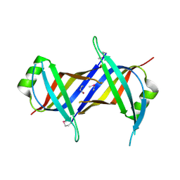
8C0T
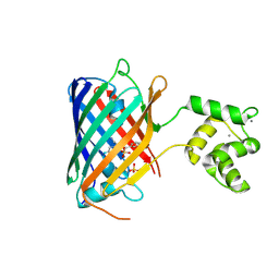 | | NRS 1.2: Fluorescent Sensors for Imaging Interstitial Calcium | | 分子名称: | CALCIUM ION, SULFATE ION, mNeonGreen,Optimized Ratiometric Calcium Sensor | | 著者 | Basquin, J, Griesbeck, O, Valiente-Gabioud, A. | | 登録日 | 2022-12-19 | | 公開日 | 2023-11-15 | | 実験手法 | X-RAY DIFFRACTION (1.28 Å) | | 主引用文献 | Fluorescent sensors for imaging of interstitial calcium.
Nat Commun, 14, 2023
|
|
1B48
 | |
1M0U
 | | Crystal Structure of the Drosophila Glutathione S-transferase-2 in Complex with Glutathione | | 分子名称: | GLUTATHIONE, GST2 gene product, SULFATE ION | | 著者 | Agianian, B, Tucker, P.A, Schouten, A, Leonard, K, Bullard, B, Gros, P. | | 登録日 | 2002-06-14 | | 公開日 | 2003-02-11 | | 最終更新日 | 2024-02-14 | | 実験手法 | X-RAY DIFFRACTION (1.75 Å) | | 主引用文献 | Structure of a Drosophila Sigma Class Glutathione S-transferase Reveals a Novel
Active Site Topography Suited for Lipid Peroxidation Products
J.Mol.Biol., 326, 2003
|
|
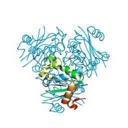
7RFR
 | | Structure of SARS-CoV-2 main protease in complex with a covalent inhibitor | | 分子名称: | (1R,2S,5S)-N-{(1S,2S)-1-(1,3-benzothiazol-2-yl)-1-hydroxy-3-[(3S)-2-oxopyrrolidin-3-yl]propan-2-yl}-3-(4-methoxy-1H-indole-2-carbonyl)-6,6-dimethyl-3-azabicyclo[3.1.0]hexane-2-carboxamide, 3C-like proteinase | | 著者 | Gajiwala, K.S, Ferre, R.A, Liu, W, Stewart, A.E. | | 登録日 | 2021-07-14 | | 公開日 | 2021-11-10 | | 最終更新日 | 2023-10-18 | | 実験手法 | X-RAY DIFFRACTION (1.626 Å) | | 主引用文献 | An oral SARS-CoV-2 M pro inhibitor clinical candidate for the treatment of COVID-19.
Science, 374, 2021
|
|
7RFU
 | | Structure of SARS-CoV-2 main protease in complex with a covalent inhibitor | | 分子名称: | (1R,2S,5S)-N-{(1S,2S)-1-(1,3-benzothiazol-2-yl)-1-hydroxy-3-[(3S)-2-oxopyrrolidin-3-yl]propan-2-yl}-3-[N-(methanesulfonyl)-L-valyl]-6,6-dimethyl-3-azabicyclo[3.1.0]hexane-2-carboxamide, 3C-like proteinase | | 著者 | Greasley, S.E, Ferre, R.A, Liu, W, Stewart, A.E. | | 登録日 | 2021-07-14 | | 公開日 | 2021-11-10 | | 最終更新日 | 2022-01-05 | | 実験手法 | X-RAY DIFFRACTION (2.498 Å) | | 主引用文献 | An oral SARS-CoV-2 M pro inhibitor clinical candidate for the treatment of COVID-19.
Science, 374, 2021
|
|
7RFW
 | | Structure of SARS-CoV-2 main protease in complex with a covalent inhibitor | | 分子名称: | (1R,2S,5S)-N-{(1E,2S)-1-imino-3-[(3S)-2-oxopyrrolidin-3-yl]propan-2-yl}-6,6-dimethyl-3-[3-methyl-N-(trifluoroacetyl)-L-valyl]-3-azabicyclo[3.1.0]hexane-2-carboxamide, 3C-like proteinase | | 著者 | Greasley, S.E, Ferre, R.A, Liu, W, Stewart, A.E. | | 登録日 | 2021-07-14 | | 公開日 | 2021-11-10 | | 最終更新日 | 2022-01-05 | | 実験手法 | X-RAY DIFFRACTION (1.729 Å) | | 主引用文献 | An oral SARS-CoV-2 M pro inhibitor clinical candidate for the treatment of COVID-19.
Science, 374, 2021
|
|
7RFS
 | | Structure of SARS-CoV-2 main protease in complex with a covalent inhibitor | | 分子名称: | (1R,2S,5S)-N-{(1E,2S)-1-imino-3-[(3S)-2-oxopyrrolidin-3-yl]propan-2-yl}-6,6-dimethyl-3-[3-methyl-N-(trifluoroacetyl)-L-valyl]-3-azabicyclo[3.1.0]hexane-2-carboxamide, 3C-like proteinase | | 著者 | Greasley, S.E, Ferre, R.A, Liu, W, Stewart, A.E. | | 登録日 | 2021-07-14 | | 公開日 | 2021-11-10 | | 最終更新日 | 2022-01-05 | | 実験手法 | X-RAY DIFFRACTION (1.91 Å) | | 主引用文献 | An oral SARS-CoV-2 M pro inhibitor clinical candidate for the treatment of COVID-19.
Science, 374, 2021
|
|


