2QLH
 
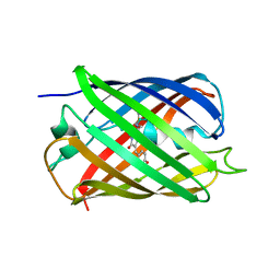 | | mPlum I65L mutant | | 分子名称: | Fluorescent protein plum | | 著者 | Shu, X, Remington, S.J. | | 登録日 | 2007-07-12 | | 公開日 | 2008-07-22 | | 最終更新日 | 2023-11-15 | | 実験手法 | X-RAY DIFFRACTION (1.9 Å) | | 主引用文献 | Structural Studies of Far-Red Emission in mPlum, a Monomeric Red Fluorescent Protein
To be Published
|
|
2QLI
 
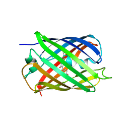 | | mPlum E16Q mutant | | 分子名称: | Fluorescent protein plum | | 著者 | Shu, X, Remington, S.J. | | 登録日 | 2007-07-12 | | 公開日 | 2008-07-22 | | 最終更新日 | 2023-11-15 | | 実験手法 | X-RAY DIFFRACTION (1.34 Å) | | 主引用文献 | Structural Studies of Far-Red Emission in mPlum, a Monomeric Red Fluorescent Protein
To be Published
|
|
2QLG
 
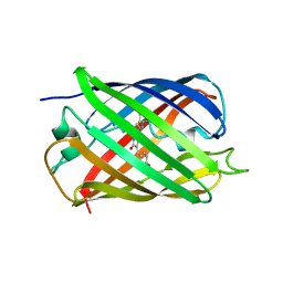 | | mPlum | | 分子名称: | Fluorescent protein plum | | 著者 | Shu, X, Remington, S.J. | | 登録日 | 2007-07-12 | | 公開日 | 2008-07-22 | | 最終更新日 | 2023-11-15 | | 実験手法 | X-RAY DIFFRACTION (1.8 Å) | | 主引用文献 | Structural Studies of Far-Red Emission in mPlum, a Monomeric Red Fluorescent Protein
To be Published
|
|
2QLE
 
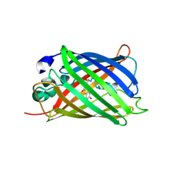 | | GFP/S205V mutant | | 分子名称: | Green fluorescent protein, IMIDAZOLE | | 著者 | Shu, X, Remington, S.J. | | 登録日 | 2007-07-12 | | 公開日 | 2008-02-12 | | 最終更新日 | 2023-11-15 | | 実験手法 | X-RAY DIFFRACTION (1.59 Å) | | 主引用文献 | An alternative excited-state proton transfer pathway in green fluorescent protein variant S205V.
Protein Sci., 16, 2007
|
|
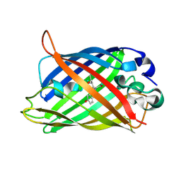
2DUF
 
 | |
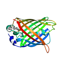
2DUI
 
 | |
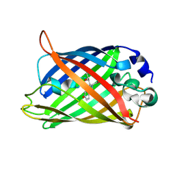
2DUH
 
 | |
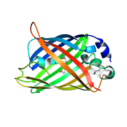
2DUE
 
 | |
2DUG
 
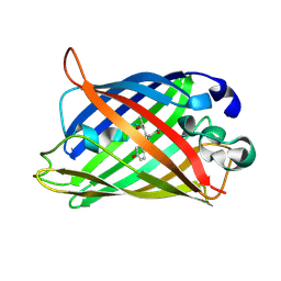 | |
2H5O
 
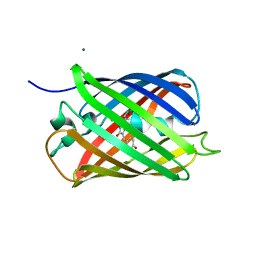 | |
2H5Q
 
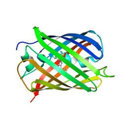 | |
2H5P
 
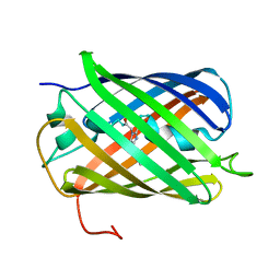 | |
2H5R
 
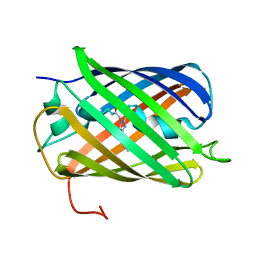 | |
428D
 
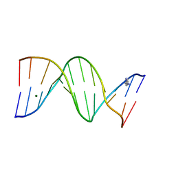 | | STRUCTURE OF THE POTASSIUM FORM OF CGCGAATTCGCG: DNA DEFORMATION BY ELECTROSTATIC COLLAPSE AROUND SMALL CATIONS | | 分子名称: | DNA (5'-D(*CP*GP*CP*GP*AP*AP*TP*TP*CP*GP*CP*G)-3'), MAGNESIUM ION, SPERMINE | | 著者 | Shui, X, Sines, C.C, McFail-Isom, L, VanDerveer, D, Williams, L.D. | | 登録日 | 1998-09-28 | | 公開日 | 1998-10-09 | | 最終更新日 | 2024-04-03 | | 実験手法 | X-RAY DIFFRACTION (1.75 Å) | | 主引用文献 | Structure of the potassium form of CGCGAATTCGCG: DNA deformation by electrostatic collapse around inorganic cations.
Biochemistry, 37, 1998
|
|
355D
 
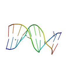 | | THE B-DNA DODECAMER AT HIGH RESOLUTION | | 分子名称: | DNA (5'-D(*CP*GP*CP*GP*AP*AP*TP*TP*CP*GP*CP*G)-3'), MAGNESIUM ION, SPERMINE | | 著者 | Hu, L.D, Shui, X, McFail-Isom, L, Williams, G.G. | | 登録日 | 1997-10-07 | | 公開日 | 1997-10-13 | | 最終更新日 | 2023-08-02 | | 実験手法 | X-RAY DIFFRACTION (1.4 Å) | | 主引用文献 | The B-DNA dodecamer at high resolution reveals a spine of water on sodium.
Biochemistry, 37, 1998
|
|
4CQH
 
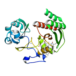 | | Structure of Infrared Fluorescent Protein IFP2.0 | | 分子名称: | 3-[2-[(Z)-[3-(2-carboxyethyl)-5-[(Z)-(4-ethenyl-3-methyl-5-oxidanylidene-pyrrol-2-ylidene)methyl]-4-methyl-pyrrol-1-ium -2-ylidene]methyl]-5-[(Z)-[(3E)-3-ethylidene-4-methyl-5-oxidanylidene-pyrrolidin-2-ylidene]methyl]-4-methyl-1H-pyrrol-3- yl]propanoic acid, BACTERIOPHYTOCHROME, SODIUM ION | | 著者 | Lafaye, C, Yu, D, Noirclerc-Savoye, M, Shu, X, Royant, A. | | 登録日 | 2014-02-17 | | 公開日 | 2014-05-28 | | 最終更新日 | 2023-12-20 | | 実験手法 | X-RAY DIFFRACTION (1.14 Å) | | 主引用文献 | An Improved Monomeric Infrared Fluorescent Protein for Neuronal and Tumour Brain Imaging.
Nat.Commun., 5, 2014
|
|
6GPU
 
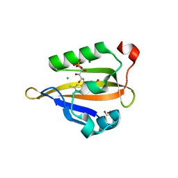 | | Crystal structure of miniSOG at 1.17A resolution | | 分子名称: | 2-AMINO-2-HYDROXYMETHYL-PROPANE-1,3-DIOL, CHLORIDE ION, COBALT (II) ION, ... | | 著者 | Lafaye, C, Signor, L, Aumonier, S, Shu, X, Gotthard, G, Royant, A. | | 登録日 | 2018-06-07 | | 公開日 | 2019-02-27 | | 最終更新日 | 2024-01-17 | | 実験手法 | X-RAY DIFFRACTION (1.17 Å) | | 主引用文献 | Tailing miniSOG: structural bases of the complex photophysics of a flavin-binding singlet oxygen photosensitizing protein.
Sci Rep, 9, 2019
|
|
6GPV
 
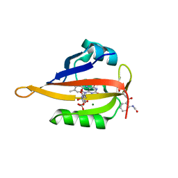 | | Crystal structure of blue-light irradiated miniSOG | | 分子名称: | CHLORIDE ION, FLAVIN MONONUCLEOTIDE, LUMICHROME, ... | | 著者 | Lafaye, C, Signor, L, Aumonier, S, Shu, X, Gotthard, G, Royant, A. | | 登録日 | 2018-06-07 | | 公開日 | 2019-02-27 | | 最終更新日 | 2024-01-17 | | 実験手法 | X-RAY DIFFRACTION (2 Å) | | 主引用文献 | Tailing miniSOG: structural bases of the complex photophysics of a flavin-binding singlet oxygen photosensitizing protein.
Sci Rep, 9, 2019
|
|
1XMZ
 
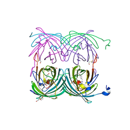 | | Crystal structure of the dark state of kindling fluorescent protein kfp from anemonia sulcata | | 分子名称: | BETA-MERCAPTOETHANOL, GFP-like non-fluorescent chromoprotein FP595 chain 1, GFP-like non-fluorescent chromoprotein FP595 chain 2 | | 著者 | Quillin, M.L, Anstrom, D.M, Shu, X, O'Leary, S, Kallio, K, Chudakov, D.M, Remington, S.J. | | 登録日 | 2004-10-04 | | 公開日 | 2005-04-19 | | 最終更新日 | 2024-07-10 | | 実験手法 | X-RAY DIFFRACTION (1.38 Å) | | 主引用文献 | Kindling Fluorescent Protein from Anemonia sulcata: Dark-State Structure at 1.38 Resolution
Biochemistry, 44, 2005
|
|
5AJG
 
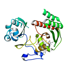 | | Structure of Infrared Fluorescent Protein IFP1.4 AT 1.11 Angstrom resolution | | 分子名称: | 3-[2-[(Z)-[3-(2-carboxyethyl)-5-[(Z)-(4-ethenyl-3-methyl-5-oxidanylidene-pyrrol-2-ylidene)methyl]-4-methyl-pyrrol-1-ium -2-ylidene]methyl]-5-[(Z)-[(3E)-3-ethylidene-4-methyl-5-oxidanylidene-pyrrolidin-2-ylidene]methyl]-4-methyl-1H-pyrrol-3- yl]propanoic acid, BACTERIOPHYTOCHROME | | 著者 | Lafaye, C, Shu, X, Royant, A. | | 登録日 | 2015-02-24 | | 公開日 | 2016-03-09 | | 最終更新日 | 2024-01-10 | | 実験手法 | X-RAY DIFFRACTION (1.11 Å) | | 主引用文献 | Structural Determinants of Improved Fluorescence in a Family of Bacteriophytochrome-Based Infrared Fluorescent Proteins: Insights from Continuum Electrostatic Calculations and Molecular Dynamics Simulations.
Biochemistry, 55, 2016
|
|
4DJB
 
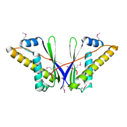 | | A Structural Basis for the Assembly and Functions of a Viral Polymer that Inactivates Multiple Tumor Suppressors | | 分子名称: | E4-ORF3 | | 著者 | Ou, H.D, Kwiatkowski, W, Deerinck, T.J, Noske, A, Blain, K.Y, Land, H.S, Soria, C, Powers, C.J, May, A.P, Shu, X, Tsien, R.Y, Fitzpatrick, J.A.J, Long, J.A, Ellisman, M.H, Choe, S, O'Shea, C.C. | | 登録日 | 2012-02-01 | | 公開日 | 2012-10-31 | | 最終更新日 | 2018-01-24 | | 実験手法 | X-RAY DIFFRACTION (2.053 Å) | | 主引用文献 | A Structural Basis for the Assembly and Functions of a Viral Polymer that Inactivates Multiple Tumor Suppressors.
Cell(Cambridge,Mass.), 151, 2012
|
|
386D
 
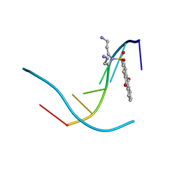 | | THREE-DIMENSIONAL STRUCTURE AND REACTIVITY OF A PHOTOCHEMICAL CLEAVAGE AGENT BOUND TO DNA | | 分子名称: | DNA (5'-D(*CP*GP*TP*AP*CP*G)-3'), N,N-BIS(3-AMINOPROPYL)-2-ANTHRAQUINONESULFONAMIDE | | 著者 | Gasper, S.M, Armitage, B, Shui, X, Hu, G.G, Yu, C, Schuster, G, Williams, L.D. | | 登録日 | 1998-03-11 | | 公開日 | 1998-03-12 | | 最終更新日 | 2024-02-21 | | 実験手法 | X-RAY DIFFRACTION (1.8 Å) | | 主引用文献 | Three-Dimensional Structure and Reactivity of a Photochemical Cleavage Agent Bound to DNA
J.Am.Chem.Soc., 120, 1998
|
|
1AGL
 
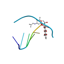 | | STRUCTURE OF A DNA-BISDAUNOMYCIN COMPLEX | | 分子名称: | 4-METHYLBENZYL-N-BIS[DAUNOMYCIN], DNA (5'-D(*CP*GP*AP*TP*CP*G)-3') | | 著者 | Hu, G.G, Shui, X, Leng, F, Priebe, W, Chaires, J.B, Williams, L.D. | | 登録日 | 1997-03-25 | | 公開日 | 1997-04-29 | | 最終更新日 | 2024-04-03 | | 実験手法 | X-RAY DIFFRACTION (2.2 Å) | | 主引用文献 | Structure of a DNA-bisdaunomycin complex.
Biochemistry, 36, 1997
|
|
2H8Q
 
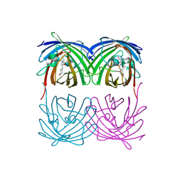 | |
1C9Z
 
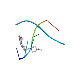 | | D232-CGTACG | | 分子名称: | 1,3-DI[[[10-METHOXY-7H-PYRIDO[4,3-C]CARBAZOL-2-IUMYL]-ETHYL]-PIPERIDIN-4-YL]-PROPANE, 5'-D(*CP*GP*TP*AP*CP*G)-3' | | 著者 | Williams, L.D. | | 登録日 | 1999-08-03 | | 公開日 | 2000-04-02 | | 最終更新日 | 2024-04-17 | | 実験手法 | X-RAY DIFFRACTION (2.4 Å) | | 主引用文献 | Effects of cationic charge on three-dimensional structures of intercalative complexes: structure of a bis-intercalated DNA complex solved by MAD phasing.
Curr.Med.Chem., 7, 2000
|
|
