7KW3
 
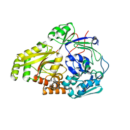
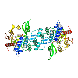
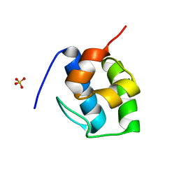 | | Non Ribosomal PCP domain | | 分子名称: | PCP domain, SULFATE ION | | 著者 | Izore, T, Ho, Y.T.C, Kaczmarski, J.A, Gavriilidou, A, Chow, K.H, Steer, D, Goode, R.J.A, Schittenhelm, R.B, Tailhades, J, Tosin, M, Challis, G.L, Krenske, E.H, Ziemert, N, Jackson, C.J, Cryle, M.J. | | 登録日 | 2020-11-29 | | 公開日 | 2021-03-24 | | 最終更新日 | 2024-04-03 | | 実験手法 | X-RAY DIFFRACTION (2.3 Å) | | 主引用文献 | Structures of a non-ribosomal peptide synthetase condensation domain suggest the basis of substrate selectivity.
Nat Commun, 12, 2021
|
|
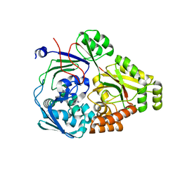
7KW0
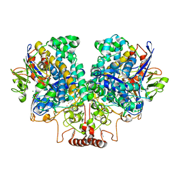
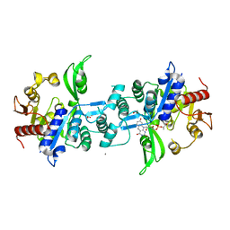
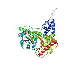 | | Non-ribosomal didomain (stabilised glycine-PCP-C) acceptor bound state | | 分子名称: | N-{2-[(2-aminoethyl)sulfanyl]ethyl}-N~3~-[(2R)-2-hydroxy-3,3-dimethyl-4-(phosphonooxy)butanoyl]-beta-alaninamide, PCP-C didomain | | 著者 | Izore, T, Ho, Y.T.C, Kaczmarski, J.A, Gavriilidou, A, Chow, K.H, Steer, D, Goode, R.J.A, Schittenhelm, R.B, Tailhades, J, Tosin, M, Challis, G.L, Krenske, E.H, Ziemert, N, Jackson, C.J, Cryle, M.J. | | 登録日 | 2020-11-29 | | 公開日 | 2021-03-24 | | 最終更新日 | 2024-04-03 | | 実験手法 | X-RAY DIFFRACTION (1.9 Å) | | 主引用文献 | Structures of a non-ribosomal peptide synthetase condensation domain suggest the basis of substrate selectivity.
Nat Commun, 12, 2021
|
|
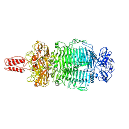
7KVW
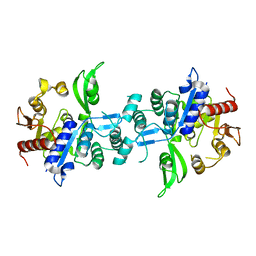
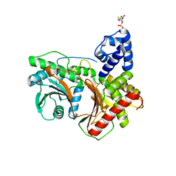 | | Non-ribosomal didomain (holo-PCP-C) acceptor bound state | | 分子名称: | 4'-PHOSPHOPANTETHEINE, PCP-C didomain | | 著者 | Izore, T, Ho, Y.T.C, Kaczmarski, J.A, Gavriilidou, A, Chow, K.H, Steer, D, Goode, R.J.A, Schittenhelm, R.B, Tailhades, J, Tosin, M, Challis, G.L, Krenske, E.H, Ziemert, N, Jackson, C.J, Cryle, M.J. | | 登録日 | 2020-11-29 | | 公開日 | 2021-03-24 | | 最終更新日 | 2024-04-03 | | 実験手法 | X-RAY DIFFRACTION (2.18 Å) | | 主引用文献 | Structures of a non-ribosomal peptide synthetase condensation domain suggest the basis of substrate selectivity.
Nat Commun, 12, 2021
|
|
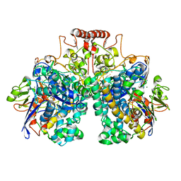
7KW2
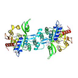
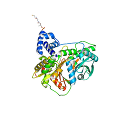 | | Non-ribosomal didomain (holo-PCP-C) acceptor bound state, R2577G | | 分子名称: | 4'-PHOSPHOPANTETHEINE, PCP-C didomain | | 著者 | Izore, T, Ho, Y.T.C, Kaczmarski, J.A, Gavriilidou, A, Chow, K.H, Steer, D, Goode, R.J.A, Schittenhelm, R.B, Tailhades, J, Tosin, M, Challis, G.L, Krenske, E.H, Ziemert, N, Jackson, C.J, Cryle, M.J. | | 登録日 | 2020-11-29 | | 公開日 | 2021-03-24 | | 最終更新日 | 2024-04-03 | | 実験手法 | X-RAY DIFFRACTION (2 Å) | | 主引用文献 | Structures of a non-ribosomal peptide synthetase condensation domain suggest the basis of substrate selectivity.
Nat Commun, 12, 2021
|
|
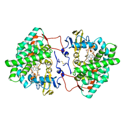
6OFQ
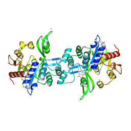
 | | ABC transporter-associated periplasmic binding protein DppA from Helicobacter pylori in complex with peptide STSA | | 分子名称: | Heme-binding protein A / AI-2 binding protein A, SER-THR-SER-ALA | | 著者 | Rahman, M.M, Machuca, M.A, Khan, M.F, Barlow, C.K, Schittenhelm, R.B, Roujeinikova, A. | | 登録日 | 2019-03-31 | | 公開日 | 2019-08-21 | | 最終更新日 | 2023-10-11 | | 実験手法 | X-RAY DIFFRACTION (1.45 Å) | | 主引用文献 | Molecular Basis of Unexpected Specificity of ABC Transporter-Associated Substrate-Binding Protein DppA from Helicobacter pylori.
J.Bacteriol., 201, 2019
|
|
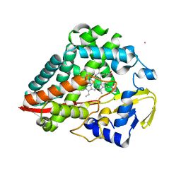
6PU3
 | | ABC transporter-associated periplasmic binding protein DppA from Helicobacter pylori | | 分子名称: | Heme-binding protein A, SER-THR-SER-ALA | | 著者 | Rahman, M.M, Machuca, M.A, Khan, M.F, Barlow, C.K, Schittenhelm, R.B, Roujeinikova, A. | | 登録日 | 2019-07-16 | | 公開日 | 2019-08-21 | | 最終更新日 | 2023-10-11 | | 実験手法 | X-RAY DIFFRACTION (1.8 Å) | | 主引用文献 | Molecular Basis of Unexpected Specificity of ABC Transporter-Associated Substrate-Binding Protein DppA from Helicobacter pylori.
J.Bacteriol., 201, 2019
|
|
7LZJ
 | |
8DQV
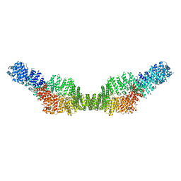
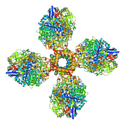
 | | The 1.52 angstrom CryoEM structure of the [NiFe]-hydrogenase Huc from Mycobacterium smegmatis - catalytic dimer (Huc2S2L) | | 分子名称: | CARBONMONOXIDE-(DICYANO) IRON, FE3-S4 CLUSTER, Hydrogenase-2, ... | | 著者 | Grinter, R, Venugopal, H, Kropp, A, Greening, C. | | 登録日 | 2022-07-20 | | 公開日 | 2023-01-04 | | 最終更新日 | 2023-03-29 | | 実験手法 | ELECTRON MICROSCOPY (1.52 Å) | | 主引用文献 | Structural basis for bacterial energy extraction from atmospheric hydrogen.
Nature, 615, 2023
|
|
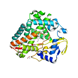
7OQ6
 | | Crystal structure of cytochrome P450 Sas16 from Streptomyces asterosporus | | 分子名称: | Cytochrome P450, PROTOPORPHYRIN IX CONTAINING FE, THIOCYANATE ION | | 著者 | Zhang, L, Zhang, S, Bechthold, A, Einsle, O. | | 登録日 | 2021-06-02 | | 公開日 | 2022-06-22 | | 最終更新日 | 2023-09-13 | | 実験手法 | X-RAY DIFFRACTION (2 Å) | | 主引用文献 | P450-mediated dehydrotyrosine formation during WS9326 biosynthesis proceeds via dehydrogenation of a specific acylated dipeptide substrate.
Acta Pharm Sin B, 13, 2023
|
|
8U3N
 | |
8UKZ
 | |
8U2M
 | |
7MP5
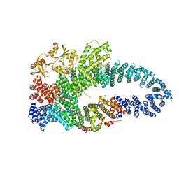 | |
7MP6
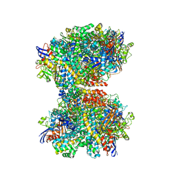
 | | Neurofibromin homodimer | | 分子名称: | Isoform I of Neurofibromin | | 著者 | Lupton, C.J, Bayly-Jones, C, Ellisdon, A.M. | | 登録日 | 2021-05-04 | | 公開日 | 2021-12-15 | | 最終更新日 | 2024-05-29 | | 実験手法 | ELECTRON MICROSCOPY (6.25 Å) | | 主引用文献 | The cryo-EM structure of the human neurofibromin dimer reveals the molecular basis for neurofibromatosis type 1.
Nat.Struct.Mol.Biol., 28, 2021
|
|
7MOC
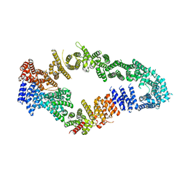 | | Neurofibromin core | | 分子名称: | Isoform I of Neurofibromin | | 著者 | Lupton, C.J, Bayly-Jones, C, Ellisdon, A.M. | | 登録日 | 2021-05-01 | | 公開日 | 2021-12-15 | | 最終更新日 | 2024-05-29 | | 実験手法 | ELECTRON MICROSCOPY (4.56 Å) | | 主引用文献 | The cryo-EM structure of the human neurofibromin dimer reveals the molecular basis for neurofibromatosis type 1.
Nat.Struct.Mol.Biol., 28, 2021
|
|
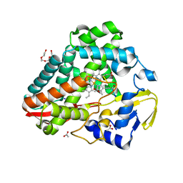
6M7L
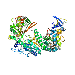 | | Complex of OxyA with the X-domain from GPA biosynthesis | | 分子名称: | PROTOPORPHYRIN IX CONTAINING FE, Putative cytochrome P450 hydroxylase, Putative non-ribosomal peptide synthetase | | 著者 | Greule, A, Izore, T, Tailhades, J, Peschke, M, Schoppet, M, Ahmed, I, Kulik, A, Adamek, M, Ziemert, N, De Voss, J, Stegmann, E, Cryle, M.J. | | 登録日 | 2018-08-20 | | 公開日 | 2019-05-22 | | 最終更新日 | 2023-10-11 | | 実験手法 | X-RAY DIFFRACTION (2.648297 Å) | | 主引用文献 | Kistamicin biosynthesis reveals the biosynthetic requirements for production of highly crosslinked glycopeptide antibiotics.
Nat Commun, 10, 2019
|
|
7UUR
 | | The 1.67 Angstrom CryoEM structure of the [NiFe]-hydrogenase Huc from Mycobacterium smegmatis - catalytic dimer (Huc2S2L) | | 分子名称: | CARBONMONOXIDE-(DICYANO) IRON, FE3-S4 CLUSTER, HYDROXIDE ION, ... | | 著者 | Grinter, R, Venugopal, H, Kropp, A, Greening, C. | | 登録日 | 2022-04-28 | | 公開日 | 2023-01-04 | | 最終更新日 | 2023-03-29 | | 実験手法 | ELECTRON MICROSCOPY (1.67 Å) | | 主引用文献 | Structural basis for bacterial energy extraction from atmospheric hydrogen.
Nature, 615, 2023
|
|
7UUS
 | | The CryoEM structure of the [NiFe]-hydrogenase Huc from Mycobacterium smegmatis - Full complex focused refinement of stalk | | 分子名称: | CARBONMONOXIDE-(DICYANO) IRON, FE3-S4 CLUSTER, Hydrogenase-2, ... | | 著者 | Grinter, R, Venugopal, H, Kropp, A, Greening, C. | | 登録日 | 2022-04-28 | | 公開日 | 2023-01-04 | | 最終更新日 | 2023-04-05 | | 実験手法 | ELECTRON MICROSCOPY (8 Å) | | 主引用文献 | Structural basis for bacterial energy extraction from atmospheric hydrogen.
Nature, 615, 2023
|
|
7UTD
 | | The 2.19-angstrom CryoEM structure of the [NiFe]-hydrogenase Huc from Mycobacterium smegmatis - Complex minus stalk | | 分子名称: | CARBONMONOXIDE-(DICYANO) IRON, FE3-S4 CLUSTER, Hydrogenase-2, ... | | 著者 | Grinter, R, Venugopal, H, Kropp, A, Greening, C. | | 登録日 | 2022-04-26 | | 公開日 | 2023-01-04 | | 最終更新日 | 2023-04-05 | | 実験手法 | ELECTRON MICROSCOPY (2.19 Å) | | 主引用文献 | Structural basis for bacterial energy extraction from atmospheric hydrogen.
Nature, 615, 2023
|
|
6UW1
 | | The crystal structure of FbiA from Mycobacterium Smegmatis, Fo bound form | | 分子名称: | 1-deoxy-1-(8-hydroxy-2,4-dioxo-3,4-dihydropyrimido[4,5-b]quinolin-10(2H)-yl)-D-ribitol, CALCIUM ION, Phosphoenolpyruvate transferase | | 著者 | Grinter, R, Gillett, D, Cordero, P.R.F, Greening, C. | | 登録日 | 2019-11-04 | | 公開日 | 2020-05-13 | | 最終更新日 | 2023-10-11 | | 実験手法 | X-RAY DIFFRACTION (2.205 Å) | | 主引用文献 | Cellular and Structural Basis of Synthesis of the Unique Intermediate Dehydro-F420-0 in Mycobacteria.
mSystems, 5, 2020
|
|
6UW7
 | | The crystal structure of FbiA from Mycobacterium smegmatis, Dehydro-F420-0 bound form | | 分子名称: | 2-[oxidanyl-[(2~{R},3~{S},4~{S})-2,3,4-tris(oxidanyl)-5-[2,4,8-tris(oxidanylidene)-1,9-dihydropyrimido[4,5-b]quinolin-10-yl]pentoxy]phosphoryl]oxyprop-2-enoic acid, CALCIUM ION, GLYCEROL, ... | | 著者 | Grinter, R, Gillett, D, Cordero, P.R.F, Izore, T, Greening, C. | | 登録日 | 2019-11-04 | | 公開日 | 2020-05-13 | | 最終更新日 | 2023-10-11 | | 実験手法 | X-RAY DIFFRACTION (2.342 Å) | | 主引用文献 | Cellular and Structural Basis of Synthesis of the Unique Intermediate Dehydro-F420-0 in Mycobacteria.
mSystems, 5, 2020
|
|
6UVX
 | | The crystal structure of FbiA from Mycobacterium Smegmatis, Apo state | | 分子名称: | CALCIUM ION, Phosphoenolpyruvate transferase | | 著者 | Grinter, R, Gillett, D, Cordero, P.R.F, Greening, C. | | 登録日 | 2019-11-04 | | 公開日 | 2020-05-13 | | 最終更新日 | 2023-10-11 | | 実験手法 | X-RAY DIFFRACTION (2.3 Å) | | 主引用文献 | Cellular and Structural Basis of Synthesis of the Unique Intermediate Dehydro-F420-0 in Mycobacteria.
mSystems, 5, 2020
|
|
6UW5
 | | The crystal structure of FbiA from Mycobacterium smegmatis, GDP and Fo bound form | | 分子名称: | 1-deoxy-1-(8-hydroxy-2,4-dioxo-3,4-dihydropyrimido[4,5-b]quinolin-10(2H)-yl)-D-ribitol, CALCIUM ION, GUANOSINE-5'-DIPHOSPHATE, ... | | 著者 | Grinter, R, Gillett, D, Cordero, P.R.F, Greening, C. | | 登録日 | 2019-11-04 | | 公開日 | 2020-05-13 | | 最終更新日 | 2023-10-11 | | 実験手法 | X-RAY DIFFRACTION (2.2 Å) | | 主引用文献 | Cellular and Structural Basis of Synthesis of the Unique Intermediate Dehydro-F420-0 in Mycobacteria.
mSystems, 5, 2020
|
|
6UW3
 | | The crystal structure of FbiA from Mycobacterium Smegmatis, GDP Bound form | | 分子名称: | CALCIUM ION, GLYCEROL, GUANOSINE-5'-DIPHOSPHATE, ... | | 著者 | Grinter, R, Gillett, D, Cordero, P.R.F, Greening, C. | | 登録日 | 2019-11-04 | | 公開日 | 2020-05-13 | | 最終更新日 | 2023-10-11 | | 実験手法 | X-RAY DIFFRACTION (2.4 Å) | | 主引用文献 | Cellular and Structural Basis of Synthesis of the Unique Intermediate Dehydro-F420-0 in Mycobacteria.
mSystems, 5, 2020
|
|
8G3I
 | |


