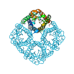
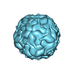
1RC2
 
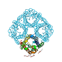 | | 2.5 Angstrom Resolution X-ray Structure of Aquaporin Z | | 分子名称: | 2-O-octyl-beta-D-glucopyranose, Aquaporin Z | | 著者 | Savage, D.F, Egea, P.F, Robles, Y.C, O'Connell III, J.D, Stroud, R.M. | | 登録日 | 2003-11-03 | | 公開日 | 2003-11-25 | | 最終更新日 | 2023-08-23 | | 実験手法 | X-RAY DIFFRACTION (2.5 Å) | | 主引用文献 | Architecture and selectivity in aquaporins: 2.5 a X-ray structure of aquaporin Z
Plos Biol., 1, 2003
|
|
2O9D
 | |
2O9G
 | |
2O9E
 | |
2O9F
 | |
3NKC
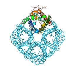 | | Crystal structure of AqpZ F43W,H174G,T183F | | 分子名称: | Aquaporin Z, octyl beta-D-glucopyranoside | | 著者 | Savage, D.F, O'Connell III, J.D, Finer-Moore, J, Stroud, R.M. | | 登録日 | 2010-06-18 | | 公開日 | 2010-11-03 | | 最終更新日 | 2023-09-06 | | 実験手法 | X-RAY DIFFRACTION (3.1 Å) | | 主引用文献 | Structural context shapes the aquaporin selectivity filter.
Proc.Natl.Acad.Sci.USA, 107, 2010
|
|
3NK5
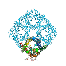 | | Crystal structure of AqpZ mutant F43W | | 分子名称: | Aquaporin Z, octyl beta-D-glucopyranoside | | 著者 | Savage, D.F, O'Connell, J.D, Stroud, R.M, Finer-Moore, J.S. | | 登録日 | 2010-06-18 | | 公開日 | 2010-08-11 | | 最終更新日 | 2024-04-03 | | 実験手法 | X-RAY DIFFRACTION (2.4 Å) | | 主引用文献 | Structural context shapes the aquaporin selectivity filter.
Proc.Natl.Acad.Sci.USA, 107, 2010
|
|
3NKA
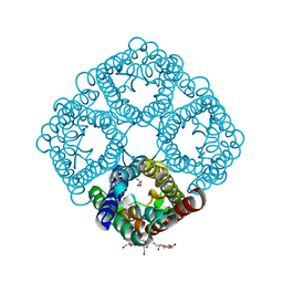 | | Crystal structure of AqpZ H174G,T183F | | 分子名称: | Aquaporin Z, GLYCEROL, octyl beta-D-glucopyranoside | | 著者 | Savage, D.F, O'Connell III, J.D, Finer-Moore, J, Stroud, R.M. | | 登録日 | 2010-06-18 | | 公開日 | 2010-11-03 | | 最終更新日 | 2023-09-06 | | 実験手法 | X-RAY DIFFRACTION (2.5 Å) | | 主引用文献 | Structural context shapes the aquaporin selectivity filter.
Proc.Natl.Acad.Sci.USA, 107, 2010
|
|
7MU1
 | |
7SMK
 | | H. neapolitanus carboxysomal rubisco/CsoSCA-peptide (1-50)complex | | 分子名称: | Carboxysome shell carbonic anhydrase, Ribulose bisphosphate carboxylase large chain, Ribulose bisphosphate carboxylase small chain | | 著者 | Blikstad, C, Dugan, E, Laughlin, T.G, Liu, M, Shoemaker, S, Remis, J, Savage, D.F. | | 登録日 | 2021-10-26 | | 公開日 | 2023-05-24 | | 最終更新日 | 2023-11-01 | | 実験手法 | ELECTRON MICROSCOPY (1.98 Å) | | 主引用文献 | Identification of a carbonic anhydrase-Rubisco complex within the alpha-carboxysome.
Proc.Natl.Acad.Sci.USA, 120, 2023
|
|
7SNV
 | | H. neapolitanus carboxysomal rubisco/CsoSCA-peptide (1-50)complex | | 分子名称: | Carboxysome shell carbonic anhydrase, Ribulose bisphosphate carboxylase large chain, Ribulose bisphosphate carboxylase small chain | | 著者 | Blikstad, C, Dugan, E, Laughlin, T.G, Liu, M, Shoemaker, S, Remis, J, Savage, D.F. | | 登録日 | 2021-10-28 | | 公開日 | 2023-05-03 | | 最終更新日 | 2023-11-01 | | 実験手法 | ELECTRON MICROSCOPY (2.07 Å) | | 主引用文献 | Identification of a carbonic anhydrase-Rubisco complex within the alpha-carboxysome.
Proc.Natl.Acad.Sci.USA, 120, 2023
|
|
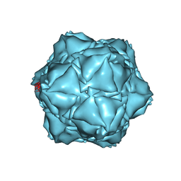
6UEW
 | |
6X8T
 | | CryoEM structure of the apo-SrpI encapasulin complex from Synechococcus elongatus PCC 7942 | | 分子名称: | Protein SrpI | | 著者 | LaFrance, B.J, Nichols, R.J, Phillips, N.R, Oltrogge, L.M, Valentin-Alvarado, L.E, Bischoff, A.J, Savage, D.F, Nogales, E. | | 登録日 | 2020-06-01 | | 公開日 | 2020-06-10 | | 最終更新日 | 2024-03-06 | | 実験手法 | ELECTRON MICROSCOPY (2.9 Å) | | 主引用文献 | Discovery and characterization of a novel family of prokaryotic nanocompartments involved in sulfur metabolism.
Elife, 10, 2021
|
|
6X8M
 | | CryoEM structure of the holo-SrpI encapsulin complex from Synechococcus elongatus PCC 7942 | | 分子名称: | Protein SrpI | | 著者 | LaFrance, B.J, Nichols, R.J, Phillips, N.R, Oltrogge, L.M, Valentin-Alvarado, L.E, Bischoff, A.J, Savage, D.F, Nogales, E. | | 登録日 | 2020-06-01 | | 公開日 | 2020-06-10 | | 最終更新日 | 2024-03-06 | | 実験手法 | ELECTRON MICROSCOPY (2.2 Å) | | 主引用文献 | Discovery and characterization of a novel family of prokaryotic nanocompartments involved in sulfur metabolism.
Elife, 10, 2021
|
|
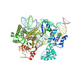
1NM2
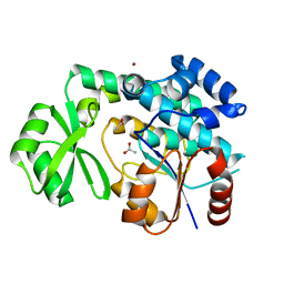 | | Malonyl-CoA:ACP Transacylase | | 分子名称: | ACETIC ACID, NICKEL (II) ION, malonyl CoA:acyl carrier protein malonyltransferase | | 著者 | Keatinge-Clay, A.T, Shelat, A.A, Savage, D.F, Tsai, S, Miercke, L.J.W, O'Connell III, J.D, Khosla, C, Stroud, R.M. | | 登録日 | 2003-01-08 | | 公開日 | 2003-01-21 | | 最終更新日 | 2023-08-16 | | 実験手法 | X-RAY DIFFRACTION (2 Å) | | 主引用文献 | Catalysis, Specificity, and ACP Docking Site of
Streptomyces coelicolor Malonyl-CoA:ACP Transacylase
Structure, 11, 2003
|
|
1RJ9
 | | Structure of the heterodimer of the conserved GTPase domains of the Signal Recognition Particle (Ffh) and Its Receptor (FtsY) | | 分子名称: | MAGNESIUM ION, PHOSPHOMETHYLPHOSPHONIC ACID GUANYLATE ESTER, Signal Recognition Protein, ... | | 著者 | Egea, P.F, Shan, S.O, Napetschnig, J, Savage, D.F, Walter, P, Stroud, R.M. | | 登録日 | 2003-11-18 | | 公開日 | 2004-01-27 | | 最終更新日 | 2024-04-03 | | 実験手法 | X-RAY DIFFRACTION (1.9 Å) | | 主引用文献 | Substrate twinning activates the signal recognition particle and its receptor
Nature, 427, 2004
|
|
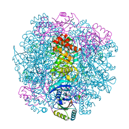
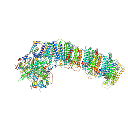
6NBY
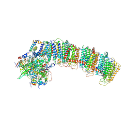 | | T.elongatus NDH (composite model) | | 分子名称: | IRON/SULFUR CLUSTER, NAD(P)H-quinone oxidoreductase chain 4 1, NAD(P)H-quinone oxidoreductase subunit 1, ... | | 著者 | Laughlin, T.G, Bayne, A, Trempe, J.-F, Savage, D.F, Davies, K.M. | | 登録日 | 2018-12-10 | | 公開日 | 2019-02-27 | | 最終更新日 | 2020-04-15 | | 実験手法 | ELECTRON MICROSCOPY (3.1 Å) | | 主引用文献 | Structure of the complex I-like molecule NDH of oxygenic photosynthesis.
Nature, 566, 2019
|
|
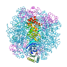
6NBQ
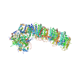 | | T.elongatus NDH (data-set 1) | | 分子名称: | IRON/SULFUR CLUSTER, NAD(P)H-quinone oxidoreductase chain 4 1, NAD(P)H-quinone oxidoreductase subunit 2, ... | | 著者 | Laughlin, T.G, Bayne, A, Trempe, J.-F, Savage, D.F, Davies, K.M. | | 登録日 | 2018-12-09 | | 公開日 | 2019-02-27 | | 最終更新日 | 2019-12-18 | | 実験手法 | ELECTRON MICROSCOPY (3.1 Å) | | 主引用文献 | Structure of the complex I-like molecule NDH of oxygenic photosynthesis.
Nature, 566, 2019
|
|
6NBX
 | | T.elongatus NDH (data-set 2) | | 分子名称: | IRON/SULFUR CLUSTER, NAD(P)H-quinone oxidoreductase chain 4 1, NAD(P)H-quinone oxidoreductase subunit 1, ... | | 著者 | Laughlin, T.G, Bayne, A, Trempe, J.-F, Savage, D.F, Davies, K.M. | | 登録日 | 2018-12-10 | | 公開日 | 2019-02-27 | | 最終更新日 | 2019-12-18 | | 実験手法 | ELECTRON MICROSCOPY (3.5 Å) | | 主引用文献 | Structure of the complex I-like molecule NDH of oxygenic photosynthesis.
Nature, 566, 2019
|
|
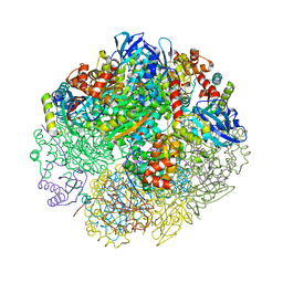
8DC2
 | | Cryo-EM structure of CasLambda (Cas12l) bound to crRNA and DNA | | 分子名称: | CasLambda, DNA NTS, DNA TS, ... | | 著者 | Al-Shayeb, B, Skopintsev, P, Soczek, K, Doudna, J. | | 登録日 | 2022-06-15 | | 公開日 | 2022-12-14 | | 最終更新日 | 2024-06-12 | | 実験手法 | ELECTRON MICROSCOPY (2.99 Å) | | 主引用文献 | Diverse virus-encoded CRISPR-Cas systems include streamlined genome editors.
Cell, 185, 2022
|
|


