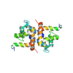
Loading
PDBj
メニューPDBj@FacebookPDBj@TwitterPDBj@YouTubewwPDB FoundationwwPDB
RCSB PDBPDBeBMRBAdv. SearchSearch help
Search by PDB author
1K2H
DownloadVisualize
BU of 1k2h by Molmil
Three-dimensional Solution Structure of apo-S100A1.
分子名称: S-100 protein, alpha chain
著者Rustandi, R.R, Baldisseri, D.M, Inman, K.G, Nizner, P, Hamilton, S.M, Landar, A, Landar, A, Zimmer, D.B, Weber, D.J.
登録日2001-09-27
公開日2002-02-13
最終更新日2024-05-01
実験手法SOLUTION NMR
主引用文献Three-dimensional solution structure of the calcium-signaling protein apo-S100A1 as determined by NMR.
Biochemistry, 41, 2002
1DT7
DownloadVisualize
BU of 1dt7 by Molmil
SOLUTION STRUCTURE OF THE C-TERMINAL NEGATIVE REGULATORY DOMAIN OF P53 IN A COMPLEX WITH CA2+-BOUND S100B(BB)
分子名称: CALCIUM ION, CELLULAR TUMOR ANTIGEN P53, S100 CALCIUM-BINDING PROTEIN
著者Rustandi, R.R, Baldisseri, D.M, Weber, D.J.
登録日2000-01-11
公開日2000-07-26
最終更新日2024-04-10
実験手法SOLUTION NMR
主引用文献Structure of the negative regulatory domain of p53 bound to S100B(betabeta).
Nat.Struct.Biol., 7, 2000
1MWN
DownloadVisualize
BU of 1mwn by Molmil
Solution NMR structure of S100B bound to the high-affinity target peptide TRTK-12
分子名称: CALCIUM ION, F-actin capping protein alpha-1 subunit, S-100 protein, ...
著者Inman, K.G, Yang, R, Rustandi, R.R, Miller, K.E, Baldisseri, D.M, Weber, D.J.
登録日2002-09-30
公開日2002-12-18
最終更新日2024-05-22
実験手法SOLUTION NMR
主引用文献Solution NMR structure of S100B bound to the high-affinity target peptide TRTK-12
J.Mol.Biol., 324, 2002
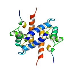
1M31
DownloadVisualize
BU of 1m31 by Molmil
Three-Dimensional Solution Structure of Apo-Mts1
分子名称: Placental calcium-binding protein
著者Vallely, K.M, Rustandi, R.R, Ellis, K.C, Varlamova, O, Bresnick, A.R, Weber, D.J.
登録日2002-06-26
公開日2002-10-30
最終更新日2024-05-22
実験手法SOLUTION NMR
主引用文献Solution structure of human Mts1 (S100A4) as determined by NMR spectroscopy.
Biochemistry, 41, 2002
1QLK
DownloadVisualize
BU of 1qlk by Molmil
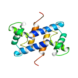
SOLUTION STRUCTURE OF CA(2+)-LOADED RAT S100B (BETABETA) NMR, 20 STRUCTURES
分子名称: CALCIUM ION, S-100 PROTEIN
著者Drohat, A.C, Baldisseri, D.M, Rustandi, R.R, Weber, D.J.
登録日1997-09-26
公開日1998-11-11
最終更新日2024-05-22
実験手法SOLUTION NMR
主引用文献Solution structure of calcium-bound rat S100B(betabeta) as determined by nuclear magnetic resonance spectroscopy,.
Biochemistry, 37, 1998
8EKK
DownloadVisualize
BU of 8ekk by Molmil
Clostridioides difficile binary toxin translocase CDTb wild-type after calcium depletion from receptor binding domain 1 (RBD1) - Class 2
分子名称: ADP-ribosyltransferase binding component, CALCIUM ION
著者Abeyawardhane, D.L, Pozharski, E.
登録日2022-09-21
公開日2023-09-27
実験手法ELECTRON MICROSCOPY (3.28 Å)
主引用文献Calcium-mediated Pore Formation of Clostridioides difficile Binary Toxin
To Be Published
8EKM
DownloadVisualize
BU of 8ekm by Molmil
Clostridioides difficile binary toxin translocase CDTb double mutant - D623A D734A
分子名称: ADP-ribosyltransferase binding component, CALCIUM ION
著者Abeyawardhane, D.L, Pozharski, E.
登録日2022-09-21
公開日2023-09-27
実験手法ELECTRON MICROSCOPY (3.56 Å)
主引用文献Calcium-mediated Pore Formation of Clostridioides difficile Binary Toxin
To Be Published
8EKL
DownloadVisualize
BU of 8ekl by Molmil
Clostridioides difficile binary toxin translocase CDTb wild-type after calcium depletion from receptor binding domain 1 (RBD1) - Class 1
分子名称: ADP-ribosyltransferase binding component, CALCIUM ION
著者Abeyawardhane, D.L, Pozharski, E.
登録日2022-09-21
公開日2023-09-27
実験手法ELECTRON MICROSCOPY (3.06 Å)
主引用文献Calcium-mediated Pore Formation of Clostridioides difficile Binary Toxin
To Be Published
6UWI
DownloadVisualize
BU of 6uwi by Molmil
Crystal structure of the Clostridium difficile translocase CDTb
分子名称: ADP-ribosyltransferase binding component, CALCIUM ION
著者Pozharski, E.
登録日2019-11-05
公開日2020-01-22
最終更新日2024-04-03
実験手法X-RAY DIFFRACTION (3.7 Å)
主引用文献Structure of the cell-binding component of theClostridium difficilebinary toxin reveals a di-heptamer macromolecular assembly.
Proc.Natl.Acad.Sci.USA, 117, 2020
6UWT
DownloadVisualize
BU of 6uwt by Molmil
Clostridium difficile binary toxin translocase CDTb tetradecamer in symmetric conformation
分子名称: ADP-ribosyltransferase binding component, CALCIUM ION
著者Xu, X, Pozharski, E, des Georges, A.
登録日2019-11-05
公開日2020-01-22
最終更新日2024-03-06
実験手法ELECTRON MICROSCOPY (3.1 Å)
主引用文献Structure of the cell-binding component of theClostridium difficilebinary toxin reveals a di-heptamer macromolecular assembly.
Proc.Natl.Acad.Sci.USA, 117, 2020
6UWR
DownloadVisualize
BU of 6uwr by Molmil
Clostridium difficile binary toxin translocase CDTb in asymmetric tetradecamer conformation
分子名称: ADP-ribosyltransferase binding component, CALCIUM ION
著者Xu, X, Pozharski, E, des Georges, A.
登録日2019-11-05
公開日2020-01-22
最終更新日2024-03-06
実験手法ELECTRON MICROSCOPY (2.8 Å)
主引用文献Structure of the cell-binding component of theClostridium difficilebinary toxin reveals a di-heptamer macromolecular assembly.
Proc.Natl.Acad.Sci.USA, 117, 2020
6UWO
DownloadVisualize
BU of 6uwo by Molmil
Crystal structure of receptor binding domain 2 from Clostridium difficile translocase CDTb
分子名称: ADP-ribosyltransferase binding component
著者Pozharski, E.
登録日2019-11-05
公開日2020-01-22
最終更新日2024-03-06
実験手法X-RAY DIFFRACTION (2.3 Å)
主引用文献Structure of the cell-binding component of theClostridium difficilebinary toxin reveals a di-heptamer macromolecular assembly.
Proc.Natl.Acad.Sci.USA, 117, 2020

223790

件を2024-08-14に公開中

PDB statisticsPDBj update infoContact PDBjnumon