5BN7
 
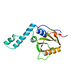
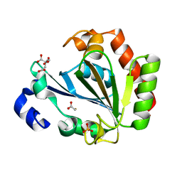
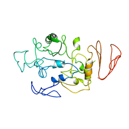 | | Crystal structure of maltodextrin glucosidase from E.coli at 3.7 A resolution | | 分子名称: | Maltodextrin glucosidase | | 著者 | Shukla, P.K, Pastor, A, Singh, A.K, Sharma, S, Singh, T.P, Chaudhuri, T.K. | | 登録日 | 2015-05-25 | | 公開日 | 2015-08-12 | | 最終更新日 | 2023-11-08 | | 実験手法 | X-RAY DIFFRACTION (3.7 Å) | | 主引用文献 | Role of N-terminal region of Escherichia coli maltodextrin glucosidase in folding and function of the protein
Biochim.Biophys.Acta, 1864, 2016
|
|
1APS
 | |
2KTM
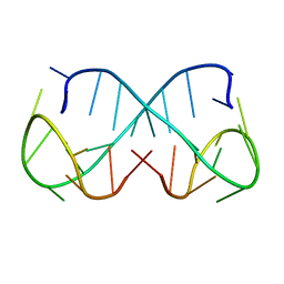
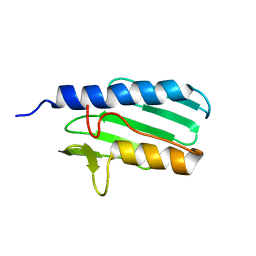
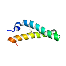 | | Solution NMR structure of H2H3 domain of ovine prion protein (residues 167-234) | | 分子名称: | Major prion protein | | 著者 | Pastore, A, Adrover, M, Pauwels, K, de Chiara, C, Prigent, S, Rezeai, H. | | 登録日 | 2010-02-04 | | 公開日 | 2010-04-07 | | 最終更新日 | 2021-11-10 | | 実験手法 | SOLUTION NMR | | 主引用文献 | Prion fibrillization is mediated by a native structural element that comprises helices H2 and H3.
J.Biol.Chem., 285, 2010
|
|
1V06
 | |
7QAB
 | |
1NCU
 | |
1NCT
 | |
1YZB
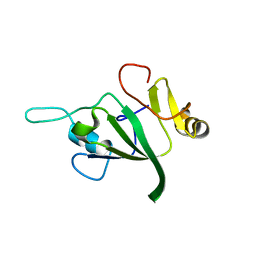
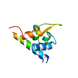
 | | Solution structure of the Josephin domain of Ataxin-3 | | 分子名称: | Machado-Joseph disease protein 1 | | 著者 | Nicastro, G, Masino, L, Menon, R.P, Knowles, P.P, McDonald, N.Q, Pastore, A. | | 登録日 | 2005-02-28 | | 公開日 | 2005-07-05 | | 最終更新日 | 2024-05-29 | | 実験手法 | SOLUTION NMR | | 主引用文献 | The solution structure of the Josephin domain of ataxin-3: Structural determinants for molecular recognition
Proc.Natl.Acad.Sci.Usa, 102, 2005
|
|
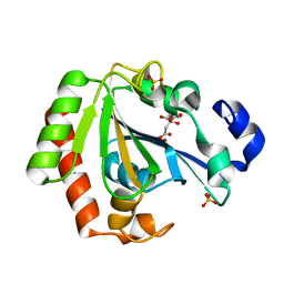
4AQP
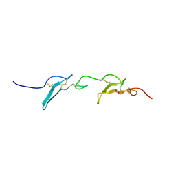
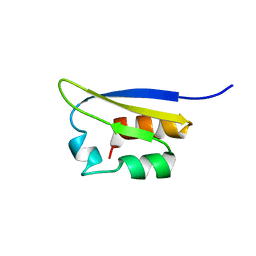
 | | The structure of the AXH domain of ataxin-1. | | 分子名称: | ATAXIN-1, DI(HYDROXYETHYL)ETHER, SODIUM ION | | 著者 | Rees, M, Chen, Y.W, de Chiara, C, Pastore, A. | | 登録日 | 2012-04-19 | | 公開日 | 2013-03-27 | | 最終更新日 | 2023-12-20 | | 実験手法 | X-RAY DIFFRACTION (2.452 Å) | | 主引用文献 | Self-Assembly and Conformational Heterogeneity of the Axh Domain of Ataxin-1: An Unusual Example of a Chameleon Fold
Biophys.J., 104, 2013
|
|
4APT
 | | The structure of the AXH domain of ataxin-1. | | 分子名称: | ATAXIN-1, SODIUM ION | | 著者 | Rees, M, Chen, Y.W, de Chiara, C, Pastore, A. | | 登録日 | 2012-04-05 | | 公開日 | 2013-03-27 | | 最終更新日 | 2023-12-20 | | 実験手法 | X-RAY DIFFRACTION (2.5 Å) | | 主引用文献 | Self-Assembly and Conformational Heterogeneity of the Axh Domain of Ataxin-1: An Unusual Example of a Chameleon Fold
Biophys.J., 104, 2013
|
|
8Q5Q
 | |
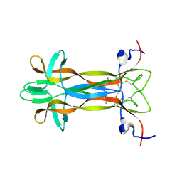
1H8B
 | | EF-hands 3,4 from alpha-actinin / Z-repeat 7 from titin | | 分子名称: | ALPHA-ACTININ 2, SKELETAL MUSCLE ISOFORM, TITIN | | 著者 | Atkinson, R.A, Joseph, C, Kelly, G, Muskett, F.W, Frenkiel, T.A, Nietlispach, D, Pastore, A. | | 登録日 | 2001-02-01 | | 公開日 | 2001-08-30 | | 最終更新日 | 2024-05-15 | | 実験手法 | SOLUTION NMR | | 主引用文献 | Ca2+-Independent Binding of an EF-Hand Domain to a Novel Motif in the Alpha-Actinin-Titin Complex
Nat.Struct.Biol., 8, 2001
|
|
1VIH
 | | NMR STUDY OF VIGILIN, REPEAT 6, MINIMIZED AVERAGE STRUCTURE | | 分子名称: | VIGILIN | | 著者 | Musco, G, Stier, G, Joseph, C, Morelli, M.A.C, Nilges, M, Gibson, T.J, Pastore, A. | | 登録日 | 1995-11-29 | | 公開日 | 1996-04-03 | | 最終更新日 | 2024-05-22 | | 実験手法 | SOLUTION NMR | | 主引用文献 | Three-dimensional structure and stability of the KH domain: molecular insights into the fragile X syndrome.
Cell(Cambridge,Mass.), 85, 1996
|
|
1ZTA
 | | THE SOLUTION STRUCTURE OF A LEUCINE-ZIPPER MOTIF PEPTIDE | | 分子名称: | LEUCINE ZIPPER MONOMER | | 著者 | Saudek, V, Pastore, A, Castiglione Morelli, M.A, Frank, R, Gausepohl, H, Gibson, T. | | 登録日 | 1990-10-11 | | 公開日 | 1993-04-15 | | 最終更新日 | 2024-05-22 | | 実験手法 | SOLUTION NMR | | 主引用文献 | The solution structure of a leucine-zipper motif peptide.
Protein Eng., 4, 1991
|
|
1VIG
 | | NMR STUDY OF VIGILIN, REPEAT 6, 40 STRUCTURES | | 分子名称: | VIGILIN | | 著者 | Musco, G, Stier, G, Joseph, C, Morelli, M.A.C, Nilges, M, Gibson, T.J, Pastore, A. | | 登録日 | 1995-11-29 | | 公開日 | 1996-04-03 | | 最終更新日 | 2024-05-22 | | 実験手法 | SOLUTION NMR | | 主引用文献 | Three-dimensional structure and stability of the KH domain: molecular insights into the fragile X syndrome.
Cell(Cambridge,Mass.), 85, 1996
|
|
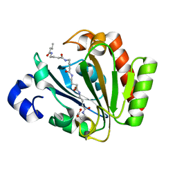
2BZT
 | | NMR structure of the bacterial protein YFHJ from E. coli | | 分子名称: | PROTEIN ISCX | | 著者 | Pastore, C, Kelly, G, Adinolfi, S, Mc Cormick, J.E, Pastore, A. | | 登録日 | 2005-08-22 | | 公開日 | 2006-12-06 | | 最終更新日 | 2024-05-15 | | 実験手法 | SOLUTION NMR | | 主引用文献 | YfhJ, a molecular adaptor in iron-sulfur cluster formation or a frataxin-like protein?
Structure, 14, 2006
|
|
1SOY
 | | Solution structure of the bacterial frataxin orthologue, CyaY | | 分子名称: | CyaY protein | | 著者 | Nair, M, Adinolfi, S, Pastore, C, Kelly, G, Temussi, P, Pastore, A. | | 登録日 | 2004-03-16 | | 公開日 | 2004-11-23 | | 最終更新日 | 2024-05-22 | | 実験手法 | SOLUTION NMR | | 主引用文献 | Solution Structure of the Bacterial Frataxin Ortholog, CyaY; Mapping the Iron Binding Sites
Structure, 12, 2004
|
|
6HRJ
 | | Apo - YndL | | 分子名称: | CITRATE ANION, SULFATE ION, YndL, ... | | 著者 | Ramaswamy, S, Rasheed, M, Morelli, C, Calvio, C, Sutton, B, Pastore, A. | | 登録日 | 2018-09-27 | | 公開日 | 2018-10-10 | | 最終更新日 | 2024-05-15 | | 実験手法 | X-RAY DIFFRACTION (1.7 Å) | | 主引用文献 | The structure of PghL hydrolase bound to its substrate poly-gamma-glutamate.
FEBS J., 285, 2018
|
|
6HRI
 | | Native YndL | | 分子名称: | CITRATE ANION, IMIDAZOLE, SULFATE ION, ... | | 著者 | Ramaswamy, S, Rasheed, M, Morelli, C, Calvio, C, Sutton, B, Pastore, A. | | 登録日 | 2018-09-27 | | 公開日 | 2018-10-10 | | 最終更新日 | 2024-05-15 | | 実験手法 | X-RAY DIFFRACTION (1.03 Å) | | 主引用文献 | The structure of PghL hydrolase bound to its substrate poly-gamma-glutamate.
FEBS J., 285, 2018
|
|
5LSD
 | | recombinant mouse Nerve Growth Factor | | 分子名称: | Beta-nerve growth factor | | 著者 | Paoletti, F, de Chiara, C, Kelly, G, Lamba, D, Cattaneo, A, Pastore, A. | | 登録日 | 2016-08-25 | | 公開日 | 2017-07-05 | | 最終更新日 | 2024-07-03 | | 実験手法 | SOLUTION NMR | | 主引用文献 | Conformational Rigidity within Plasticity Promotes Differential Target Recognition of Nerve Growth Factor.
Front Mol Biosci, 3, 2016
|
|
1TNM
 | |
1TNN
 | |
5ONJ
 | | YnDL in Complex with 5 amino acid (PGA) complex | | 分子名称: | (2R,3S,4R,5R,6R)-6-((1R,2R,3S,4R,6S)-4,6-DIAMINO-2,3-DIHYDROXYCYCLOHEXYLOXY)-5-AMINO-2-(AMINOMETHYL)-TETRAHYDRO-2H-PYRAN-3,4-DIOL, YndL, ZINC ION | | 著者 | Ramaswamy, S, Rasheed, M, Morelli, C, Calvio, C, Sutton, B, Pastore, A. | | 登録日 | 2017-08-03 | | 公開日 | 2018-08-29 | | 最終更新日 | 2024-01-17 | | 実験手法 | X-RAY DIFFRACTION (1.7 Å) | | 主引用文献 | The structure of PghL hydrolase bound to its substrate poly-gamma-glutamate.
Febs J., 285, 2018
|
|
1LY7
 | | The solution structure of the the c-terminal domain of frataxin, the protein responsible for friedreich ataxia | | 分子名称: | frataxin | | 著者 | Musco, G, Stier, G, Kolmerer, B, Adinolfi, S, Martin, S, Frenkiel, T, Gibson, T, Pastore, A. | | 登録日 | 2002-06-07 | | 公開日 | 2002-06-26 | | 最終更新日 | 2024-05-22 | | 実験手法 | SOLUTION NMR | | 主引用文献 | Towards a structural understanding of Friedreich's
ataxia: the solution structure of frataxin
Structure Fold.Des., 8, 2000
|
|
1TIU
 | | TITIN, IG REPEAT 27, NMR, 24 STRUCTURES | | 分子名称: | TITIN, I27 | | 著者 | Improta, S, Politou, A.S, Pastore, A. | | 登録日 | 1996-02-02 | | 公開日 | 1996-07-11 | | 最終更新日 | 2024-05-22 | | 実験手法 | SOLUTION NMR | | 主引用文献 | Immunoglobulin-like modules from titin I-band: extensible components of muscle elasticity.
Structure, 4, 1996
|
|


