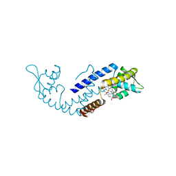
6LKT
 
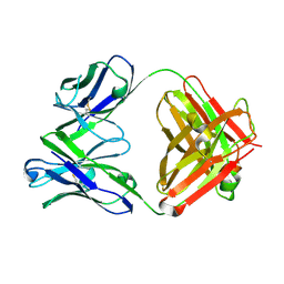 | | Crystal structure of the Fab fragment of murine monoclonal antibody KH-1 against Human herpesvirus 6B | | 分子名称: | antibody Fab Fragment L-chain, antibody Fab fragment H chain | | 著者 | Nishimura, M, Novita, B.D, Kato, T, Tjan, L.H, Wang, B, Wakata, A, Poetranto, A.L, Kawabata, A, Tang, H, Aoshi, T, Mori, Y. | | 登録日 | 2019-12-20 | | 公開日 | 2020-06-17 | | 最終更新日 | 2023-11-22 | | 実験手法 | X-RAY DIFFRACTION (1.8 Å) | | 主引用文献 | Structural basis for the interaction of human herpesvirus 6B tetrameric glycoprotein complex with the cellular receptor, human CD134.
Plos Pathog., 16, 2020
|
|
2PA2
 | | Crystal structure of human Ribosomal protein L10 core domain | | 分子名称: | 60S ribosomal protein L10, POTASSIUM ION | | 著者 | Nishimura, M, Kaminishi, T, Takemoto, C, Kawazoe, M, Yoshida, T, Tanaka, A, Sugano, S, Shirouzu, M, Ohkubo, T, Yokoyama, S, Kobayashi, Y, RIKEN Structural Genomics/Proteomics Initiative (RSGI) | | 登録日 | 2007-03-27 | | 公開日 | 2008-03-11 | | 最終更新日 | 2023-10-25 | | 実験手法 | X-RAY DIFFRACTION (2.5 Å) | | 主引用文献 | Crystal Structure of Human Ribosomal Protein L10 Core Domain Reveals Eukaryote-Specific Motifs in Addition to the Conserved Fold
J.Mol.Biol., 377, 2008
|
|
8IHL
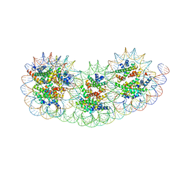 | | Overlapping tri-nucleosome | | 分子名称: | DNA (353-MER), Histone H2A type 1-B/E, Histone H2B type 1-J, ... | | 著者 | Nishimura, M, Fujii, T, Tanaka, H, Maehara, K, Nozawa, K, Takizawa, Y, Ohkawa, Y, Kurumizaka, H. | | 登録日 | 2023-02-23 | | 公開日 | 2024-01-17 | | 最終更新日 | 2024-01-24 | | 実験手法 | ELECTRON MICROSCOPY (7.64 Å) | | 主引用文献 | Genome-wide mapping and cryo-EM structural analyses of the overlapping tri-nucleosome composed of hexasome-hexasome-octasome moieties.
Commun Biol, 7, 2024
|
|
6LTG
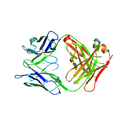 | | Crystal structure of the Fab fragment of murine monoclonal antibody OHV-3 against Human herpesvirus 6B | | 分子名称: | MAGNESIUM ION, antibody Fab fragment H-chain, antibody Fab fragment L-chain | | 著者 | Nishimura, M, Novita, B.D, Kato, T, Tjan, L.H, Wang, B, Wakata, A, Poetranto, A.L, Kawabata, A, Tang, H, Aoshi, T, Mori, Y. | | 登録日 | 2020-01-22 | | 公開日 | 2020-06-17 | | 最終更新日 | 2023-11-29 | | 実験手法 | X-RAY DIFFRACTION (1.63 Å) | | 主引用文献 | Structural basis for the interaction of human herpesvirus 6B tetrameric glycoprotein complex with the cellular receptor, human CD134.
Plos Pathog., 16, 2020
|
|
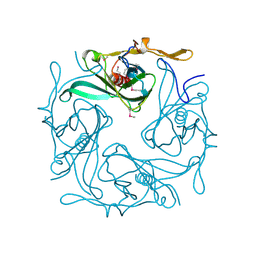
1GCQ
 | | CRYSTAL STRUCTURE OF VAV AND GRB2 SH3 DOMAINS | | 分子名称: | (4R)-2-METHYLPENTANE-2,4-DIOL, GROWTH FACTOR RECEPTOR-BOUND PROTEIN 2, VAV PROTO-ONCOGENE | | 著者 | Nishida, M, Nagata, K, Hachimori, Y, Ogura, K, Inagaki, F. | | 登録日 | 2000-08-08 | | 公開日 | 2001-08-08 | | 最終更新日 | 2023-12-27 | | 実験手法 | X-RAY DIFFRACTION (1.68 Å) | | 主引用文献 | Novel recognition mode between Vav and Grb2 SH3 domains.
EMBO J., 20, 2001
|
|
1GCP
 | | CRYSTAL STRUCTURE OF VAV SH3 DOMAIN | | 分子名称: | VAV PROTO-ONCOGENE | | 著者 | Nishida, M, Nagata, K, Hachimori, Y, Ogura, K, Inagaki, F. | | 登録日 | 2000-08-08 | | 公開日 | 2001-08-08 | | 最終更新日 | 2023-10-25 | | 実験手法 | X-RAY DIFFRACTION (2.1 Å) | | 主引用文献 | Novel recognition mode between Vav and Grb2 SH3 domains.
EMBO J., 20, 2001
|
|
7DVU
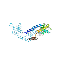 | | Crystal structure of heme sensor protein PefR in complex with heme and cyanide | | 分子名称: | CYANIDE ION, HTH marR-type domain-containing protein, PROTOPORPHYRIN IX CONTAINING FE | | 著者 | Nishinaga, M, Nagai, S, Nishitani, Y, Sugimoto, H, Shiro, Y, Sawai, H. | | 登録日 | 2021-01-15 | | 公開日 | 2021-09-29 | | 最終更新日 | 2023-11-29 | | 実験手法 | X-RAY DIFFRACTION (2.1 Å) | | 主引用文献 | Heme controls the structural rearrangement of its sensor protein mediating the hemolytic bacterial survival.
Commun Biol, 4, 2021
|
|
7DVT
 | | Crystal structure of heme sensor protein PefR in complex with heme and carbon monoxide | | 分子名称: | CARBON MONOXIDE, HTH marR-type domain-containing protein, PROTOPORPHYRIN IX CONTAINING FE | | 著者 | Nishinaga, M, Nagai, S, Nishitani, Y, Sugimoto, H, Shiro, Y, Sawai, H. | | 登録日 | 2021-01-15 | | 公開日 | 2021-09-29 | | 最終更新日 | 2023-11-29 | | 実験手法 | X-RAY DIFFRACTION (2.09 Å) | | 主引用文献 | Heme controls the structural rearrangement of its sensor protein mediating the hemolytic bacterial survival.
Commun Biol, 4, 2021
|
|
7DVR
 | | Crystal structure of heme sensor protein PefR from Streptococcus agalactiae in complex with heme | | 分子名称: | COBALT (II) ION, HTH marR-type domain-containing protein, PROTOPORPHYRIN IX CONTAINING FE | | 著者 | Nishinaga, M, Nagai, S, Nishitani, Y, Sugimoto, H, Shiro, Y, Sawai, H. | | 登録日 | 2021-01-15 | | 公開日 | 2021-09-29 | | 最終更新日 | 2024-05-29 | | 実験手法 | X-RAY DIFFRACTION (1.7 Å) | | 主引用文献 | Heme controls the structural rearrangement of its sensor protein mediating the hemolytic bacterial survival.
Commun Biol, 4, 2021
|
|
7DVV
 | | Heme sensor protein PefR from Streptococcus agalactiae bound to operator DNA (28-mer) | | 分子名称: | DNA (28-MER), HTH marR-type domain-containing protein | | 著者 | Nishinaga, M, Nagai, S, Nishitani, Y, Sugimoto, H, Shiro, Y, Sawai, H. | | 登録日 | 2021-01-15 | | 公開日 | 2021-09-29 | | 最終更新日 | 2023-11-29 | | 実験手法 | X-RAY DIFFRACTION (2.49 Å) | | 主引用文献 | Heme controls the structural rearrangement of its sensor protein mediating the hemolytic bacterial survival.
Commun Biol, 4, 2021
|
|
3VKW
 | | Crystal Structure of the Superfamily 1 Helicase from Tomato Mosaic Virus | | 分子名称: | Replicase large subunit, SULFATE ION | | 著者 | Nishikiori, M, Sugiyama, S, Xiang, H, Niiyama, M, Ishibashi, K, Inoue, T, Ishikawa, M, Matsumura, H, Katoh, E. | | 登録日 | 2011-11-22 | | 公開日 | 2012-07-11 | | 最終更新日 | 2024-03-20 | | 実験手法 | X-RAY DIFFRACTION (1.9 Å) | | 主引用文献 | Crystal structure of the superfamily 1 helicase from tomato mosaic virus
J.Virol., 86, 2012
|
|
7F47
 | | Cryo-EM structure of Rhizobium etli MprF | | 分子名称: | (1R)-2-{[(S)-{[(2S)-2,3-dihydroxypropyl]oxy}(hydroxy)phosphoryl]oxy}-1-[(hexadecanoyloxy)methyl]ethyl (9Z)-octadec-9-enoate, Hypothetical conserved protein, [(2R)-1-[[(2R)-3-[(2S)-2,6-bis(azanyl)hexanoyl]oxy-2-oxidanyl-propoxy]-oxidanyl-phosphoryl]oxy-3-hexadecanoyloxy-propan-2-yl] (E)-octadec-9-enoate | | 著者 | Nishimura, M, Hirano, H, Kobayashi, K, Gill, C.P, Phan, C.N.K, Kise, Y, Kusakizako, T, Yamashita, K, Ito, Y, Roy, H, Nishizawa, T, Nureki, O. | | 登録日 | 2021-06-17 | | 公開日 | 2022-06-22 | | 最終更新日 | 2024-06-12 | | 実験手法 | ELECTRON MICROSCOPY (2.99 Å) | | 主引用文献 | Cryo-EM structure of Rhizobium etli MprF
To Be Published
|
|
1N9P
 | |
1ZDV
 | | Solution Structure of the type 1 pilus assembly platform FimD(25-139) | | 分子名称: | Outer membrane usher protein fimD | | 著者 | Nishiyama, M, Horst, R, Herrmann, T, Vetsch, M, Bettendorff, P, Ignatov, O, Grutter, M, Wuthrich, K, Glockshuber, R, Capitani, G. | | 登録日 | 2005-04-15 | | 公開日 | 2005-06-14 | | 最終更新日 | 2024-05-22 | | 実験手法 | SOLUTION NMR | | 主引用文献 | Structural basis of chaperone-subunit complex recognition by the type 1 pilus assembly platform FimD.
Embo J., 24, 2005
|
|
1ZDX
 | | Solution Structure of the type 1 pilus assembly platform FimD(25-125) | | 分子名称: | Outer membrane usher protein fimD | | 著者 | Nishiyama, M, Horst, R, Herrmann, T, Vetsch, M, Bettendorff, P, Ignatov, O, Grutter, M, Wuthrich, K, Glockshuber, R, Capitani, G. | | 登録日 | 2005-04-15 | | 公開日 | 2005-06-14 | | 最終更新日 | 2024-05-22 | | 実験手法 | SOLUTION NMR | | 主引用文献 | Structural basis of chaperone-subunit complex recognition by the type 1 pilus assembly platform FimD.
Embo J., 24, 2005
|
|
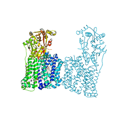
1IWP
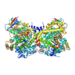 | | Glycerol Dehydratase-cyanocobalamin Complex of Klebsiella pneumoniae | | 分子名称: | COBALAMIN, Glycerol Dehydratase Alpha subunit, Glycerol Dehydratase Beta subunit, ... | | 著者 | Yamanishi, M, Yunoki, M, Tobimatsu, T, Toraya, T. | | 登録日 | 2002-05-28 | | 公開日 | 2002-10-02 | | 最終更新日 | 2023-10-25 | | 実験手法 | X-RAY DIFFRACTION (2.1 Å) | | 主引用文献 | The crystal structure of coenzyme B12-dependent glycerol dehydratase in complex with cobalamin and propane-1,2-diol.
Eur.J.Biochem., 269, 2002
|
|
2QKS
 | |
1WKI
 | | solution structure of ribosomal protein L16 from thermus thermophilus HB8 | | 分子名称: | LSU ribosomal protein L16P | | 著者 | Nishimura, M, Yoshida, T, Shirouzu, M, Terada, T, Kuramitsu, S, Yokoyama, S, Ohkubo, T, Kobayashi, Y, RIKEN Structural Genomics/Proteomics Initiative (RSGI) | | 登録日 | 2004-05-31 | | 公開日 | 2004-12-14 | | 最終更新日 | 2024-05-01 | | 実験手法 | SOLUTION NMR | | 主引用文献 | Solution Structure of Ribosomal Protein L16 from Thermus thermophilus HB8
J.Mol.Biol., 344, 2004
|
|
1ZE3
 | | Crystal Structure of the Ternary Complex of FIMD (N-Terminal Domain) with FIMC and the Pilin Domain of FIMH | | 分子名称: | 1,2-ETHANEDIOL, Chaperone protein fimC, FimH protein, ... | | 著者 | Nishiyama, M, Horst, R, Eidam, O, Herrmann, T, Ignatov, O, Vetsch, M, Bettendorff, P, Jelesarov, I, Grutter, M.G, Wuthrich, K, Glockshuber, R, Capitani, G. | | 登録日 | 2005-04-17 | | 公開日 | 2005-06-14 | | 最終更新日 | 2023-08-23 | | 実験手法 | X-RAY DIFFRACTION (1.84 Å) | | 主引用文献 | Structural basis of chaperone-subunit complex recognition by the type 1 pilus assembly platform FimD.
Embo J., 24, 2005
|
|
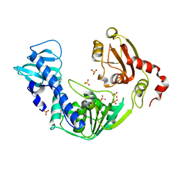
1A0F
 | | CRYSTAL STRUCTURE OF GLUTATHIONE S-TRANSFERASE FROM ESCHERICHIA COLI COMPLEXED WITH GLUTATHIONESULFONIC ACID | | 分子名称: | GLUTATHIONE S-TRANSFERASE, GLUTATHIONE SULFONIC ACID | | 著者 | Nishida, M, Harada, S, Noguchi, S, Inoue, H, Takahashi, K, Satow, Y. | | 登録日 | 1997-11-29 | | 公開日 | 1999-01-13 | | 最終更新日 | 2024-02-07 | | 実験手法 | X-RAY DIFFRACTION (2.1 Å) | | 主引用文献 | Three-dimensional structure of Escherichia coli glutathione S-transferase complexed with glutathione sulfonate: catalytic roles of Cys10 and His106.
J.Mol.Biol., 281, 1998
|
|
5WX8
 | | Human herpesvirus 6A immediate early protein 2 C-terminal domain | | 分子名称: | Immediate-early protein 2 | | 著者 | Nishimura, M, Wang, J, Wakata, A, Sakamoto, K, Mori, Y. | | 登録日 | 2017-01-06 | | 公開日 | 2017-08-02 | | 最終更新日 | 2017-12-06 | | 実験手法 | X-RAY DIFFRACTION (2.5 Å) | | 主引用文献 | Crystal Structure of the DNA-Binding Domain of Human Herpesvirus 6A Immediate Early Protein 2.
J. Virol., 91, 2017
|
|
3A4U
 | | Crystal structure of MCFD2 in complex with carbohydrate recognition domain of ERGIC-53 | | 分子名称: | CALCIUM ION, GLYCEROL, Multiple coagulation factor deficiency protein 2, ... | | 著者 | Nishio, M, Kamiya, Y, Mizushima, T, Wakatsuki, S, Sasakawa, H, Yamamoto, K, Uchiyama, S, Noda, M, McKay, A.R, Fukui, K, Hauri, H.P, Kato, K. | | 登録日 | 2009-07-17 | | 公開日 | 2010-01-05 | | 最終更新日 | 2023-11-01 | | 実験手法 | X-RAY DIFFRACTION (1.84 Å) | | 主引用文献 | Structural basis for the cooperative interplay between the two causative gene products of combined factor V and factor VIII deficiency.
Proc.Natl.Acad.Sci.USA, 107, 2010
|
|
3AUJ
 | | Structure of diol dehydratase complexed with glycerol | | 分子名称: | CALCIUM ION, COBALAMIN, Diol dehydrase alpha subunit, ... | | 著者 | Yamanishi, M, Kinoshita, K, Fukuoka, M, Shibata, T, Tobimatsu, T, Toraya, T. | | 登録日 | 2011-02-07 | | 公開日 | 2012-02-22 | | 最終更新日 | 2023-11-01 | | 実験手法 | X-RAY DIFFRACTION (2.1 Å) | | 主引用文献 | Redesign of coenzyme B(12) dependent diol dehydratase to be resistant to the mechanism-based inactivation by glycerol and act on longer chain 1,2-diols
Febs J., 279, 2012
|
|
7Y00
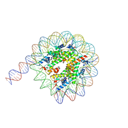 | | Cryo-EM structure of the nucleosome containing 169 base-pair DNA with a p53 target sequence | | 分子名称: | DNA (169-MER), Histone H2A type 1-B/E, Histone H2B type 1-J, ... | | 著者 | Nishimura, M, Nozawa, K, Takizawa, Y, Kurumizaka, H. | | 登録日 | 2022-06-03 | | 公開日 | 2022-10-19 | | 最終更新日 | 2024-07-03 | | 実験手法 | ELECTRON MICROSCOPY (3.96 Å) | | 主引用文献 | Structural basis for p53 binding to its nucleosomal target DNA sequence.
Pnas Nexus, 1, 2022
|
|
7XZY
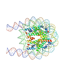 | | Cryo-EM structure of the nucleosome containing 193 base-pair DNA with a p53 target sequence | | 分子名称: | DNA (193-MER), Histone H2A type 1-B/E, Histone H2B type 1-J, ... | | 著者 | Nishimura, M, Nozawa, K, Takizawa, Y, Kurumizaka, H. | | 登録日 | 2022-06-03 | | 公開日 | 2022-10-19 | | 最終更新日 | 2024-07-03 | | 実験手法 | ELECTRON MICROSCOPY (3.97 Å) | | 主引用文献 | Structural basis for p53 binding to its nucleosomal target DNA sequence.
Pnas Nexus, 1, 2022
|
|


