Loading
PDBj
メニューPDBj@FacebookPDBj@TwitterPDBj@YouTubewwPDB FoundationwwPDB
RCSB PDBPDBeBMRBAdv. SearchSearch help
Search by PDB author
3ZY1
DownloadVisualize
BU of 3zy1 by Molmil
Crystal structure of the human p63 tetramerization domain
分子名称: TUMOR PROTEIN 63
著者Natan, E, Joerger, A.C.
登録日2011-08-16
公開日2011-11-30
最終更新日2024-05-08
実験手法X-RAY DIFFRACTION (2.15 Å)
主引用文献Structure and Kinetic Stability of the P63 Tetramerization Domain.
J.Mol.Biol., 415, 2012
2XWR
DownloadVisualize
BU of 2xwr by Molmil
Crystal structure of the DNA-binding domain of human p53 with extended N terminus
分子名称: CELLULAR TUMOR ANTIGEN P53, ZINC ION
著者Joerger, A.C, Natan, E, Fersht, A.R.
登録日2010-11-04
公開日2011-03-30
最終更新日2024-05-08
実験手法X-RAY DIFFRACTION (1.68 Å)
主引用文献Interaction of the P53 DNA-Binding Domain with its N-Terminal Extension Modulates the Stability of the P53 Tetramer.
J.Mol.Biol., 409, 2011
3ZY0
DownloadVisualize
BU of 3zy0 by Molmil
Crystal structure of a truncated variant of the human p63 tetramerization domain lacking the C-terminal helix
分子名称: TUMOR PROTEIN P63
著者Joerger, A.C.
登録日2011-08-16
公開日2011-11-30
最終更新日2019-05-08
実験手法X-RAY DIFFRACTION (1.9 Å)
主引用文献Structure and Kinetic Stability of the P63 Tetramerization Domain.
J.Mol.Biol., 415, 2012
2YBG
DownloadVisualize
BU of 2ybg by Molmil
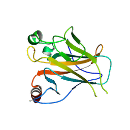
Structure of Lys120-acetylated p53 core domain
分子名称: CELLULAR TUMOR ANTIGEN P53, ZINC ION
著者Arbely, E, Allen, M.D, Joerger, A.C, Fersht, A.R.
登録日2011-03-08
公開日2011-05-04
最終更新日2011-07-13
実験手法X-RAY DIFFRACTION (1.9 Å)
主引用文献Acetylation of Lysine 120 of P53 Endows DNA- Binding Specificity at Effective Physiological Salt Concentration.
Proc.Natl.Acad.Sci.USA, 108, 2011
2WQI
DownloadVisualize
BU of 2wqi by Molmil
Crystal structure of the human p73 tetramerization domain
分子名称: TUMOR PROTEIN P73
著者Joerger, A.C.
登録日2009-08-21
公開日2009-10-06
最終更新日2024-05-08
実験手法X-RAY DIFFRACTION (1.7 Å)
主引用文献Structural Evolution of P53, P63, and P73: Implication for Heterotetramer Formation.
Proc.Natl.Acad.Sci.USA, 106, 2009
2WTT
DownloadVisualize
BU of 2wtt by Molmil
Structure of the human p73 tetramerization domain (crystal form II)
分子名称: TUMOR PROTEIN P73
著者Joerger, A.C.
登録日2009-09-21
公開日2009-10-13
最終更新日2023-12-20
実験手法X-RAY DIFFRACTION (2.3 Å)
主引用文献Structural Evolution of P53, P63, and P73: Implication for Heterotetramer Formation.
Proc.Natl.Acad.Sci.USA, 106, 2009
2WQJ
DownloadVisualize
BU of 2wqj by Molmil
Crystal structure of a truncated variant of the human p73 tetramerization domain
分子名称: TUMOR PROTEIN P73
著者Joerger, A.C.
登録日2009-08-21
公開日2009-10-06
最終更新日2024-05-08
実験手法X-RAY DIFFRACTION (2 Å)
主引用文献Structural Evolution of P53, P63, and P73: Implication for Heterotetramer Formation.
Proc.Natl.Acad.Sci.USA, 106, 2009

225946

件を2024-10-09に公開中

PDB statisticsPDBj update infoContact PDBjnumon