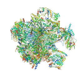
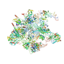
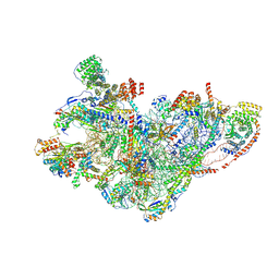
7QH6
 
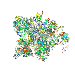 | | Cryo-EM structure of the human mtLSU assembly intermediate upon MRM2 depletion - class 1 | | 分子名称: | 16S ribosomal RNA, 39S ribosomal protein L13, mitochondrial, ... | | 著者 | Rebelo-Guiomar, P, Pellegrino, S, Dent, K.C, Warren, A.J, Minczuk, M. | | 登録日 | 2021-12-10 | | 公開日 | 2022-03-02 | | 最終更新日 | 2024-07-17 | | 実験手法 | ELECTRON MICROSCOPY (3.08 Å) | | 主引用文献 | A late-stage assembly checkpoint of the human mitochondrial ribosome large subunit.
Nat Commun, 13, 2022
|
|
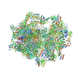
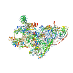
7QH7
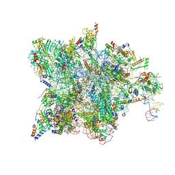 | | Cryo-EM structure of the human mtLSU assembly intermediate upon MRM2 depletion - class 4 | | 分子名称: | 16S ribosomal RNA, 39S ribosomal protein L10, mitochondrial, ... | | 著者 | Rebelo-Guiomar, P, Pellegrino, S, Dent, K.C, Warren, A.J, Minczuk, M. | | 登録日 | 2021-12-10 | | 公開日 | 2022-05-18 | | 実験手法 | ELECTRON MICROSCOPY (2.89 Å) | | 主引用文献 | A late-stage assembly checkpoint of the human mitochondrial ribosome large subunit.
Nat Commun, 13, 2022
|
|
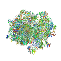
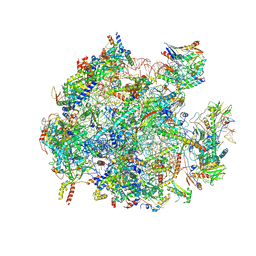
7A5H
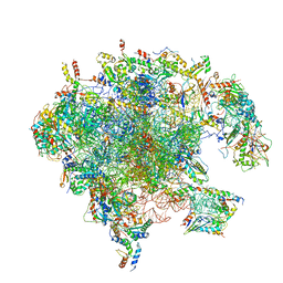 | | Structure of the split human mitoribosomal large subunit with rescue factors mtRF-R and MTRES1 | | 分子名称: | 16S rRNA, 39S ribosomal protein L10, mitochondrial, ... | | 著者 | Desai, N, Yang, H, Chandrasekaran, V, Kazi, R, Minczuk, M, Ramakrishnan, V. | | 登録日 | 2020-08-21 | | 公開日 | 2020-12-23 | | 最終更新日 | 2023-03-01 | | 実験手法 | ELECTRON MICROSCOPY (3.3 Å) | | 主引用文献 | Elongational stalling activates mitoribosome-associated quality control.
Science, 370, 2020
|
|
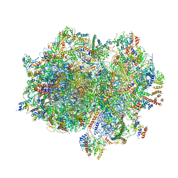
7A5F
 | | Structure of the stalled human mitoribosome with P- and E-site mt-tRNAs | | 分子名称: | 12S rRNA, 16S rRNA, 28S ribosomal protein S10, ... | | 著者 | Desai, N, Yang, H, Chandrasekaran, V, Kazi, R, Minczuk, M, Ramakrishnan, V. | | 登録日 | 2020-08-21 | | 公開日 | 2020-12-23 | | 実験手法 | ELECTRON MICROSCOPY (4.4 Å) | | 主引用文献 | Elongational stalling activates mitoribosome-associated quality control.
Science, 370, 2020
|
|
7A5J
 | | Structure of the split human mitoribosomal large subunit with P-and E-site mt-tRNAs | | 分子名称: | 16S rRNA, 39S ribosomal protein L10, mitochondrial, ... | | 著者 | Desai, N, Yang, H, Chandrasekaran, V, Kazi, R, Minczuk, M, Ramakrishnan, V. | | 登録日 | 2020-08-21 | | 公開日 | 2020-12-23 | | 最終更新日 | 2023-03-15 | | 実験手法 | ELECTRON MICROSCOPY (3.1 Å) | | 主引用文献 | Elongational stalling activates mitoribosome-associated quality control.
Science, 370, 2020
|
|
7A5I
 | | Structure of the human mitoribosome with A- P-and E-site mt-tRNAs | | 分子名称: | 12S rRNA, 16S rRNA, 28S ribosomal protein S10, ... | | 著者 | Desai, N, Yang, H, Chandrasekaran, V, Kazi, R, Minczuk, M, Ramakrishnan, V. | | 登録日 | 2020-08-21 | | 公開日 | 2020-12-23 | | 実験手法 | ELECTRON MICROSCOPY (3.7 Å) | | 主引用文献 | Elongational stalling activates mitoribosome-associated quality control.
Science, 370, 2020
|
|
7A5G
 | | Structure of the elongating human mitoribosome bound to mtEF-Tu.GMPPCP and A/T mt-tRNA | | 分子名称: | 12S rRNA, 16S rRNA, 28S ribosomal protein S10, ... | | 著者 | Desai, N, Yang, H, Chandrasekaran, V, Kazi, R, Minczuk, M, Ramakrishnan, V. | | 登録日 | 2020-08-21 | | 公開日 | 2020-12-23 | | 実験手法 | ELECTRON MICROSCOPY (4.33 Å) | | 主引用文献 | Elongational stalling activates mitoribosome-associated quality control.
Science, 370, 2020
|
|
7A5K
 | | Structure of the human mitoribosome in the post translocation state bound to mtEF-G1 | | 分子名称: | 12S rRNA, 16S rRNA, 28S ribosomal protein S10, ... | | 著者 | Desai, N, Yang, H, Chandrasekaran, V, Kazi, R, Minczuk, M, Ramakrishnan, V. | | 登録日 | 2020-08-21 | | 公開日 | 2020-12-23 | | 最終更新日 | 2022-12-07 | | 実験手法 | ELECTRON MICROSCOPY (3.7 Å) | | 主引用文献 | Elongational stalling activates mitoribosome-associated quality control.
Science, 370, 2020
|
|
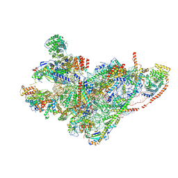
8QU1
 | | mt-LSU assembly intermediate in GTPBP8 knock-out cells, state 1 | | 分子名称: | 16S ribosomal RNA, 39S ribosomal protein L10, mitochondrial, ... | | 著者 | Valentin Gese, G, Cipullo, M, Rorbach, J, Hallberg, B.M. | | 登録日 | 2023-10-13 | | 公開日 | 2024-06-26 | | 最終更新日 | 2024-07-17 | | 実験手法 | ELECTRON MICROSCOPY (2.74 Å) | | 主引用文献 | GTPBP8 plays a role in mitoribosome formation in human mitochondria.
Nat Commun, 15, 2024
|
|
8QRL
 | | mt-SSU assembly intermediate in GTPBP8 knock-out cells, state 2 | | 分子名称: | 12S mitochondrial rRNA, 12S rRNA N4-methylcytidine (m4C) methyltransferase, 28S ribosomal protein S10, ... | | 著者 | Valentin Gese, G, Cipullo, M, Rorbach, J, Hallberg, B.M. | | 登録日 | 2023-10-09 | | 公開日 | 2024-06-26 | | 最終更新日 | 2024-07-17 | | 実験手法 | ELECTRON MICROSCOPY (3.34 Å) | | 主引用文献 | GTPBP8 plays a role in mitoribosome formation in human mitochondria.
Nat Commun, 15, 2024
|
|
8QRK
 | | mt-SSU assembly intermediate in GTPBP8 knock-out cells, state 1 | | 分子名称: | 12S mitochondrial rRNA, 28S ribosomal protein S10, mitochondrial, ... | | 著者 | Valentin Gese, G, Cipullo, M, Rorbach, J, Hallberg, B.M. | | 登録日 | 2023-10-09 | | 公開日 | 2024-06-26 | | 最終更新日 | 2024-07-17 | | 実験手法 | ELECTRON MICROSCOPY (6.69 Å) | | 主引用文献 | GTPBP8 plays a role in mitoribosome formation in human mitochondria.
Nat Commun, 15, 2024
|
|
8QU5
 | | mt-LSU assembly intermediate in GTPBP8 knock-out cells, state 2 | | 分子名称: | 16S ribosomal RNA, 39S ribosomal protein L10, mitochondrial, ... | | 著者 | Valentin Gese, G, Cipullo, M, Rorbach, J, Hallberg, B.M. | | 登録日 | 2023-10-13 | | 公開日 | 2024-06-26 | | 最終更新日 | 2024-07-17 | | 実験手法 | ELECTRON MICROSCOPY (2.42 Å) | | 主引用文献 | GTPBP8 plays a role in mitoribosome formation in human mitochondria.
Nat Commun, 15, 2024
|
|
8QRN
 | | mt-SSU in GTPBP8 knock-out cells, state 4 | | 分子名称: | 12S mitochondrial rRNA, 28S ribosomal protein S10, mitochondrial, ... | | 著者 | Valentin Gese, G, Cipullo, M, Rorbach, J, Hallberg, B.M. | | 登録日 | 2023-10-09 | | 公開日 | 2024-06-26 | | 最終更新日 | 2024-07-17 | | 実験手法 | ELECTRON MICROSCOPY (2.98 Å) | | 主引用文献 | GTPBP8 plays a role in mitoribosome formation in human mitochondria.
Nat Commun, 15, 2024
|
|
8QRM
 | | mt-SSU assembly intermediate in GTPBP8 knock-out cells, state 3 | | 分子名称: | 12S mitochondrial rRNA, 28S ribosomal protein S10, mitochondrial, ... | | 著者 | Valentin Gese, G, Cipullo, M, Rorbach, J, Hallberg, B.M. | | 登録日 | 2023-10-09 | | 公開日 | 2024-06-26 | | 最終更新日 | 2024-07-17 | | 実験手法 | ELECTRON MICROSCOPY (3.05 Å) | | 主引用文献 | GTPBP8 plays a role in mitoribosome formation in human mitochondria.
Nat Commun, 15, 2024
|
|
6K1D
 | |
6K1C
 | |
6K18
 | | Crystal structure of EXD2 exonuclease domain soaked in Mn | | 分子名称: | Exonuclease 3'-5' domain-containing protein 2, MANGANESE (II) ION | | 著者 | Park, J, Lee, C. | | 登録日 | 2019-05-10 | | 公開日 | 2019-05-22 | | 最終更新日 | 2023-11-22 | | 実験手法 | X-RAY DIFFRACTION (2.303 Å) | | 主引用文献 | The structure of human EXD2 reveals a chimeric 3' to 5' exonuclease domain that discriminates substrates via metal coordination.
Nucleic Acids Res., 47, 2019
|
|
6K1A
 | | Crystal structure of EXD2 exonuclease domain soaked in Mn and Mg | | 分子名称: | Exonuclease 3'-5' domain-containing protein 2, MAGNESIUM ION, MANGANESE (II) ION | | 著者 | Park, J, Lee, C. | | 登録日 | 2019-05-10 | | 公開日 | 2019-05-22 | | 最終更新日 | 2023-11-22 | | 実験手法 | X-RAY DIFFRACTION (2.602 Å) | | 主引用文献 | The structure of human EXD2 reveals a chimeric 3' to 5' exonuclease domain that discriminates substrates via metal coordination.
Nucleic Acids Res., 47, 2019
|
|
6K17
 | | Crystal structure of EXD2 exonuclease domain | | 分子名称: | Exonuclease 3'-5' domain-containing protein 2, SODIUM ION | | 著者 | Park, J, Lee, C. | | 登録日 | 2019-05-10 | | 公開日 | 2019-05-22 | | 最終更新日 | 2023-11-22 | | 実験手法 | X-RAY DIFFRACTION (1.602 Å) | | 主引用文献 | The structure of human EXD2 reveals a chimeric 3' to 5' exonuclease domain that discriminates substrates via metal coordination.
Nucleic Acids Res., 47, 2019
|
|
6K1E
 | |
6K1B
 | |
6K19
 | | Crystal structure of EXD2 exonuclease domain soaked in Mg | | 分子名称: | Exonuclease 3'-5' domain-containing protein 2, MAGNESIUM ION | | 著者 | Park, J, Lee, C. | | 登録日 | 2019-05-10 | | 公開日 | 2019-05-22 | | 最終更新日 | 2023-11-22 | | 実験手法 | X-RAY DIFFRACTION (2.202 Å) | | 主引用文献 | The structure of human EXD2 reveals a chimeric 3' to 5' exonuclease domain that discriminates substrates via metal coordination.
Nucleic Acids Res., 47, 2019
|
|


