Loading
PDBj
メニューPDBj@FacebookPDBj@TwitterPDBj@YouTubewwPDB FoundationwwPDB
RCSB PDBPDBeBMRBAdv. SearchSearch help
Search by PDB author
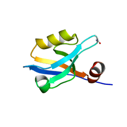
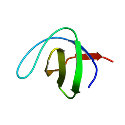
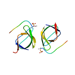
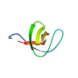
1BK2
DownloadVisualize
BU of 1bk2 by Molmil
A-SPECTRIN SH3 DOMAIN D48G MUTANT
分子名称: A-SPECTRIN
著者Martinez, J.C, Pisabarro, M.T, Serrano, L.
登録日1998-07-14
公開日1999-02-16
最終更新日2024-05-22
実験手法X-RAY DIFFRACTION (2.01 Å)
主引用文献Obligatory steps in protein folding and the conformational diversity of the transition state.
Nat.Struct.Biol., 5, 1998
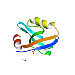
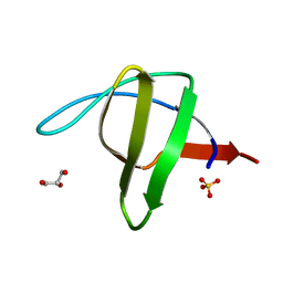
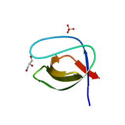
1PWT
DownloadVisualize
BU of 1pwt by Molmil
THERMODYNAMIC ANALYSIS OF ALPHA-SPECTRIN SH3 AND TWO OF ITS CIRCULAR PERMUTANTS WITH DIFFERENT LOOP LENGTHS: DISCERNING THE REASONS FOR RAPID FOLDING IN PROTEINS
分子名称: ALPHA SPECTRIN
著者Martinez, J.C, Viguera, A.R, Berisio, R, Wilmanns, M, Mateo, P.L, Filmonov, V.V, Serrano, L.
登録日1998-10-06
公開日1999-05-11
最終更新日2024-05-22
実験手法X-RAY DIFFRACTION (1.77 Å)
主引用文献Thermodynamic analysis of alpha-spectrin SH3 and two of its circular permutants with different loop lengths: discerning the reasons for rapid folding in proteins.
Biochemistry, 38, 1999
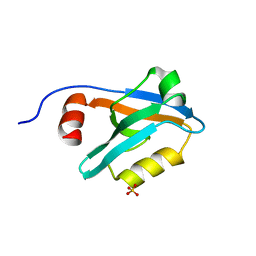
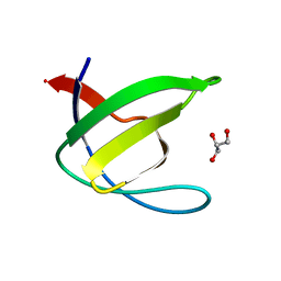
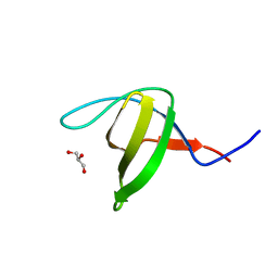
2F0R
DownloadVisualize
BU of 2f0r by Molmil
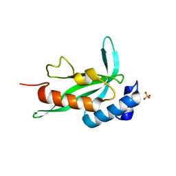
Crystallographic structure of human Tsg101 UEV domain
分子名称: SULFATE ION, Tumor susceptibility gene 101 protein
著者Camara-Artigas, A, Luque, I, Palencia, A, Martinez, J.C, Mateo, P.L.
登録日2005-11-13
公開日2006-03-28
最終更新日2023-08-23
実験手法X-RAY DIFFRACTION (2.26 Å)
主引用文献Structure of human TSG101 UEV domain.
Acta Crystallogr.,Sect.D, 62, 2006
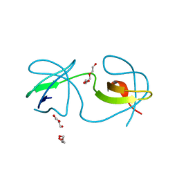
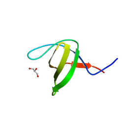
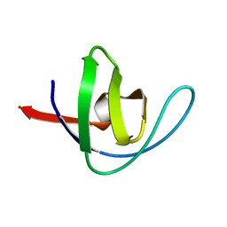
2JMC
DownloadVisualize
BU of 2jmc by Molmil
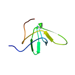
Chimer between Spc-SH3 and P41
分子名称: Spectrin alpha chain, brain and P41 peptide chimera
著者van Nuland, N.A.J, Candel, A.M, Martinez, J.C, Conejero-Lara, F, Bruix, M.
登録日2006-11-02
公開日2007-04-24
最終更新日2023-12-20
実験手法SOLUTION NMR
主引用文献The high-resolution NMR structure of a single-chain chimeric protein mimicking a SH3-peptide complex
Febs Lett., 581, 2007
6XVM
DownloadVisualize
BU of 6xvm by Molmil
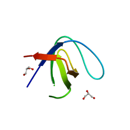
Crystal structure of c-Src SH3 domain without ATCUN motif: monomer 2
分子名称: GLYCEROL, Proto-oncogene tyrosine-protein kinase Src
著者Camara-Artigas, A, Plaza-Garrido, M.
登録日2020-01-22
公開日2020-04-22
最終更新日2024-01-24
実験手法X-RAY DIFFRACTION (0.9 Å)
主引用文献The effect of an engineered ATCUN motif on the structure and biophysical properties of the SH3 domain of c-Src tyrosine kinase.
J.Biol.Inorg.Chem., 25, 2020
6XVN
DownloadVisualize
BU of 6xvn by Molmil
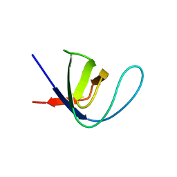
Crystal structure of c-Src SH3 domain without ATCUN motif: monomer 1
分子名称: Proto-oncogene tyrosine-protein kinase Src
著者Camara-Artigas, A, Plaza-Garrido, M.
登録日2020-01-22
公開日2020-04-22
最終更新日2024-01-24
実験手法X-RAY DIFFRACTION (1.7 Å)
主引用文献The effect of an engineered ATCUN motif on the structure and biophysical properties of the SH3 domain of c-Src tyrosine kinase.
J.Biol.Inorg.Chem., 25, 2020
7ZX2
DownloadVisualize
BU of 7zx2 by Molmil
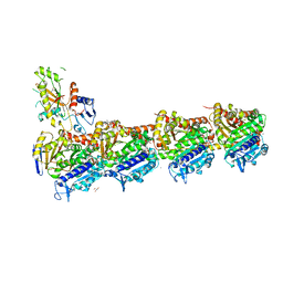
Tubulin-Pelophen B complex
分子名称: (3R,4S,7S,9S,11S)-3,4,11-trihydroxy-7-((R,Z)-4-(hydroxymethyl)hex-2-en-2-yl)-9-methoxy-12,12-dimethyl-6-oxa-1(1,3)-benzenacyclododecaphan-5-one, 2-(N-MORPHOLINO)-ETHANESULFONIC ACID, CALCIUM ION, ...
著者Estevez-Gallego, J, Diaz, J.F, Van der Eycken, J, Oliva, M.A.
登録日2022-05-20
公開日2022-11-23
最終更新日2024-02-07
実験手法X-RAY DIFFRACTION (2.5 Å)
主引用文献Chemical modulation of microtubule structure through the laulimalide/peloruside site.
Structure, 31, 2023
8A0L
DownloadVisualize
BU of 8a0l by Molmil
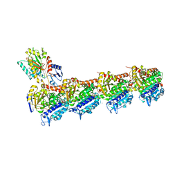
Tubulin-CW1-complex
分子名称: (3~{S},4~{R},8~{S},10~{S},12~{S},14~{S})-14-[(~{Z},4~{R})-4-(hydroxymethyl)hex-2-en-2-yl]-4,12-dimethoxy-9,9-dimethyl-3,8,10-tris(oxidanyl)-1-oxacyclotetradecan-2-one, 2-(N-MORPHOLINO)-ETHANESULFONIC ACID, CALCIUM ION, ...
著者Prota, A.E, Diaz, J.F, Steinmetz, M.O, Oliva, M.A.
登録日2022-05-28
公開日2022-12-14
最終更新日2024-01-31
実験手法X-RAY DIFFRACTION (1.9981 Å)
主引用文献Chemical modulation of microtubule structure through the laulimalide/peloruside site.
Structure, 31, 2023
8AH5
DownloadVisualize
BU of 8ah5 by Molmil
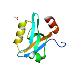
Crystal Structure of the third PDZ domain of PSD-95 protein in the space group P212121 at pH 4.6
分子名称: ACETATE ION, cDNA FLJ50577, highly similar to Discs large homolog 4
著者Camara-Artigas, A, Salinas Garcia, M.C.
登録日2022-07-20
公開日2023-03-08
最終更新日2024-02-07
実験手法X-RAY DIFFRACTION (1.25 Å)
主引用文献pH-Driven Polymorphic Behaviour of the Third PDZ Domain of PSD95: The Role of Electrostatic Interactions
Crystals, 2023
8AH6
DownloadVisualize
BU of 8ah6 by Molmil
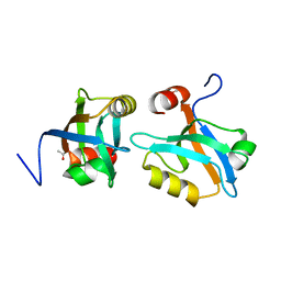
Crystal Structure of the third PDZ domain of PSD-95 protein in the space group P21 at pH 4.0
分子名称: ACETATE ION, cDNA FLJ50577, highly similar to Discs large homolog 4
著者Camara-Artigas, A, Salinas Garcia, M.C.
登録日2022-07-20
公開日2023-03-08
最終更新日2024-02-07
実験手法X-RAY DIFFRACTION (1.63 Å)
主引用文献pH-Driven Polymorphic Behaviour of the Third PDZ Domain of PSD95: The Role of Electrostatic Interactions
Crystals, 2023
8AH8
DownloadVisualize
BU of 8ah8 by Molmil
Crystal Structure of the third PDZ domain of PSD-95 protein in the space group P3121 at pH 3.7
分子名称: cDNA FLJ50577, highly similar to Discs large homolog 4
著者Camara-Artigas, A, Salinas Garcia, M.C.
登録日2022-07-20
公開日2023-03-08
最終更新日2024-02-07
実験手法X-RAY DIFFRACTION (1.5 Å)
主引用文献pH-Driven Polymorphic Behaviour of the Third PDZ Domain of PSD95: The Role of Electrostatic Interactions
Crystals, 2023
8AH4
DownloadVisualize
BU of 8ah4 by Molmil
Crystal Structure of the third PDZ domain of PSD-95 protein in the space group P3112 at pH 4.0
分子名称: ACETATE ION, cDNA FLJ50577, highly similar to Discs large homolog 4
著者Camara-Artigas, A, Salinas Garcia, M.C.
登録日2022-07-20
公開日2023-03-08
最終更新日2024-02-07
実験手法X-RAY DIFFRACTION (1.48 Å)
主引用文献pH-Driven Polymorphic Behaviour of the Third PDZ Domain of PSD95: The Role of Electrostatic Interactions
Crystals, 2023
8AH7
DownloadVisualize
BU of 8ah7 by Molmil
Crystal Structure of the third PDZ domain of PSD-95 protein in the space group P212121 at pH 4.0
分子名称: SULFATE ION, cDNA FLJ50577, highly similar to Discs large homolog 4
著者Camara-Artigas, A, Salinas-Garcia, M.C.
登録日2022-07-20
公開日2023-03-08
最終更新日2024-02-07
実験手法X-RAY DIFFRACTION (1.25 Å)
主引用文献pH-Driven Polymorphic Behaviour of the Third PDZ Domain of PSD95: The Role of Electrostatic Interactions
Crystals, 2023
6XVO
DownloadVisualize
BU of 6xvo by Molmil
Crystal structure of the intertwined dimer of the c-Src SH3 domain without ATCUN motif
分子名称: DI(HYDROXYETHYL)ETHER, Proto-oncogene tyrosine-protein kinase Src, TRIETHYLENE GLYCOL
著者Camara-Artigas, A, Plaza-Garrido, M.
登録日2020-01-22
公開日2020-04-22
最終更新日2024-01-24
実験手法X-RAY DIFFRACTION (1.7 Å)
主引用文献The effect of an engineered ATCUN motif on the structure and biophysical properties of the SH3 domain of c-Src tyrosine kinase.
J.Biol.Inorg.Chem., 25, 2020
6XX4
DownloadVisualize
BU of 6xx4 by Molmil
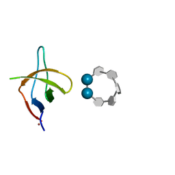
Crystal structure of the c-Src SH3 domain H122R-Q128E mutant in complex with Ni(II) at pH 7.5 co-crystallized with methyl beta-cyclodextrin
分子名称: Cyclic 2,3-di-O-methyl-alpha-D-glucopyranose-(1-4)-2-O-methyl-alpha-D-glucopyranose-(1-4)-2,6-di-O-methyl-alpha-D-glucopyranose-(1-4)-2-O-methyl-alpha-D-glucopyranose-(1-4)-alpha-D-glucopyranose-(1-4)-alpha-D-glucopyranose-(1-4)-3-O-methyl-alpha-D-glucopyranose, NICKEL (II) ION, Proto-oncogene tyrosine-protein kinase Src
著者Camara-Artigas, A.
登録日2020-01-26
公開日2020-04-22
最終更新日2024-01-24
実験手法X-RAY DIFFRACTION (1.05 Å)
主引用文献The effect of an engineered ATCUN motif on the structure and biophysical properties of the SH3 domain of c-Src tyrosine kinase.
J.Biol.Inorg.Chem., 25, 2020
6XX3
DownloadVisualize
BU of 6xx3 by Molmil
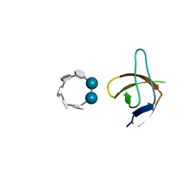
Crystal structure of the c-Src SH3 domain H122R-Q128E mutant in complex with Cu(II) at pH 6.5 co-crystallized with methyl beta-cyclodextrin
分子名称: COPPER (II) ION, Cyclic 2,3-di-O-methyl-alpha-D-glucopyranose-(1-4)-2-O-methyl-alpha-D-glucopyranose-(1-4)-2,6-di-O-methyl-alpha-D-glucopyranose-(1-4)-2-O-methyl-alpha-D-glucopyranose-(1-4)-alpha-D-glucopyranose-(1-4)-alpha-D-glucopyranose-(1-4)-3-O-methyl-alpha-D-glucopyranose, Proto-oncogene tyrosine-protein kinase Src
著者Camara-Artigas, A.
登録日2020-01-26
公開日2020-04-22
最終更新日2024-01-24
実験手法X-RAY DIFFRACTION (1.36 Å)
主引用文献The effect of an engineered ATCUN motif on the structure and biophysical properties of the SH3 domain of c-Src tyrosine kinase.
J.Biol.Inorg.Chem., 25, 2020
6XX5
DownloadVisualize
BU of 6xx5 by Molmil
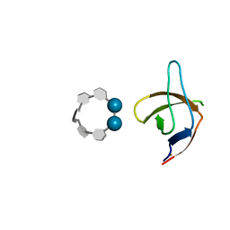
Crystal structure of the c-Src SH3 domain H122R-Q128K mutant in complex with Ni(II) at pH 7.5 co-crystallized with methyl beta-cyclodextrin
分子名称: Cyclic 2,3-di-O-methyl-alpha-D-glucopyranose-(1-4)-2-O-methyl-alpha-D-glucopyranose-(1-4)-2,6-di-O-methyl-alpha-D-glucopyranose-(1-4)-2-O-methyl-alpha-D-glucopyranose-(1-4)-alpha-D-glucopyranose-(1-4)-alpha-D-glucopyranose-(1-4)-3-O-methyl-alpha-D-glucopyranose, NICKEL (II) ION, Proto-oncogene tyrosine-protein kinase Src
著者Camara-Artigas, A.
登録日2020-01-26
公開日2020-05-27
最終更新日2024-01-24
実験手法X-RAY DIFFRACTION (1.3 Å)
主引用文献The effect of an engineered ATCUN motif on the structure and biophysical properties of the SH3 domain of c-Src tyrosine kinase.
J.Biol.Inorg.Chem., 25, 2020
6XX2
DownloadVisualize
BU of 6xx2 by Molmil
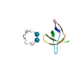
Crystal structure of the c-Src SH3 domain H122R-Q128K mutant in complex with Cu(II) at pH 7.5 co-crystallized with methyl beta-cyclodextrin
分子名称: COPPER (II) ION, Cyclic 2,3-di-O-methyl-alpha-D-glucopyranose-(1-4)-2-O-methyl-alpha-D-glucopyranose-(1-4)-2,6-di-O-methyl-alpha-D-glucopyranose-(1-4)-2-O-methyl-alpha-D-glucopyranose-(1-4)-alpha-D-glucopyranose-(1-4)-alpha-D-glucopyranose-(1-4)-3-O-methyl-alpha-D-glucopyranose, Proto-oncogene tyrosine-protein kinase Src
著者Camara-Artigas, A.
登録日2020-01-26
公開日2020-04-22
最終更新日2024-01-24
実験手法X-RAY DIFFRACTION (1.25 Å)
主引用文献The effect of an engineered ATCUN motif on the structure and biophysical properties of the SH3 domain of c-Src tyrosine kinase.
J.Biol.Inorg.Chem., 25, 2020
1QKX
DownloadVisualize
BU of 1qkx by Molmil
Alpha-spectrin Src Homology 3 domain, N47A mutant in the distal loop.
分子名称: SPECTRIN ALPHA CHAIN
著者Vega, M.C, Martinez, J, Serrano, L.
登録日1999-08-16
公開日2000-12-15
最終更新日2023-12-13
実験手法X-RAY DIFFRACTION (1.8 Å)
主引用文献Thermodynamic and structural characterization of Asn and Ala residues in the disallowed II' region of the Ramachandran plot.
Protein Sci., 9, 2000
1QKW
DownloadVisualize
BU of 1qkw by Molmil
Alpha-spectrin Src Homology 3 domain, N47G mutant in the distal loop.
分子名称: ALPHA II SPECTRIN, GLYCEROL, SULFATE ION
著者Vega, M.C, Martinez, J, Serrano, L.
登録日1999-08-16
公開日2000-08-18
最終更新日2023-12-13
実験手法X-RAY DIFFRACTION (2 Å)
主引用文献Thermodynamic and structural characterization of Asn and Ala residues in the disallowed II' region of the Ramachandran plot.
Protein Sci., 9, 2000
3EG0
DownloadVisualize
BU of 3eg0 by Molmil
Crystal structure of the N114T mutant of ABL-SH3 domain
分子名称: GLYCEROL, Proto-oncogene tyrosine-protein kinase ABL1
著者Camara-Artigas, A.
登録日2008-09-10
公開日2009-09-15
最終更新日2023-08-30
実験手法X-RAY DIFFRACTION (2.3 Å)
主引用文献Role of interfacial water molecules in proline-rich ligand recognition by the Src homology 3 domain of Abl.
J.Biol.Chem., 285, 2010
3EG2
DownloadVisualize
BU of 3eg2 by Molmil
Crystal structure of the N114Q mutant of ABL-SH3 domain
分子名称: GLYCEROL, Proto-oncogene tyrosine-protein kinase ABL1
著者Camara-Artigas, A.
登録日2008-09-10
公開日2009-09-15
最終更新日2023-08-30
実験手法X-RAY DIFFRACTION (1.8 Å)
主引用文献Role of interfacial water molecules in proline-rich ligand recognition by the Src homology 3 domain of Abl.
J.Biol.Chem., 285, 2010
3EG1
DownloadVisualize
BU of 3eg1 by Molmil
Crystal structure of the N114Q mutant of ABL-SH3 domain complexed with a designed high-affinity peptide ligand: implications for SH3-ligand interactions
分子名称: Proto-oncogene tyrosine-protein kinase ABL1, SULFATE ION, p41 peptide
著者Camara-Artigas, A.
登録日2008-09-10
公開日2009-09-15
最終更新日2023-08-30
実験手法X-RAY DIFFRACTION (1.85 Å)
主引用文献Role of interfacial water molecules in proline-rich ligand recognition by the Src homology 3 domain of Abl.
J.Biol.Chem., 285, 2010
3EGU
DownloadVisualize
BU of 3egu by Molmil
Crystal structure of the N114A mutant of ABL-SH3 domain
分子名称: GLYCEROL, Proto-oncogene tyrosine-protein kinase ABL1, SULFATE ION
著者Camara-Artigas, A.
登録日2008-09-11
公開日2009-09-15
最終更新日2024-02-21
実験手法X-RAY DIFFRACTION (2.25 Å)
主引用文献Role of interfacial water molecules in proline-rich ligand recognition by the Src homology 3 domain of Abl.
J.Biol.Chem., 285, 2010
3EG3
DownloadVisualize
BU of 3eg3 by Molmil
Crystal structure of the N114A mutant of ABL-SH3 domain
分子名称: GLYCEROL, Proto-oncogene tyrosine-protein kinase ABL1
著者Camara-Artigas, A.
登録日2008-09-10
公開日2009-09-15
最終更新日2023-08-30
実験手法X-RAY DIFFRACTION (1.4 Å)
主引用文献Role of interfacial water molecules in proline-rich ligand recognition by the Src homology 3 domain of Abl.
J.Biol.Chem., 285, 2010


123>

224572

件を2024-09-04に公開中

PDB statisticsPDBj update infoContact PDBjnumon