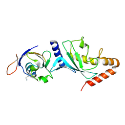
5FQ2
 
 | |
6Q9Z
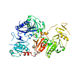 | | Crystal structure of the pathological G167R variant of calcium-free human gelsolin, | | 分子名称: | GLYCEROL, Gelsolin, SULFATE ION | | 著者 | Boni, F, Scalone, E, Milani, M, Eloise, M, de Rosa, M. | | 登録日 | 2018-12-18 | | 公開日 | 2019-11-27 | | 最終更新日 | 2024-01-24 | | 実験手法 | X-RAY DIFFRACTION (3.8 Å) | | 主引用文献 | The structure of N184K amyloidogenic variant of gelsolin highlights the role of the H-bond network for protein stability and aggregation properties.
Eur.Biophys.J., 49, 2020
|
|
6QBF
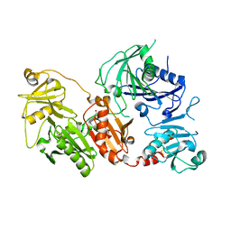 | | Crystal structure of the pathological D187N variant of calcium-free human gelsolin. | | 分子名称: | GLYCEROL, Gelsolin, SODIUM ION, ... | | 著者 | Scalone, E, Boni, F, Milani, M, Eloise, M, de Rosa, M. | | 登録日 | 2018-12-21 | | 公開日 | 2019-11-27 | | 最終更新日 | 2024-01-24 | | 実験手法 | X-RAY DIFFRACTION (3.499 Å) | | 主引用文献 | The structure of N184K amyloidogenic variant of gelsolin highlights the role of the H-bond network for protein stability and aggregation properties.
Eur.Biophys.J., 49, 2020
|
|
6Q9R
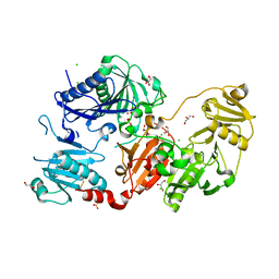 | | Crystal structure of the pathological N184K variant of calcium-free human gelsolin | | 分子名称: | 2-AMINO-2-HYDROXYMETHYL-PROPANE-1,3-DIOL, CHLORIDE ION, GLYCEROL, ... | | 著者 | Scalone, E, Boni, F, Milani, M, Eloise, M, de Rosa, M. | | 登録日 | 2018-12-18 | | 公開日 | 2019-11-27 | | 最終更新日 | 2024-01-24 | | 実験手法 | X-RAY DIFFRACTION (2.73 Å) | | 主引用文献 | The structure of N184K amyloidogenic variant of gelsolin highlights the role of the H-bond network for protein stability and aggregation properties.
Eur.Biophys.J., 49, 2020
|
|


