Loading
PDBj
メニューPDBj@FacebookPDBj@TwitterPDBj@YouTubewwPDB FoundationwwPDB
RCSB PDBPDBeBMRBAdv. SearchSearch help
Search by PDB author
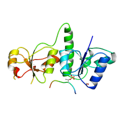
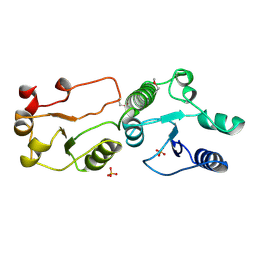
3AL2
DownloadVisualize
BU of 3al2 by Molmil
Crystal Structure of TopBP1 BRCT7/8
分子名称: DNA topoisomerase 2-binding protein 1, SULFATE ION
著者Leung, C.C, Glover, J.N.
登録日2010-07-22
公開日2010-12-01
最終更新日2017-10-11
実験手法X-RAY DIFFRACTION (2 Å)
主引用文献Molecular basis of BACH1/FANCJ recognition by TopBP1 in DNA replication checkpoint control
J.Biol.Chem., 286, 2011
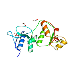
3AL3
DownloadVisualize
BU of 3al3 by Molmil
Crystal Structure of TopBP1 BRCT7/8-BACH1 peptide complex
分子名称: DNA topoisomerase 2-binding protein 1, FORMIC ACID, Peptide of Fanconi anemia group J protein
著者Leung, C.C, Glover, J.N.
登録日2010-07-22
公開日2010-12-01
最終更新日2023-11-01
実験手法X-RAY DIFFRACTION (2.15 Å)
主引用文献Molecular basis of BACH1/FANCJ recognition by TopBP1 in DNA replication checkpoint control
J.Biol.Chem., 286, 2011
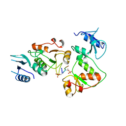
3UEN
DownloadVisualize
BU of 3uen by Molmil
Crystal structure of TopBP1 BRCT4/5 domains
分子名称: DNA topoisomerase 2-binding protein 1, GLYCEROL, THIOCYANATE ION
著者Leung, C.C, Glover, J.N.M.
登録日2011-10-31
公開日2013-07-03
最終更新日2024-02-28
実験手法X-RAY DIFFRACTION (1.9 Å)
主引用文献Structural insights into recognition of MDC1 by TopBP1 in DNA replication checkpoint control.
Structure, 21, 2013
3UEO
DownloadVisualize
BU of 3ueo by Molmil
Crystal structure of TopBP1 BRCT4/5 domains in complex with a phospho-peptide
分子名称: DNA topoisomerase 2-binding protein 1, phospho-peptide
著者Leung, C.C, Glover, J.N.M.
登録日2011-10-31
公開日2013-07-03
最終更新日2024-10-30
実験手法X-RAY DIFFRACTION (2.6 Å)
主引用文献Structural insights into recognition of MDC1 by TopBP1 in DNA replication checkpoint control.
Structure, 21, 2013
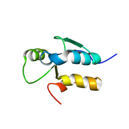
3JVE
DownloadVisualize
BU of 3jve by Molmil
Crystal Structure of the Sixth BRCT Domain of TopBP1
分子名称: DNA topoisomerase 2-binding protein 1
著者Leung, C.C, Kellogg, E, Baker, D, Glover, J.N.M.
登録日2009-09-16
公開日2010-01-19
最終更新日2024-02-21
実験手法X-RAY DIFFRACTION (1.34 Å)
主引用文献Insights from the crystal structure of the sixth BRCT domain of topoisomerase IIbeta binding protein 1.
Protein Sci., 19, 2010
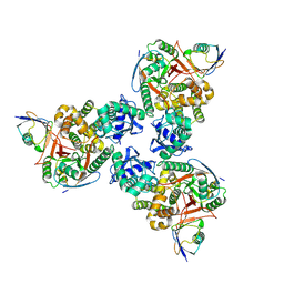
4DDG
DownloadVisualize
BU of 4ddg by Molmil
Crystal structure of human OTUB1/UbcH5b~Ub/Ub
分子名称: Polyubiquitin-C, Ubiquitin-conjugating enzyme E2 D2, Ubiquitin thioesterase OTUB1
著者Juang, Y.C, Sanches, M, Sicheri, F.
登録日2012-01-18
公開日2012-02-22
最終更新日2023-09-13
実験手法X-RAY DIFFRACTION (3.2987 Å)
主引用文献OTUB1 Co-opts Lys48-Linked Ubiquitin Recognition to Suppress E2 Enzyme Function.
Mol.Cell, 45, 2012
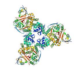
4DDI
DownloadVisualize
BU of 4ddi by Molmil
Crystal structure of human OTUB1/UbcH5b~Ub/Ub
分子名称: Polyubiquitin-C, Ubiquitin-conjugating enzyme E2 D2, Ubiquitin thioesterase OTUB1
著者Juang, Y.C, Sanches, M, Sicheri, F.
登録日2012-01-18
公開日2012-02-22
最終更新日2017-11-15
実験手法X-RAY DIFFRACTION (3.802 Å)
主引用文献OTUB1 Co-opts Lys48-Linked Ubiquitin Recognition to Suppress E2 Enzyme Function.
Mol.Cell, 45, 2012
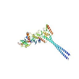
4ORH
DownloadVisualize
BU of 4orh by Molmil
Crystal structure of RNF8 bound to the UBC13/MMS2 heterodimer
分子名称: E3 ubiquitin-protein ligase RNF8, Ubiquitin-conjugating enzyme E2 N, Ubiquitin-conjugating enzyme E2 variant 2, ...
著者Campbell, S.J, Edwards, R.A, Glover, J.N.M.
登録日2014-02-11
公開日2014-02-26
最終更新日2024-02-28
実験手法X-RAY DIFFRACTION (4.802 Å)
主引用文献Molecular insights into the function of RING finger (RNF)-containing proteins hRNF8 and hRNF168 in Ubc13/Mms2-dependent ubiquitylation.
J.Biol.Chem., 287, 2012
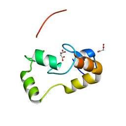
3L11
DownloadVisualize
BU of 3l11 by Molmil
Crystal Structure of the Ring Domain of RNF168
分子名称: E3 ubiquitin-protein ligase RNF168, MALONATE ION, ZINC ION
著者Neculai, D, Yermekbayeva, L, Crombet, L, Weigelt, J, Bountra, C, Edwards, A.M, Arrowsmith, C.H, Bochkarev, A, Dhe-Paganon, S, Structural Genomics Consortium (SGC)
登録日2009-12-10
公開日2010-01-19
最終更新日2023-09-06
実験手法X-RAY DIFFRACTION (2.12 Å)
主引用文献Molecular insights into the function of RING finger (RNF)-containing proteins hRNF8 and hRNF168 in Ubc13/Mms2-dependent ubiquitylation.
J.Biol.Chem., 287, 2012

227111

件を2024-11-06に公開中

PDB statisticsPDBj update infoContact PDBjnumon