2FMC
 
 | |
2K6A
 | |
1MM2
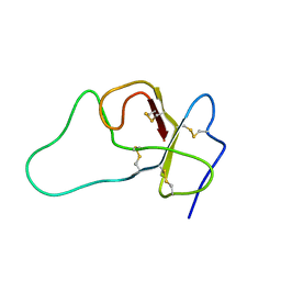
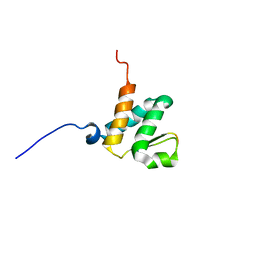
 | | Solution structure of the 2nd PHD domain from Mi2b | | 分子名称: | Mi2-beta, ZINC ION | | 著者 | Kwan, A.H.Y, Gell, D.A, Verger, A, Crossley, M, Matthews, J.M, Mackay, J.P. | | 登録日 | 2002-09-02 | | 公開日 | 2003-07-22 | | 最終更新日 | 2024-05-29 | | 実験手法 | SOLUTION NMR | | 主引用文献 | Engineering a Protein Scaffold from a PHD Finger
structure, 11, 2003
|
|
1MM3
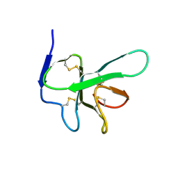
 | | Solution structure of the 2nd PHD domain from Mi2b with C-terminal loop replaced by corresponding loop from WSTF | | 分子名称: | Mi2-beta(Chromodomain helicase-DNA-binding protein 4) and transcription factor WSTF, ZINC ION | | 著者 | Kwan, A.H.Y, Gell, D.A, Verger, A, Crossley, M, Matthews, J.M, Mackay, J.P. | | 登録日 | 2002-09-02 | | 公開日 | 2003-07-22 | | 最終更新日 | 2024-05-29 | | 実験手法 | SOLUTION NMR | | 主引用文献 | Engineering a Protein Scaffold from a PHD Finger
structure, 11, 2003
|
|
1M36
 | |
1Y03
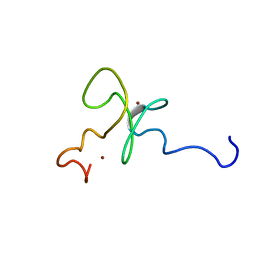
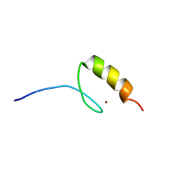
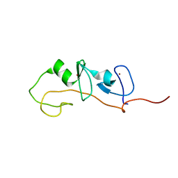
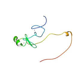
 | | Solution structure of a recombinant type I sculpin antifreeze protein | | 分子名称: | Antifreeze peptide SS-3 | | 著者 | Kwan, A.H.Y, Fairley, K, Anderberg, P.I, Liew, C.W, Harding, M.M, Mackay, J.P. | | 登録日 | 2004-11-14 | | 公開日 | 2005-03-15 | | 最終更新日 | 2024-05-29 | | 実験手法 | SOLUTION NMR | | 主引用文献 | Solution structure of a recombinant type I sculpin antifreeze protein
Biochemistry, 44, 2005
|
|
1Y04
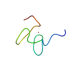
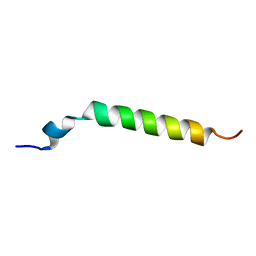
 | | Solution structure of a recombinant type I sculpin antifreeze protein | | 分子名称: | Antifreeze peptide SS-3 | | 著者 | Kwan, A.H.Y, Fairley, K, Anderberg, P.I, Liew, C.W, Harding, M.M, Mackay, J.P. | | 登録日 | 2004-11-14 | | 公開日 | 2005-03-15 | | 最終更新日 | 2024-05-29 | | 実験手法 | SOLUTION NMR | | 主引用文献 | Solution structure of a recombinant type I sculpin antifreeze protein
Biochemistry, 44, 2005
|
|
5K6P
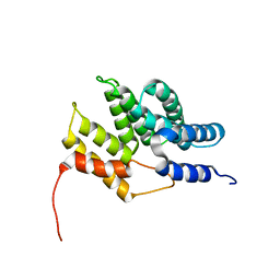
 | | The NMR structure of the m domain tri-helix bundle and C2 of human cardiac Myosin Binding Protein C | | 分子名称: | Myosin-binding protein C, cardiac-type | | 著者 | Michie, K.A, Kwan, A.H, Tung, C.S, Guss, J.M, Trewhella, J. | | 登録日 | 2016-05-25 | | 公開日 | 2016-11-09 | | 最終更新日 | 2024-05-15 | | 実験手法 | SOLUTION NMR | | 主引用文献 | A Highly Conserved Yet Flexible Linker Is Part of a Polymorphic Protein-Binding Domain in Myosin-Binding Protein C.
Structure, 24, 2016
|
|
1J2O
 | | Structure of FLIN2, a complex containing the N-terminal LIM domain of LMO2 and ldb1-LID | | 分子名称: | Fusion of Rhombotin-2 and LIM domain-binding protein 1, ZINC ION | | 著者 | Deane, J.E, Mackay, J.P, Kwan, A.H, Sum, E.Y, Visvader, J.E, Matthews, J.M. | | 登録日 | 2003-01-08 | | 公開日 | 2003-05-13 | | 最終更新日 | 2023-12-27 | | 実験手法 | SOLUTION NMR | | 主引用文献 | Structural basis for the recognition of ldb1 by the N-terminal LIM domains of LMO2 and LMO4
EMBO J., 22, 2003
|
|
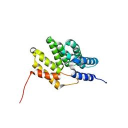
6CRL
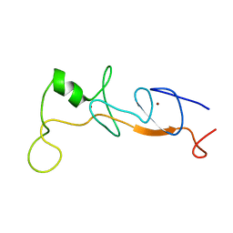
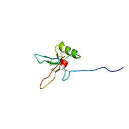
 | | HusA haemophore from Porphyromonas gingivalis | | 分子名称: | hypothetical protein PG_2227 | | 著者 | Gell, D.A, Kwan, A.H, Horne, J, Hugrass, B.M, Collins, D.A.T. | | 登録日 | 2018-03-19 | | 公開日 | 2018-10-10 | | 最終更新日 | 2024-05-01 | | 実験手法 | SOLUTION NMR | | 主引用文献 | Structural properties of a haemophore facilitate targeted elimination of the pathogen Porphyromonas gingivalis.
Nat Commun, 9, 2018
|
|
1PM4
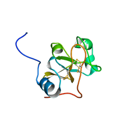
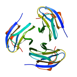 | | Crystal structure of Yersinia pseudotuberculosis-derived mitogen (YPM) | | 分子名称: | YPM | | 著者 | Donadini, R, Liew, C.W, Kwan, A.H, Mackay, J.P, Fields, B.A. | | 登録日 | 2003-06-09 | | 公開日 | 2004-01-27 | | 最終更新日 | 2011-07-13 | | 実験手法 | X-RAY DIFFRACTION (1.755 Å) | | 主引用文献 | Crystal and Solution Structures of a Superantigen from Yersinia pseudotuberculosis Reveal a Jelly-Roll Fold.
Structure, 12, 2004
|
|
1POQ
 | | Solution Structure of a Superantigen from Yersinia pseudotuberculosis | | 分子名称: | YPM | | 著者 | Donadini, R, Liew, C.W, Kwan, A.H, Mackay, J.P, Fields, B.A. | | 登録日 | 2003-06-16 | | 公開日 | 2004-01-27 | | 最終更新日 | 2022-03-02 | | 実験手法 | SOLUTION NMR | | 主引用文献 | Crystal and Solution Structures of a Superantigen from Yersinia pseudotuberculosis Reveal a Jelly-Roll Fold.
Structure, 12, 2004
|
|
6BGG
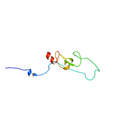
 | | Solution NMR structures of the BRD3 ET domain in complex with a CHD4 peptide | | 分子名称: | Bromodomain-containing protein 3, CHD4 | | 著者 | Wai, D.C.C, Szyszka, T.N, Campbell, A.E, Kwong, C, Wilkinson-White, L, Silva, A.P.G, Low, J.K.K, Kwan, A.H, Gamsjaeger, R, Lu, B, Vakoc, C.R, Blobel, G.A, Mackay, J.P. | | 登録日 | 2017-10-28 | | 公開日 | 2018-03-21 | | 最終更新日 | 2024-05-15 | | 実験手法 | SOLUTION NMR | | 主引用文献 | The BRD3 ET domain recognizes a short peptide motif through a mechanism that is conserved across chromatin remodelers and transcriptional regulators.
J. Biol. Chem., 293, 2018
|
|
6BQS
 | | HusA haemophore from Porphyromonas gingivalis | | 分子名称: | hypothetical protein PG_2227 | | 著者 | Gell, D.A, Kwan, A.H, Horne, J, Hugrass, B.M, Collins, D.A.T. | | 登録日 | 2017-11-28 | | 公開日 | 2018-10-10 | | 最終更新日 | 2024-05-01 | | 実験手法 | SOLUTION NMR | | 主引用文献 | Structural properties of a haemophore facilitate targeted elimination of the pathogen Porphyromonas gingivalis.
Nat Commun, 9, 2018
|
|
1M3V
 | | FLIN4: Fusion of the LIM binding domain of Ldb1 and the N-terminal LIM domain of LMO4 | | 分子名称: | ZINC ION, fusion of the LIM interacting domain of ldb1 and the N-terminal LIM domain of LMO4 | | 著者 | Deane, J.E, Mackay, J.P, Kwan, A.H.Y, Sum, E.Y, Visvader, J.E, Matthews, J.M. | | 登録日 | 2002-06-30 | | 公開日 | 2003-05-13 | | 最終更新日 | 2024-05-15 | | 実験手法 | SOLUTION NMR | | 主引用文献 | Structural basis for the recognition of ldb1 by the N-terminal LIM domains of LMO2 and LMO4
EMBO J., 22, 2003
|
|
2LXD
 | | Backbone 1H, 13C, and 15N Chemical Shift Assignments for LMO2(LIM2)-Ldb1(LID) | | 分子名称: | Rhombotin-2,LIM domain-binding protein 1, ZINC ION | | 著者 | Dastmalchi, S, Wilkinson-White, L, Kwan, A.H, Gamsjaeger, R, Mackay, J.P, Matthews, J.M. | | 登録日 | 2012-08-20 | | 公開日 | 2012-09-12 | | 最終更新日 | 2024-05-15 | | 実験手法 | SOLUTION NMR | | 主引用文献 | Solution structure of a tethered Lmo2(LIM2) /Ldb1(LID) complex.
Protein Sci., 21, 2012
|
|
4AOG
 | |
2L4Z
 | | NMR structure of fusion of CtIP (641-685) to LMO4-LIM1 (18-82) | | 分子名称: | DNA endonuclease RBBP8,LIM domain transcription factor LMO4, ZINC ION | | 著者 | Liew, C, Stokes, P.H, Kwan, A.H, Matthews, J.M. | | 登録日 | 2010-10-22 | | 公開日 | 2011-10-26 | | 最終更新日 | 2024-05-29 | | 実験手法 | SOLUTION NMR | | 主引用文献 | Structural Basis of the Interaction of the Breast Cancer Oncogene LMO4 with the Tumour Suppressor CtIP/RBBP8.
J.Mol.Biol., 425, 2013
|
|
2LSH
 | |
2L75
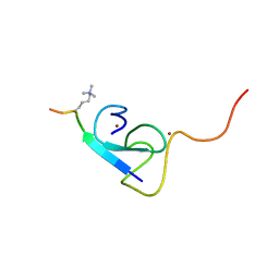 | |
2JYD
 | | Structure of the fifth zinc finger of Myelin Transcription Factor 1 | | 分子名称: | F5 domain of Myelin transcription factor 1, ZINC ION | | 著者 | Gamsjaeger, R, Swanton, M.K, Kobus, F.J, Lehtomaki, E, Lowry, J.A, Kwan, A.H, Matthews, J.M, Mackay, J.P. | | 登録日 | 2007-12-12 | | 公開日 | 2008-01-15 | | 最終更新日 | 2024-05-29 | | 実験手法 | SOLUTION NMR | | 主引用文献 | Structural and biophysical analysis of the DNA binding properties of myelin transcription factor 1.
J.Biol.Chem., 283, 2008
|
|
2JX1
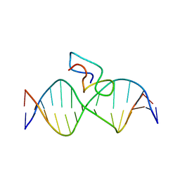 | | Structure of the fifth zinc finger of Myelin Transcription Factor 1 in complex with RARE DNA | | 分子名称: | DNA (5'-D(*DAP*DCP*DCP*DGP*DAP*DAP*DAP*DGP*DTP*DTP*DCP*DAP*DC)-3'), DNA (5'-D(*DGP*DTP*DGP*DAP*DAP*DCP*DTP*DTP*DTP*DCP*DGP*DGP*DT)-3'), Myelin transcription factor 1 | | 著者 | Gamsjaeger, R, Swanton, M.K, Kobus, F.J, Lehtomaki, E, Lowry, J.A, Kwan, A.H, Matthews, J.M, Mackay, J.P. | | 登録日 | 2007-11-01 | | 公開日 | 2007-12-11 | | 最終更新日 | 2024-05-29 | | 実験手法 | SOLUTION NMR | | 主引用文献 | Structure of the fifth zinc finger of Myelin Transcription Factor 1 in complex with RARE DNA
To be Published
|
|
2MF8
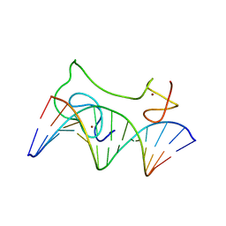 | | HADDOCK model of MyT1 F4F5 - DNA complex | | 分子名称: | DNA (5'-D(*AP*CP*CP*GP*AP*AP*AP*GP*TP*TP*CP*AP*C)-3'), DNA (5'-D(*GP*TP*GP*AP*AP*CP*TP*TP*TP*CP*GP*GP*T)-3'), Myelin transcription factor 1, ... | | 著者 | Gamsjaeger, R, O'Connell, M.R, Cubeddu, L, Shepherd, N.E, Lowry, J.A, Kwan, A.H, Vandevenne, M, Swanton, M.K, Matthews, J.M, Mackay, J.P. | | 登録日 | 2013-10-08 | | 公開日 | 2013-11-06 | | 最終更新日 | 2024-05-15 | | 実験手法 | SOLUTION NMR | | 主引用文献 | A structural analysis of DNA binding by myelin transcription factor 1 double zinc fingers.
J.Biol.Chem., 288, 2013
|
|
2LFN
 | | Identification of the key regions that drive functional amyloid formation by the fungal hydrophobin EAS | | 分子名称: | Hydrophobin | | 著者 | Macindoe, I, Kwan, A.H, Morris, V.K, Mackay, J.P, Sunde, M. | | 登録日 | 2011-07-06 | | 公開日 | 2012-01-25 | | 最終更新日 | 2023-06-14 | | 実験手法 | SOLUTION NMR | | 主引用文献 | Self-assembly of functional, amphipathic amyloid monolayers by the fungal hydrophobin EAS
Proc.Natl.Acad.Sci.USA, 109, 2012
|
|
2L5U
 | |


