1H9H
 
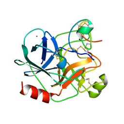 | | COMPLEX OF EETI-II WITH PORCINE TRYPSIN | | 分子名称: | CALCIUM ION, TRYPSIN, TRYPSIN INHIBITOR II | | 著者 | Kraetzner, R, Wentzel, A, Kolmar, H, Uson, I. | | 登録日 | 2001-03-12 | | 公開日 | 2004-07-26 | | 最終更新日 | 2023-12-13 | | 実験手法 | X-RAY DIFFRACTION (1.5 Å) | | 主引用文献 | Structure of Ecballium Elaterium Trypsin Inhibitor II (Eeti-II): A Rigid Molecular Scaffold
Acta Crystallogr.,Sect.D, 61, 2005
|
|
1H9I
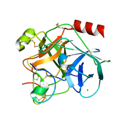 | | COMPLEX OF EETI-II MUTANT WITH PORCINE TRYPSIN | | 分子名称: | CALCIUM ION, TRYPSIN, TRYPSIN INHIBITOR II | | 著者 | Kraetzner, R, Wentzel, A, Kolmar, H, Uson, I. | | 登録日 | 2001-03-12 | | 公開日 | 2004-07-26 | | 最終更新日 | 2023-12-13 | | 実験手法 | X-RAY DIFFRACTION (1.9 Å) | | 主引用文献 | Structure of Ecballium Elaterium Trypsin Inhibitor II (Eeti-II): A Rigid Molecular Scaffold
Acta Crystallogr.,Sect.D, 61, 2005
|
|
1W7Z
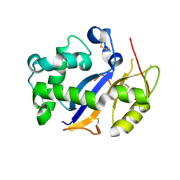
 | | Crystal structure of the free (uncomplexed) Ecballium elaterium trypsin inhibitor (EETI-II) | | 分子名称: | FORMIC ACID, SODIUM ION, TRYPSIN INHIBITOR II | | 著者 | Kraetzner, R, Debreczeni, J.E, Pape, T, Kolmar, H, Schneider, T.R, Uson, I, Scheldrick, G.M. | | 登録日 | 2004-09-14 | | 公開日 | 2005-11-01 | | 最終更新日 | 2023-12-13 | | 実験手法 | X-RAY DIFFRACTION (1.67 Å) | | 主引用文献 | Structure of Ecballium Elaterium Trypsin Inhibitor II (Eeti-II): A Rigid Molecular Scaffold
Acta Crystallogr.,Sect.D, 61, 2005
|
|
3EE6
 | | Crystal Structure Analysis of Tripeptidyl peptidase -I | | 分子名称: | 2-acetamido-2-deoxy-beta-D-glucopyranose, CALCIUM ION, CHLORIDE ION, ... | | 著者 | Pal, A, Kraetzner, R, Grapp, M, Gruene, T, Schreiber, K, Granborg, M, Urlaub, H, Asif, A.R, Becker, S, Gartner, J, Sheldrick, G.M, Steinfeld, R. | | 登録日 | 2008-09-04 | | 公開日 | 2008-11-25 | | 最終更新日 | 2020-07-29 | | 実験手法 | X-RAY DIFFRACTION (2.35 Å) | | 主引用文献 | Structure of tripeptidyl-peptidase I provides insight into the molecular basis of late infantile neuronal ceroid lipofuscinosis
J.Biol.Chem., 284, 2009
|
|
3T0O
 | | Crystal Structure Analysis of Human RNase T2 | | 分子名称: | 2-acetamido-2-deoxy-beta-D-glucopyranose, Ribonuclease T2 | | 著者 | Thorn, A, Kraetzner, R, Steinfeld, R, Sheldrick, G. | | 登録日 | 2011-07-20 | | 公開日 | 2012-07-11 | | 最終更新日 | 2023-09-13 | | 実験手法 | X-RAY DIFFRACTION (1.59 Å) | | 主引用文献 | Structure and activity of the only human RNase T2.
Nucleic Acids Res., 40, 2012
|
|
4MIV
 | | Crystal Structure of Sulfamidase, Crystal Form L | | 分子名称: | 2-acetamido-2-deoxy-beta-D-glucopyranose, 2-acetamido-2-deoxy-beta-D-glucopyranose-(1-4)-2-acetamido-2-deoxy-beta-D-glucopyranose, CALCIUM ION, ... | | 著者 | Sidhu, N.S, Uson, I, Schreiber, K, Proepper, K, Becker, S, Sheldrick, G.M, Gaertner, J, Kraetzner, R, Steinfeld, R. | | 登録日 | 2013-09-02 | | 公開日 | 2014-05-14 | | 最終更新日 | 2021-06-02 | | 実験手法 | X-RAY DIFFRACTION (2.4 Å) | | 主引用文献 | Structure of sulfamidase provides insight into the molecular pathology of mucopolysaccharidosis IIIA.
Acta Crystallogr.,Sect.D, 70, 2014
|
|
4MHX
 | | Crystal Structure of Sulfamidase | | 分子名称: | 2-acetamido-2-deoxy-beta-D-glucopyranose, 2-acetamido-2-deoxy-beta-D-glucopyranose-(1-4)-2-acetamido-2-deoxy-beta-D-glucopyranose, CALCIUM ION, ... | | 著者 | Sidhu, N.S, Uson, I, Schreiber, K, Proepper, K, Becker, S, Gaertner, J, Kraetzner, R, Steinfeld, R, Sheldrick, G.M. | | 登録日 | 2013-08-30 | | 公開日 | 2014-05-14 | | 最終更新日 | 2021-06-02 | | 実験手法 | X-RAY DIFFRACTION (2 Å) | | 主引用文献 | Structure of sulfamidase provides insight into the molecular pathology of mucopolysaccharidosis IIIA.
Acta Crystallogr.,Sect.D, 70, 2014
|
|


