5JK4
 
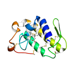
 | | Phosphate-Binding Protein from Stenotrophomonas maltophilia. | | 分子名称: | Alkaline phosphatase, PHOSPHATE ION | | 著者 | Keegan, R, Waterman, D, Hopper, D, Coates, L, Guo, J, Coker, A.R, Erskine, P.T, Wood, S.P, Cooper, J.B. | | 登録日 | 2016-04-25 | | 公開日 | 2016-05-04 | | 最終更新日 | 2024-01-10 | | 実験手法 | X-RAY DIFFRACTION (1.1 Å) | | 主引用文献 | The 1.1 angstrom resolution structure of a periplasmic phosphate-binding protein from Stenotrophomonas maltophilia: a crystallization contaminant identified by molecular replacement using the entire Protein Data Bank.
Acta Crystallogr D Struct Biol, 72, 2016
|
|
4P9G
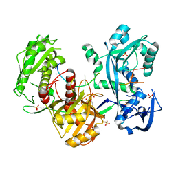
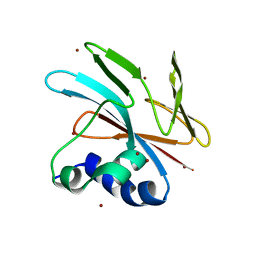
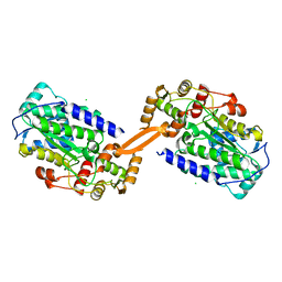
 | | Structure of the 2,4'-dihydroxyacetophenone dioxygenase from Alcaligenes sp. | | 分子名称: | 2,4'-dihydroxyacetophenone dioxygenase, CARBONATE ION, FE (III) ION, ... | | 著者 | Keegan, R, Lebedev, A, Erskine, P, Guo, J, Wood, S.P, Hopper, D.J, Cooper, J.B. | | 登録日 | 2014-04-03 | | 公開日 | 2014-09-10 | | 最終更新日 | 2023-12-20 | | 実験手法 | X-RAY DIFFRACTION (2.2 Å) | | 主引用文献 | Structure of the 2,4'-dihydroxyacetophenone dioxygenase from Alcaligenes sp. 4HAP
Acta Crystallogr.,Sect.D, 70, 2014
|
|
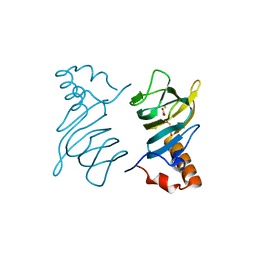
7M6C
 | | Crystal structure of PLA2 from snake venom of peruvian Bothrops atrox | | 分子名称: | Basic phospholipase A2, LAURIC ACID | | 著者 | Leonardo, D.A, Chojnowski, G, Simpkin, A, Seifert-Davila, W, Vivas-Ruiz, D.E, Keegan, R, Rigden, D. | | 登録日 | 2021-03-25 | | 公開日 | 2022-01-12 | | 最終更新日 | 2023-10-18 | | 実験手法 | X-RAY DIFFRACTION (1.95 Å) | | 主引用文献 | findMySequence: a neural-network-based approach for identification of unknown proteins in X-ray crystallography and cryo-EM
Iucrj, 9, 2022
|
|
5G5T
 | | Structure of the Argonaute protein from Methanocaldcoccus janaschii in complex with guide DNA | | 分子名称: | ARGONAUTE, GUIDE DNA, MAGNESIUM ION, ... | | 著者 | Schneider, S, Oellig, C.A, Keegan, R, Grohmann, D, Zander, A, Willkomm, S. | | 登録日 | 2016-06-03 | | 公開日 | 2017-02-08 | | 最終更新日 | 2024-01-10 | | 実験手法 | X-RAY DIFFRACTION (2.85 Å) | | 主引用文献 | Structural and mechanistic insights into an archaeal DNA-guided Argonaute protein.
Nat Microbiol, 2, 2017
|
|
5G5S
 | | Structure of the Argonaute protein from Methanocaldcoccus janaschii | | 分子名称: | 2-AMINO-2-HYDROXYMETHYL-PROPANE-1,3-DIOL, ARGONAUTE, MAGNESIUM ION | | 著者 | Schneider, S, Oellig, C.A, Keegan, R, Grohmann, D, Zander, A, Willkomm, S. | | 登録日 | 2016-06-03 | | 公開日 | 2017-02-08 | | 最終更新日 | 2024-05-08 | | 実験手法 | X-RAY DIFFRACTION (2.29 Å) | | 主引用文献 | Structural and mechanistic insights into an archaeal DNA-guided Argonaute protein.
Nat Microbiol, 2, 2017
|
|
5FD5
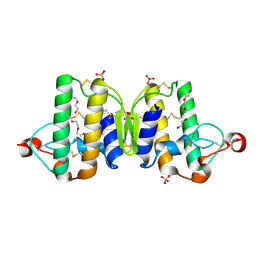
 | | manganese uptake regulator | | 分子名称: | 1,2-ETHANEDIOL, Ferric uptake regulation protein, SULFATE ION | | 著者 | Bellini, D, Lebedev, A, Keegan, R, Walsh, M.A. | | 登録日 | 2015-12-15 | | 公開日 | 2016-12-28 | | 最終更新日 | 2024-05-08 | | 実験手法 | X-RAY DIFFRACTION (1.91 Å) | | 主引用文献 | Structure of an apo metal-free manganese uptake regulator, mur
To Be Published
|
|
9FGP
 | |
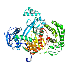
5OT1
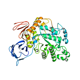 | | The type III pullulan hydrolase from Thermococcus kodakarensis | | 分子名称: | CALCIUM ION, Pullulanase type II, GH13 family | | 著者 | Guo, J, Coker, A.R, Wood, S.P, Cooper, J.B, Keegan, R, Ahmad, N, Muhammad, M.A, Rashid, N, Akhtar, M. | | 登録日 | 2017-08-18 | | 公開日 | 2018-04-18 | | 最終更新日 | 2024-01-17 | | 実験手法 | X-RAY DIFFRACTION (2.8 Å) | | 主引用文献 | Structure and function of the type III pullulan hydrolase from Thermococcus kodakarensis.
Acta Crystallogr D Struct Biol, 74, 2018
|
|
4N5B
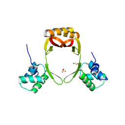
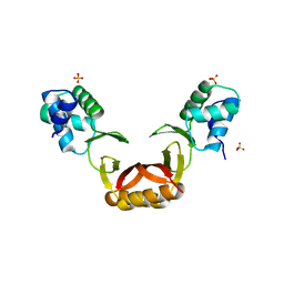
 | | Crystal structure of the Nipah virus phosphoprotein tetramerization domain | | 分子名称: | IMIDAZOLE, Phosphoprotein | | 著者 | Bruhn, J.F, Barnett, K, Bibby, J, Thomas, J, Keegan, R, Rigden, D, Bornholdt, Z.A, Saphire, E.O. | | 登録日 | 2013-10-09 | | 公開日 | 2013-11-27 | | 最終更新日 | 2023-09-20 | | 実験手法 | X-RAY DIFFRACTION (2.2 Å) | | 主引用文献 | Crystal structure of the nipah virus phosphoprotein tetramerization domain.
J.Virol., 88, 2014
|
|
8AEP
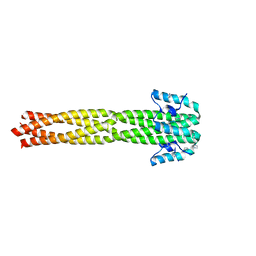
 | | Reductase domain of the carboxylate reductase of Neurospora crassa | | 分子名称: | Acetyl-CoA synthetase-like protein, CHLORIDE ION, SULFATE ION | | 著者 | Daniel, B, Schrufer, A, Marlene, L, Sagmeister, T, Pavkov-Keller, T. | | 登録日 | 2022-07-13 | | 公開日 | 2023-03-08 | | 最終更新日 | 2024-02-07 | | 実験手法 | X-RAY DIFFRACTION (2.3 Å) | | 主引用文献 | Structure of the Reductase Domain of a Fungal Carboxylic Acid Reductase and Its Substrate Scope in Thioester and Aldehyde Reduction.
Acs Catalysis, 12, 2022
|
|
3V9M
 | |
5FD6
 | |


