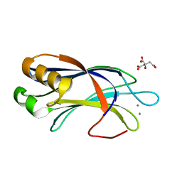
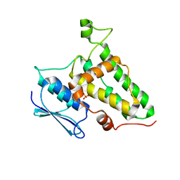
3US9
 
 | | Crystal Structure of the NCX1 Intracellular Tandem Calcium Binding Domains(CBD12) | | 分子名称: | CALCIUM ION, Sodium/calcium exchanger 1 | | 著者 | Giladi, M, Sasson, Y, Hirsch, J.A, Khananshvili, D. | | 登録日 | 2011-11-23 | | 公開日 | 2012-07-11 | | 最終更新日 | 2023-11-08 | | 実験手法 | X-RAY DIFFRACTION (2.68 Å) | | 主引用文献 | A common Ca2+-driven interdomain module governs eukaryotic NCX regulation.
Plos One, 7, 2012
|
|
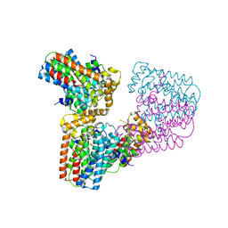
7PAY
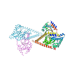 | | Structure of the human heterotetrameric cis-prenyltransferase complex in complex with magnesium and GGsPP | | 分子名称: | Dehydrodolichyl diphosphate synthase complex subunit DHDDS, Dehydrodolichyl diphosphate synthase complex subunit NUS1, MAGNESIUM ION, ... | | 著者 | Giladi, M, Lisnyansky Bar-El, M, Haitin, Y. | | 登録日 | 2021-07-30 | | 公開日 | 2022-06-01 | | 最終更新日 | 2024-01-31 | | 実験手法 | X-RAY DIFFRACTION (2.4 Å) | | 主引用文献 | Structural basis for long-chain isoprenoid synthesis by cis -prenyltransferases.
Sci Adv, 8, 2022
|
|
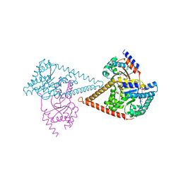
7PB1
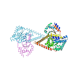 | | Structure of the human heterotetrameric cis-prenyltransferase complex in complex with magnesium, GGPP and IsPP | | 分子名称: | 3-methylbut-3-enylsulfanyl(phosphonooxy)phosphinic acid, Dehydrodolichyl diphosphate synthase complex subunit DHDDS, Dehydrodolichyl diphosphate synthase complex subunit NUS1, ... | | 著者 | Giladi, M, Lisnyansky Bar-El, M, Haitin, Y. | | 登録日 | 2021-07-30 | | 公開日 | 2022-06-01 | | 最終更新日 | 2024-01-31 | | 実験手法 | X-RAY DIFFRACTION (2.59 Å) | | 主引用文献 | Structural basis for long-chain isoprenoid synthesis by cis -prenyltransferases.
Sci Adv, 8, 2022
|
|
7PAX
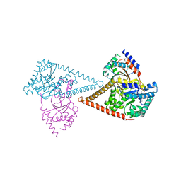 | | Structure of the human heterotetrameric cis-prenyltransferase complex in complex with magnesium, FsPP and IPP | | 分子名称: | 3-METHYLBUT-3-ENYL TRIHYDROGEN DIPHOSPHATE, Dehydrodolichyl diphosphate synthase complex subunit DHDDS, Dehydrodolichyl diphosphate synthase complex subunit NUS1, ... | | 著者 | Giladi, M, Lisnyansky Bar-El, M, Haitin, Y. | | 登録日 | 2021-07-30 | | 公開日 | 2022-06-01 | | 最終更新日 | 2024-01-31 | | 実験手法 | X-RAY DIFFRACTION (2 Å) | | 主引用文献 | Structural basis for long-chain isoprenoid synthesis by cis -prenyltransferases.
Sci Adv, 8, 2022
|
|
7PB0
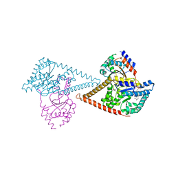 | | Structure of the human heterotetrameric cis-prenyltransferase complex in complex with magnesium, GGsPP and IsPP | | 分子名称: | 3-methylbut-3-enylsulfanyl(phosphonooxy)phosphinic acid, Dehydrodolichyl diphosphate synthase complex subunit DHDDS, Dehydrodolichyl diphosphate synthase complex subunit NUS1, ... | | 著者 | Giladi, M, Lisnyansky Bar-El, M, Haitin, Y. | | 登録日 | 2021-07-30 | | 公開日 | 2022-06-01 | | 最終更新日 | 2024-01-31 | | 実験手法 | X-RAY DIFFRACTION (2.301 Å) | | 主引用文献 | Structural basis for long-chain isoprenoid synthesis by cis -prenyltransferases.
Sci Adv, 8, 2022
|
|
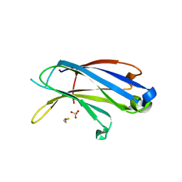
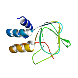
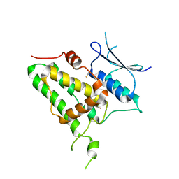
4LDC
 | | Crystal Structure of DOC2B C2B domain | | 分子名称: | CALCIUM ION, CITRATE ANION, Double C2-like domain-containing protein beta | | 著者 | Giladi, M, Almagor, L, Hirsch, J.A. | | 登録日 | 2013-06-24 | | 公開日 | 2013-09-11 | | 最終更新日 | 2024-02-28 | | 実験手法 | X-RAY DIFFRACTION (1.264 Å) | | 主引用文献 | The C2B Domain Is the Primary Ca(2+) Sensor in DOC2B: A Structural and Functional Analysis.
J.Mol.Biol., 425, 2013
|
|
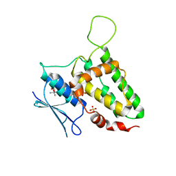
4LCV
 | | Crystal Structure of DOC2B C2A domain | | 分子名称: | BETA-MERCAPTOETHANOL, CALCIUM ION, CITRATE ANION, ... | | 著者 | Giladi, M, Almagor, L, Hirsch, J.A. | | 登録日 | 2013-06-23 | | 公開日 | 2013-09-11 | | 最終更新日 | 2013-11-13 | | 実験手法 | X-RAY DIFFRACTION (2 Å) | | 主引用文献 | The C2B Domain Is the Primary Ca(2+) Sensor in DOC2B: A Structural and Functional Analysis.
J.Mol.Biol., 425, 2013
|
|
6ERY
 | |
6ERZ
 | | The crystal structure of mouse chloride intracellular channel protein 6 | | 分子名称: | 2-AMINO-2-HYDROXYMETHYL-PROPANE-1,3-DIOL, Chloride intracellular channel protein 6, SULFATE ION | | 著者 | Ferofontov, A, Giladi, M, Haitin, Y. | | 登録日 | 2017-10-19 | | 公開日 | 2018-05-16 | | 最終更新日 | 2024-01-17 | | 実験手法 | X-RAY DIFFRACTION (1.923 Å) | | 主引用文献 | Inherent flexibility of CLIC6 revealed by crystallographic and solution studies.
Sci Rep, 8, 2018
|
|
6R4V
 | | Crystal structure of human geranylgeranyl diphosphate synthase bound to ibandronate | | 分子名称: | GLYCEROL, Geranylgeranyl pyrophosphate synthase, IBANDRONATE, ... | | 著者 | Lisnyansky, M, Giladi, M, Haitin, Y. | | 登録日 | 2019-03-24 | | 公開日 | 2019-09-18 | | 最終更新日 | 2024-01-24 | | 実験手法 | X-RAY DIFFRACTION (2.202 Å) | | 主引用文献 | Metal Coordination Is Crucial for Geranylgeranyl Diphosphate Synthase-Bisphosphonate Interactions: A Crystallographic and Computational Analysis.
Mol.Pharmacol., 96, 2019
|
|
8Q4I
 | |
8Q4J
 | |
6SYG
 | |
6G31
 | | Crystal structure of human geranylgeranyl diphosphate synthase mutant D188Y bound to zoledronate | | 分子名称: | Geranylgeranyl pyrophosphate synthase, MAGNESIUM ION, ZOLEDRONIC ACID | | 著者 | Lisnyansky, M, Kapelushnik, N, Ben-Bassat, A, Marom, M, Loewenstein, A, Khananshvili, D, Giladi, M, Haitin, Y. | | 登録日 | 2018-03-24 | | 公開日 | 2018-10-17 | | 最終更新日 | 2024-01-17 | | 実験手法 | X-RAY DIFFRACTION (3 Å) | | 主引用文献 | Reduced Activity of Geranylgeranyl Diphosphate Synthase Mutant Is Involved in Bisphosphonate-Induced Atypical Fractures.
Mol. Pharmacol., 94, 2018
|
|
6G32
 | | Crystal structure of human geranylgeranyl diphosphate synthase mutant D188Y | | 分子名称: | GLYCEROL, Geranylgeranyl pyrophosphate synthase | | 著者 | Lisnyansky, M, Kapelushnik, N, Ben-Bassat, A, Marom, M, Loewenstein, A, Khananshvili, D, Giladi, M, Haitin, Y. | | 登録日 | 2018-03-24 | | 公開日 | 2018-10-17 | | 最終更新日 | 2024-01-17 | | 実験手法 | X-RAY DIFFRACTION (3.281 Å) | | 主引用文献 | Reduced Activity of Geranylgeranyl Diphosphate Synthase Mutant Is Involved in Bisphosphonate-Induced Atypical Fractures.
Mol. Pharmacol., 94, 2018
|
|
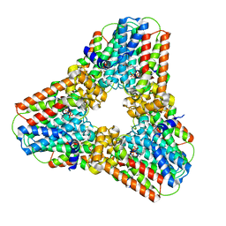
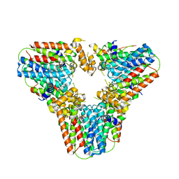
6Z1N
 | | Structure of the human heterotetrameric cis-prenyltransferase complex | | 分子名称: | Dehydrodolichyl diphosphate synthase complex subunit DHDDS, Dehydrodolichyl diphosphate synthase complex subunit NUS1, FARNESYL DIPHOSPHATE, ... | | 著者 | Lisnyansky Bar-El, M, Haitin, Y, Giladi, M. | | 登録日 | 2020-05-14 | | 公開日 | 2020-10-21 | | 最終更新日 | 2024-01-24 | | 実験手法 | X-RAY DIFFRACTION (2.3 Å) | | 主引用文献 | Structural basis of heterotetrameric assembly and disease mutations in the human cis-prenyltransferase complex.
Nat Commun, 11, 2020
|
|
6Y2H
 | |
5O44
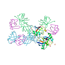 | | Crystal structure of unbranched mixed tri-Ubiquitin chain containing K48 and K63 linkages. | | 分子名称: | MAGNESIUM ION, Polyubiquitin-B, SULFATE ION, ... | | 著者 | Padala, P, Isupov, M.N, Wiener, R. | | 登録日 | 2017-05-26 | | 公開日 | 2017-11-08 | | 最終更新日 | 2024-01-17 | | 実験手法 | X-RAY DIFFRACTION (3.14 Å) | | 主引用文献 | The Crystal Structure and Conformations of an Unbranched Mixed Tri-Ubiquitin Chain Containing K48 and K63 Linkages.
J. Mol. Biol., 429, 2017
|
|


